Double balloon enteroscopy is a new method allowing the exploration of the whole intestine by the oral or anal route,1 with the possibility of endoscopic intervention. We describe here the first case of enduring paralytic ileus following this technique.
Case report
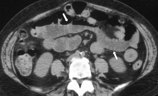
A 47 old woman was referred to our unit for chronic and obscure undiagnosed gastrointestinal bleeding. Unremarkable conventional upper and lower endoscopies were performed twice. Small bowel follow through studies, abdominal computed tomography (CT), and pushed enteroscopy were also normal. A capsule enteroscopy was performed showing three angiodysplasias in the distal jejunum, all measuring 2–3 mm. To reach them, we performed a double balloon enteroscopy which showed two of the three lesions. Electrocoagulation with an argon plasma coagulator (50 W, 1.5 l/min) was performed on both lesions. Twelve uneventful hours followed the procedure, after which nausea and vomiting occurred. Abdominal examination showed abdominal meteorism with diffuse moderate pain and no focal tenderness. No fever or other clinical signs apart from those of intestinal occlusion were observed. An abdominal CT showed localised and moderate dilation of a few intestinal loops with normal proximal and distal ileum (fig 1 ▶). No complications such as pneumoperitoneum, abscess, intestinal haematoma, or intussusceptions were observed. Oral intake was stopped and intravenous fluids given. The clinical state remained stable but transit remained totally interrupted without passage of stool or flatus, and 2–3 litres of gastric aspirate was obtained per day. As the intestinal occlusion had not resolved by day 7, erythromycin 3 mg/kg/day was given intravenously in two doses, of 30 minutes each, with disappearance of signs of occlusion within 48 hours. Total recovery was observed after four days, allowing discharge with no further events over the following five months.
Figure 1.
Abdominal computed tomography: dilated (white arrows) small bowel loops.
Discussion
Double balloon enteroscopy was first reported by Yamamoto et al in 2001 in a series of four patients, with insertion of the endoscope as far as 30–50 cm distal to the ligament of Treitz in three cases and to the ileocaecal valve in one, without any complications.2 Since then, two larger series of 1231 and 62 patients3 have been reported, with two and no complications recorded, respectively. In the former,1 a total of 178 procedures were performed. The first complication was multiple perforations in a patient with intestinal lymphoma thought to be due to chemotherapy whereas the second complication was of spontaneously resolving postoperative fever and abdominal pain in a patient with Crohn’s disease. Elsewhere, a total of eight papers4,5,6,7,8,9,10,11 reported 20 patients having DBE with only one recorded complication (post polypectomy sepsis).10 Two large series of 62 and 125 patients, respectively, have been published in abstract form,12,13 with no complications in the former and two in the latter. The two reported complications were intra-abdominal abscess and mild pancreatitis thought to be due to balloon inflation near the ampulla of Vater.
Two aspects are of interest in this case. Firstly, this is, to our knowledge, the first case of small bowel ileus following double balloon enteroscopy. This is of interest because the ileus appeared without the diagnosis of perforation, abscess, gastrointestinal haemorrhage, or haematoma following the procedure, indicating isolated motility impairment. Indeed, plasma argon coagulation is not known to induce an ileus lasting seven days without other complications.14 Therefore, it could be that the ileus was caused by the double balloon technique itself due to the pronounced stretching of the small bowel and possibly the mesentery.1 Secondly, it should be emphasised that conservative medical management should be tried first, and that treatment may include prokinetic agents such as erythromycin which improve motility impairment disorders.15
Conflict of interest: None declared.
References
- 1.Yamamoto H, Kita H, Sunada K, et al. Clinical outcomes of double-balloon endoscopy for the diagnosis and treatment of small-intestinal diseases. Clin Gastroenterol Hepatol 2004;2:1010–16. [DOI] [PubMed] [Google Scholar]
- 2.Yamamoto H, Sekine Y, Sato Y, et al. Total enteroscopy with a nonsurgical steerable double-balloon method. Gastrointest Endosc 2001;53:216–20. [DOI] [PubMed] [Google Scholar]
- 3.Sunada K, Yamamoto H, Kita H, et al. Clinical outcomes of enteroscopy using the double-balloon method for strictures of the small intestine. World J Gastroenterol 2005;11:1087–9. [DOI] [PMC free article] [PubMed] [Google Scholar]
- 4.May A, Nachbar L, Wardak A, et al. Double-balloon enteroscopy: preliminary experience in patients with obscure gastrointestinal bleeding or chronic abdominal pain. Endoscopy 2003;35:985–91. [DOI] [PubMed] [Google Scholar]
- 5.Miyata T, Yamamoto H, Kita H, et al. A case of inflammatory fibroid polyp causing small-bowel intussusception in which retrograde double-balloon enteroscopy was useful for the preoperative diagnosis. Endoscopy 2004;36:344–7. [DOI] [PubMed] [Google Scholar]
- 6.Shinozaki S, Yamamoto H, Kita H, et al. Direct observation with double-balloon enteroscopy of an intestinal intramural hematoma resulting in anticoagulant ileus. Dig Dis Sci 2004;49:902–5. [DOI] [PubMed] [Google Scholar]
- 7.Nishimura M, Yamamoto H, Kita H, et al. Gastrointestinal stromal tumor in the jejunum: diagnosis and control of bleeding with electrocoagulation by using double-balloon enteroscopy. J Gastroenterol 2004;39:1001–4. [DOI] [PubMed] [Google Scholar]
- 8.Yoshida N, Wakabayashi N, Nomura K, et al. Ileal mucosa-associated lymphoid tissue lymphoma showing several ulcer scars detected using double-balloon endoscopy. Endoscopy 2004;36:1022–4. [DOI] [PubMed] [Google Scholar]
- 9.Kuno A, Yamamoto H, Kita H, et al. Double-balloon enteroscopy through a Roux-en-Y anastomosis for EMR of an early carcinoma in the afferent duodenal limb. Gastrointest Endosc 2004;60:1032–4. [DOI] [PubMed] [Google Scholar]
- 10.Ohmiya N, Taguchi A, Shirai K, et al. Endoscopic resection of Peutz-Jeghers polyps throughout the small intestine at double-balloon enteroscopy without laparotomy. Gastrointest Endosc 2005;61:140–7. [DOI] [PubMed] [Google Scholar]
- 11.Hayashi Y, Yamamoto H, Kita H, et al. Endoscopic resection of elevated lesions of the small bowel by using double-balloon endoscopy. Gastrointest Endosc 2005;61:AB166. [Google Scholar]
- 12.Landi B, May A, Gasbarrini A, et al. Expérience européenne de l’entéroscopie double-ballon: faisabilité, indications et résultats. Gastroenterol Clin Biol 2005;29:A21. [Google Scholar]
- 13.Heine G, Hadithi M, Groenen MJ, et al. Double balloon enteroscopy: the Dutch experience. Indications, yields and complications in a series of 125 cases. Gastrointest Endosc 2005;61:AB166. [Google Scholar]
- 14.Ginsberg GG, Barkun AN, Bosco JJ, et al. The argon plasma coagulator. Gastrointest Endosc 2002;55:807–10. [DOI] [PubMed] [Google Scholar]
- 15.Emmanuel AV, Shand AG, Kamm MA. Erythromycin for the treatment of chronic intestinal pseudo-obstruction: description of six cases with a positive response. Aliment Pharmacol Ther 2004;19:687–94. [DOI] [PubMed] [Google Scholar]


