

Abstract
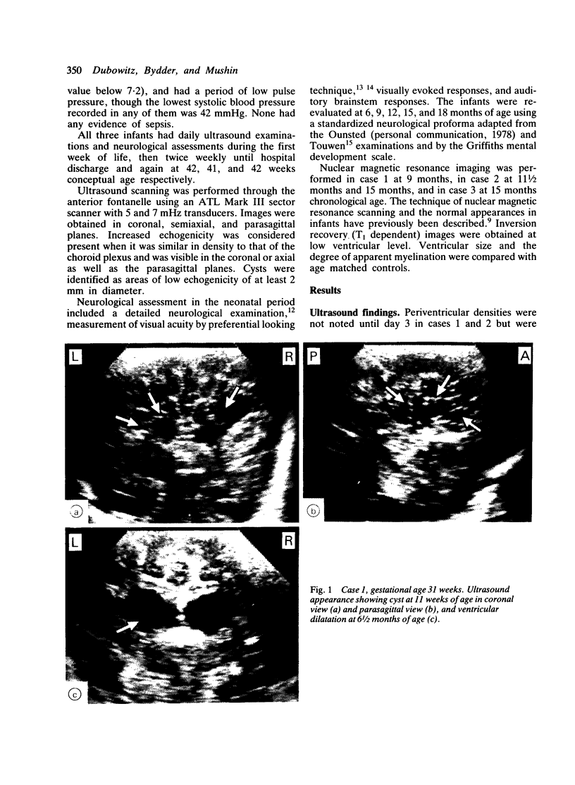
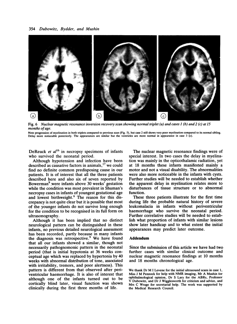
The evolution of severe periventricular leukomalacia was followed by ultrasonography in three newborn infants, and the subsequent myelination of the brain was assessed by nuclear magnetic resonance imaging. Four stages of periventricular leukomalacia could be identified by ultrasonography; (1) initial congestion, followed by (2) relative normalisation, (3) development of cysts, and (4) resolution of cysts but development of ventricular enlargement. All infants exhibited abnormal neurological signs from 36 weeks conceptual age and had unequivocal signs of cerebral palsy by 6 to 9 months of age. One infant became cortically blind but the other two seemed to have normal vision. Nuclear magnetic resonance imaging showed some abnormality of the ventricular system and delayed myelination in all three infants. The delay was most noticeable in the opticothalamic region, which was also the site of the most extensive lesions observed on ultrasonography. Progress in myelination was observed in the infants where a repeat scan was performed.
Full text
PDF


Images in this article


Selected References
These references are in PubMed. This may not be the complete list of references from this article.
- BANKER B. Q., LARROCHE J. C. Periventricular leukomalacia of infancy. A form of neonatal anoxic encephalopathy. Arch Neurol. 1962 Nov;7:386–410. doi: 10.1001/archneur.1962.04210050022004. [DOI] [PubMed] [Google Scholar]
- Bowerman R. A., Donn S. M., DiPietro M. A., D'Amato C. J., Hicks S. P. Periventricular leukomalacia in the pre-term newborn infant: sonographic and clinical features. Radiology. 1984 May;151(2):383–388. doi: 10.1148/radiology.151.2.6709907. [DOI] [PubMed] [Google Scholar]
- DeReuck J., Chattha A. S., Richardson E. P., Jr Pathogenesis and evolution of periventricular leukomalacia in infancy. Arch Neurol. 1972 Sep;27(3):229–236. doi: 10.1001/archneur.1972.00490150037007. [DOI] [PubMed] [Google Scholar]
- Fantz R. L., Fagan J. F., 3rd Visual attention to size and number of pattern details by term and preterm infants during the first six months. Child Dev. 1975 Mar;46(1):3–18. [PubMed] [Google Scholar]
- Hill A., Melson G. L., Clark H. B., Volpe J. J. Hemorrhagic periventricular leukomalacia: diagnosis by real time ultrasound and correlation with autopsy findings. Pediatrics. 1982 Mar;69(3):282–284. [PubMed] [Google Scholar]
- Levene M. I., Wigglesworth J. S., Dubowitz V. Hemorrhagic periventricular leukomalacia in the neonate: a real-time ultrasound study. Pediatrics. 1983 May;71(5):794–797. [PubMed] [Google Scholar]
- Morante A., Dubowitz L. M., Leven M., Dubowitz V. The development of visual function in normal and neurologically abnormal preterm and fullterm infants. Dev Med Child Neurol. 1982 Dec;24(6):771–784. doi: 10.1111/j.1469-8749.1982.tb13698.x. [DOI] [PubMed] [Google Scholar]
- Nwaesei C. G., Pape K. E., Martin D. J., Becker L. E., Fitz C. R. Periventricular infarction diagnosed by ultrasound: a postmortem correlation. J Pediatr. 1984 Jul;105(1):106–110. doi: 10.1016/s0022-3476(84)80372-8. [DOI] [PubMed] [Google Scholar]
- Shuman R. M., Selednik L. J. Periventricular leukomalacia. A one-year autopsy study. Arch Neurol. 1980 Apr;37(4):231–235. doi: 10.1001/archneur.1980.00500530069011. [DOI] [PubMed] [Google Scholar]
- Young R. S., Hernandez M. J., Yagel S. K. Selective reduction of blood flow to white matter during hypotension in newborn dogs: a possible mechanism of periventricular leukomalacia. Ann Neurol. 1982 Nov;12(5):445–448. doi: 10.1002/ana.410120506. [DOI] [PubMed] [Google Scholar]