Abstract
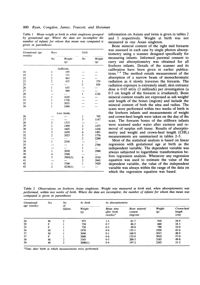
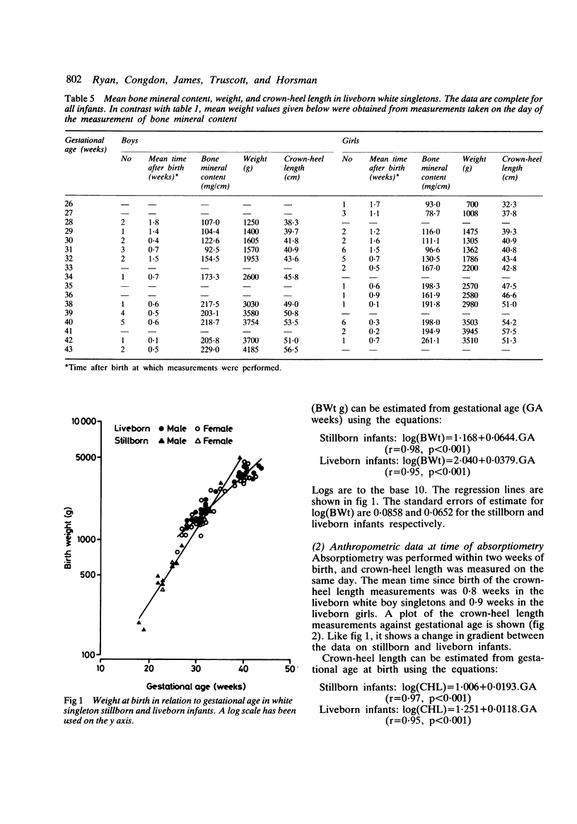
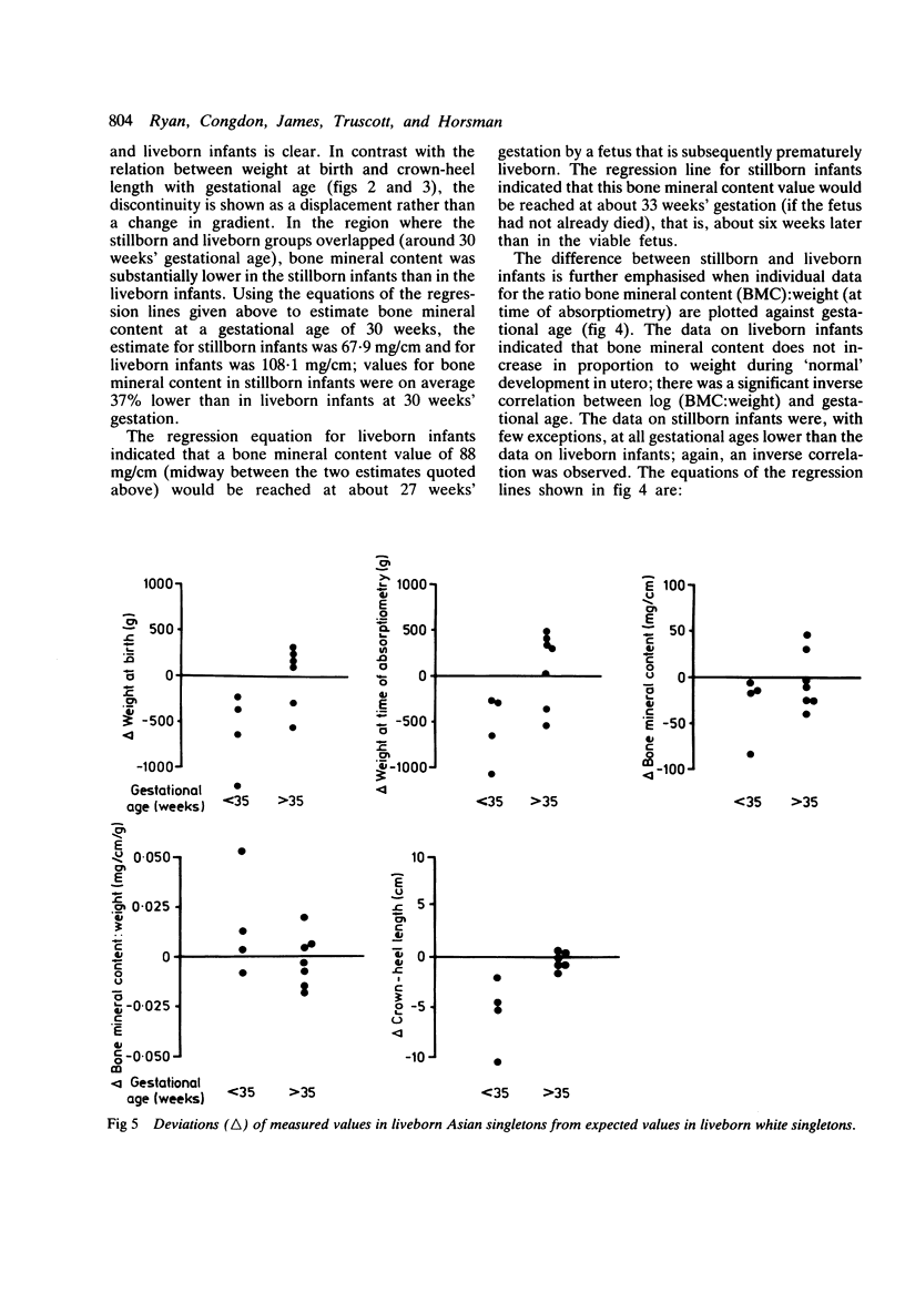
Bone mineral content of the mid forearm was measured by photon absorptiometry in 73 white singletons (36 boys, 37 girls) born between 18 and 43 weeks' gestation. Results obtained within two weeks of birth for liveborn infants were used to establish bone mineral deposition curves approximating normal in utero development. Results for stillborn infants indicated a retardation of bone mineralisation relative to liveborn infants. This has important implications concerning previous estimates of daily calcium needs for preterm infants. Eleven liveborn Asian singletons and nine pairs of white twins were examined similarly. With respect to bone mineral content, weight, and crown-heel length, Asians with gestational ages over 35 weeks and twins were closely comparable with their white singleton peers. Asians under 35 weeks tended to be smaller than white singletons of comparable age.
Full text
PDF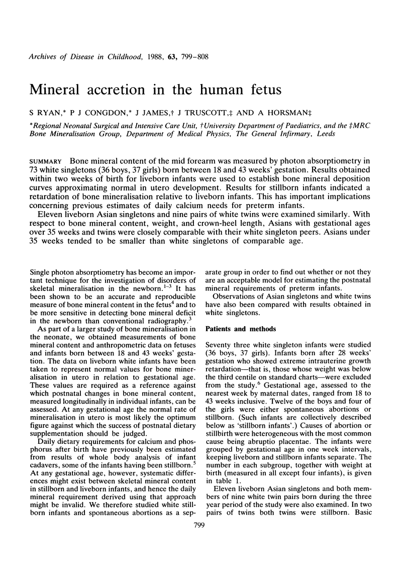





Selected References
These references are in PubMed. This may not be the complete list of references from this article.
- Congdon P., Horsman A., Kirby P. A., Dibble J., Bashir T. Mineral content of the forearms of babies born to Asian and white mothers. Br Med J (Clin Res Ed) 1983 Apr 16;286(6373):1233–1235. doi: 10.1136/bmj.286.6373.1233. [DOI] [PMC free article] [PubMed] [Google Scholar]
- Forbes G. B. Letter: Calcium accumulation by the human fetus. Pediatrics. 1976 Jun;57(6):976–977. [PubMed] [Google Scholar]
- Gairdner D., Pearson J. A growth chart for premature and other infants. Arch Dis Child. 1971 Dec;46(250):783–787. doi: 10.1136/adc.46.250.783. [DOI] [PMC free article] [PubMed] [Google Scholar]
- Greer F. R., Lane J., Weiner S., Mazess R. B. An accurate and reproducible absorptiometric technique for determining bone mineral content in newborn infants. Pediatr Res. 1983 Apr;17(4):259–262. doi: 10.1203/00006450-198304000-00005. [DOI] [PubMed] [Google Scholar]
- James J. R., Congdon P. J., Truscott J., Horsman A., Arthur R. Osteopenia of prematurity. Arch Dis Child. 1986 Sep;61(9):871–876. doi: 10.1136/adc.61.9.871. [DOI] [PMC free article] [PubMed] [Google Scholar]
- James J. R., Truscott J., Congdon P. J., Horsman A. Measurement of bone mineral content in the human fetus by photon absorptiometry. Early Hum Dev. 1986 Apr;13(2):169–181. doi: 10.1016/0378-3782(86)90005-8. [DOI] [PubMed] [Google Scholar]
- Minton S. D., Steichen J. J., Tsang R. C. Bone mineral content in term and preterm appropriate-for-gestational-age infants. J Pediatr. 1979 Dec;95(6):1037–1042. doi: 10.1016/s0022-3476(79)80305-4. [DOI] [PubMed] [Google Scholar]
- Shaw J. C. Evidence for defective skeletal mineralization in low-birthweight infants: the absorption of calcium and fat. Pediatrics. 1976 Jan;57(1):16–25. [PubMed] [Google Scholar]
- Steichen J. J., Gratton T. L., Tsang R. C. Osteopenia of prematurity: the cause and possible treatment. J Pediatr. 1980 Mar;96(3 Pt 2):528–534. doi: 10.1016/s0022-3476(80)80861-4. [DOI] [PubMed] [Google Scholar]
- Vyhmeister N. R., Linkhart T. A., Hay S., Baylink D. J., Ghosh B. Measurement of bone mineral content in the term and preterm infant. Am J Dis Child. 1987 May;141(5):506–510. doi: 10.1001/archpedi.1987.04460050048028. [DOI] [PubMed] [Google Scholar]
- WIDDOWSON E. M., SPRAY C. M. Chemical development in utero. Arch Dis Child. 1951 Jun;26(127):205–214. doi: 10.1136/adc.26.127.205. [DOI] [PMC free article] [PubMed] [Google Scholar]
- Ziegler E. E., O'Donnell A. M., Nelson S. E., Fomon S. J. Body composition of the reference fetus. Growth. 1976 Dec;40(4):329–341. [PubMed] [Google Scholar]


