

Abstract
The occurrence of penile schwannoma is very rare. A 41-year-old man presented with multiple penile tumors and pain on erection. The largest tumor causing pain was excised. Pathology was characteristic of benign schwannoma. We recommend that penile schwannomas be excised if the tumors cause pain or are malignant.
Introduction
Schwannoma, a type of neurogenic tumor, originates from Schwann cells and may occur in any region of the body; however, it is extremely rare on the penis. Only 21 cases have been reported in the literature. We present a case of multifocal penile schwannomas and report on the clinical features of this unusual condition.
Case Report
A 41-year-old man presented to our hospital in December 2005 with multifocal penile masses (Figure1). The tumors were located on the penile shaft and had been enlarging gradually for the past 5 years. He had had feelings of compression on erection for about 1 year. Both he and his wife had experienced pain during sexual intercourse, causing them to refrain from sex for the previous 6 months. He had moderate erectile dysfunction; the International Index of Erectile Function (IIEF) for the patient was 14. There was neither history of penile trauma nor any sexual transmitted disease in his medical record.
Figure 1.
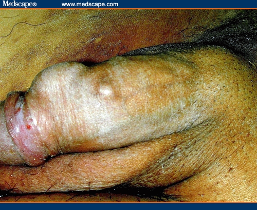
Multifocal penile tumors located at the dorsum of the penis.
On physical examination, we found 5 painless tumors over the dorsal penile shaft. They were elastic in consistency and had a smooth surface. The largest was approximately 2 cm in diameter and was located near the coronal sulcus at the dorsum of the penis (Figure 1). There was neither penile curvature on erection nor bilateral inguinal lymphadenopathy. Results of blood biochemistry studies and urinalysis were normal.
The largest tumor located near the coronal sulcus was excised in December 2005. We made a circular incision on the coronal sulcus and then peeled back the shaft skin; a well-capsulated tumor located in the Buck's fascia was noted. The tumor was divided carefully in order to preserve the nerve branches. The cut surface of the tumor was homogeneously gray. Hematoxylin-eosin staining of the tumor revealed benign morphology with areas of densely packed spindle cells, termed Antoni A tissue, intermixed with loose myxoid regions, termed Antoni B tissue (Figure 2).
Figure 2.

Microscopic pattern of schwannoma. (A) Palisading nuclear regions termed Verocay bodies (Antoni A area). (B) Looser, myxoid regions (Antoni B area; hematoxylin-eosin, 200X).
Postoperatively, there was no numbness or tenderness of the wound on the penile shaft; however, the patient stated at follow-up that he had lost all sexual desire (IIEF=0). We advised that he be evaluated for erectile dysfunction at our outpatient department, but he refused because he was not concerned.
Discussion
Most schwannomas are benign. Only 4 malignant penile schwannomas have been reported in the literature; 3 of them were associated with Recklinghausen's disease. No cases of benign penile schwannoma have been reported to be associated with hereditary diseases.[1]
If the schwannoma is at or near the glans penis, it may interfere with sexual intercourse. Surgical excision of the tumor is indicated if the tumor induces dyspareunia. There is no strong evidence about the relationship between schwannoma and erectile dysfunction.[2–4] Our patient's erectile dysfunction (preoperative IIEF = 14) worsened postoperatively (IIEF = 0); however, because there were no surgical complications, such as numbness, tenderness, or scarring on the penile shaft, it is difficult to say whether his penile schwannoma was a major or minor factor in his erectile dysfunction. Therefore, the underlying causes of erectile dysfunction in this patient should be evaluated further.
The differential diagnosis of superficial tumor in the penis should include lipoma, atheroma, fibroma, Peyronie's disease, fibrosis from autoinjection, and schwannoma.[5] In the literature, most penile schwannomas are located at the dorsum of the penis in the Buck's fascia, which is not characteristic of Peyronie's disease or injection-related fibrosis.[5,6] Lipoma and atheroma can be differentiated from penile schwannoma because the former are softer and more superficial. In addition to excision biopsy of the tumor, imaging studies, such as ultrasonography and magnetic resonance imaging, can be informative diagnostic modalities.
Conclusion
Schwannomas of the penis are extremely rare but should be included in the differential diagnosis for solid penile tumors. Because most penile schwannomas are benign, we recommend surgical excision only if the tumor causes pain or is growing rapidly. There is no strong evidence linking penile schwannomas with erectile dysfunction. Thus, more cases and studies are required to further clarify the clinical characteristics of these tumors.
Contributor Information
Wayne Young Liu, Department of Urology, China Medical University Hospital, Taichung, Taiwan.
Chang Chao-Hsiang, Department of Urology, China Medical University Hospital, Taichung, Taiwan.
Gaun-Chin Tseng, Department of Pathology, China Medical University Hospital, Taichung, Taiwan.
References
- 1.Sato D, Kase T, Tajima M, et al. Penile schwannoma. Int J Urol. 2001;8:87–89. doi: 10.1046/j.1442-2042.2001.00248.x. [DOI] [PubMed] [Google Scholar]
- 2.Algaba F, Chivite A, Rodriguez-Villalba R, et al. Schwannoma of the penis. J Androl. 2003;24:651–652. doi: 10.1002/j.1939-4640.2003.tb02722.x. [DOI] [PubMed] [Google Scholar]
- 3.Chan WP, Chiang SS, Huang AH, et al. Penile frenulum neurilemoma: a rare and unusual genitourinary tract tumor. J Urol. 1990;144:136–137. doi: 10.1016/s0022-5347(17)39394-1. [DOI] [PubMed] [Google Scholar]
- 4.Kandeel FR, Koussa KT, Swerdloff RS. Male sexual function and its disorders: physiology, pathophysiology, clinical investigation, and treatment. Endocr Rev. 2001;22:342–388. doi: 10.1210/edrv.22.3.0430. [DOI] [PubMed] [Google Scholar]
- 5.Dehner LP, Smith BH. Soft tissue tumor of the penis. A clinicopathologic study of 46 cases. Cancer. 1970;25:1431–1447. doi: 10.1002/1097-0142(197006)25:6<1431::aid-cncr2820250624>3.0.co;2-b. [DOI] [PubMed] [Google Scholar]
- 6.Mayersak JS, Viviano CJ, Barbiarz JW. Schwannoma of the penis. J Urol. 1995;15:1931–1932. [PubMed] [Google Scholar]
- 7.Jung DC, Hwang SI, Jung SI, et al. Neurilemoma of the glans penis: ultrasonography and magnetic resonance imaging findings. J Comput Assist Tomogr. 2006;30:68–69. doi: 10.1097/01.rct.0000193815.42051.c1. [DOI] [PubMed] [Google Scholar]