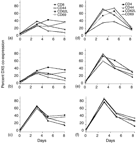
Figure 4.
Expression of the DX5 marker on naïve CD4 and CD8 splenocytes stimulated in vitro. Splenocytes were isolated from A/J mice, CD8 (a, b, c) and CD4 (d, e, f) populations isolated by immunomagentic bead selection, and then cultured with irradiated K562-based aAPC loaded with anti-CD3 (a, d), anti-CD3 and anti-CD28 (b, e) or irradiated K562/CD137L aAPC coated with anti-CD3 and anti-CD28 (c, f). Cells were stained for CD4 or CD8 (according to the population isolated, closed squares), CD44 (open triangles), CD62L (closed triangles), CD69 (open squares), and DX5 on days 3, 5 and 8. The percentage of cells coexpressing DX5 and CD4 (d, e, f) or DX5 and CD8 (a, b, c) and one of the other three markers as detected by three-colour flow cytometric analysis is plotted against days in culture.

