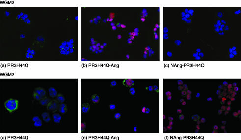
Figure 4.
Detection of PR3-toxin-induced apoptosis by TUNEL technology, staining apoptotic cells red. To preserve fluorescence, cytospin preparations were coverslipped with mounting medium containing DAPI. WGM2 cells treated with PR3H44Q (a), PR3H44Q-Ang (b) and NAng-PR3H44Q (c); WGM3 cells treated with PR3H44Q (d), PR3H44Q-Ang (e) and NAng-PR3H44Q (f).

