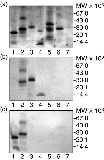
Figure 2.
Specificity of anti-human invariant chain 2C12 and 5F6 mAbs determined by Western blot analysis. Antigens were subjected to SDS–PAGE (a) and transferred onto PVDF membrane (b, c) for the reaction with 2C12 mAb(b) and 5F6 mAb(c). All samples but one (lane 3), were reduced with 0·01 m DTT and exposed to 100° in the presence of SDS for 5 min prior to SDS–PAGE. Samples: (lane 1) recombinant p31 Ii (lane 2) recombinant p41 Ii (lane 3) isolated non-covalent p41 fragment/cathepsin L complex (non-reduced) (lane 4) isolated non-covalent p41 fragment/cathepsin L complex (reduced) (lane 5) recombinant procathepsin S (lane 6) cathepsin H (lane 7) recombinant cathepsin L. Bands at higher molecular masses (lane 2) represent oligomers of recombinant p41 Ii. Cathepsin L from the complex was under reducing conditions (lane 4) separated to heavy chain (below 30 000 MW) and light chain (band below inhibitory p41 fragment, i.e. below 14 000 MW) compared to single-chain recombinant cathepsin L (lane 7). Pro-cathepsin S, its mature form and pro-region are also visible under reducing conditions (lane 5).

