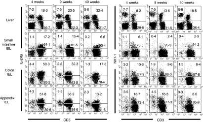
Figure 2.
Phenotypic characterization of lymphocytes in immunofluorescence tests. Two-colour staining for CD3 and IL-2Rβ and that for CD3 and NK1.1 were conducted in mice at the ages of 4, 9 and 40 weeks. Numbers in the figure represent the percentages of fluorescence-positive cells in corresponding areas. Data representative of three experiments are depicted.

