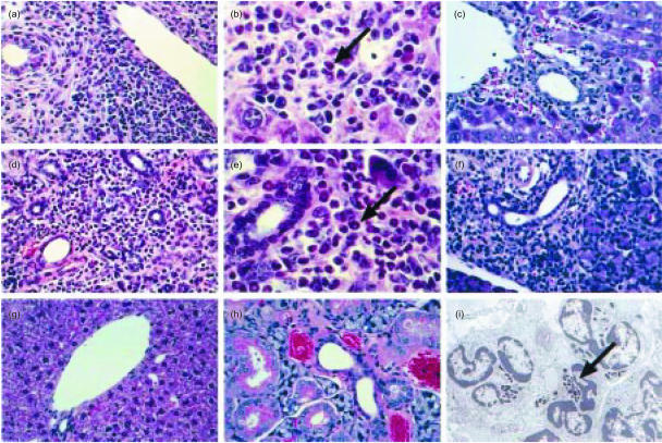
Figure 3.
Photomicrographs showing the histopathology of graft-versus-host disease (GvHD) in the salivary gland and liver. (a) Liver from an interferon-γ (IFN-γ) gene knockout (gko) graft recipient (magnification ×340). (b) Liver from an IFN-γ gko graft recipient (magnification ×680). (c) Liver from a wild-type graft recipient (magnification ×340). (d) Salivary gland from an IFN-γ gko graft recipient (magnification ×340). (e) Salivary gland from an IFN-γ gko graft recipient (magnification ×680). (f) Salivary gland from a wild-type graft recipient (magnification ×340). (g) Liver from a B6D2F1 control mouse (magnification ×340). (h) Salivary gland from a B6D2F1 control mouse (magnification ×680). (i) Electronmicrograph showing eosinophils in the salivary gland of an IFN-γ gko graft recipient (magnification ×6000). Note the characteristic crystalloid cores within the granules (arrow). Arrows indicate eosinophils in (b) and (e). Sections from IFN-γ gko graft recipients were collected on day 22 postinduction. Those from wild-type graft recipients were collected on day 8 postinduction. B6D2F1 = (C57BL/6 × DBA/2)F1 hybrid.

