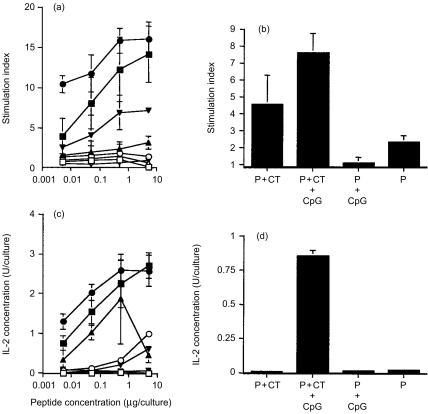
Figure 1.
Proliferation (top panel) and IL-2 secretion (bottom panel) of splenocytes of mice immunized onto bare skin with 100 µg HA:307-319 peptide plus 50 µg CT (▪), or 100 µg HA:307-319 peptide formulated with 50 µg CT and 100 µg CpG ODN 1826 (•), or 100 µg HA:307-319 peptide given with 100 µg CpG ODN 1826 (▴), or 100 µg HA:307-319 peptide given alone (▾). Splenocyte cultures were restimulated in vitro with various concentrations of HA:307-319 peptide (a, c) or with 3 × 10³ p.f.u. of heat-inactivated influenza virus (b, d), for four days. Data are presented as SI from hexaplicate cultures in the presence of peptide or from triplicate cultures in the presence of virus ±SD. Background proliferation values were 109, 251, and 126 c.p.m for the above groups, respectively. For IL-2 secretion, supernatants were collected after 72 h culture and tested for IL-2 secretion using the CTLL-2 IL-2-dependent cell line. Data are presented as IL-2 Units/culture from hexaplicate cultures in the presence of peptide or from triplicate cultures in the presence of virus ±SD. (□, ○, ▵, ▿) control NP:55-69 peptide.

