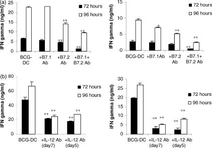
Figure 6.
IFN-γ secretion as determined by solid-phase ELISA in co-culture supernatants. (a) Autologous peripheral blood lymphocytes were co-cultured with BCG-exposed DC at a ratio of 5 : 1 for 72 and 96 hr in the presence or absence of blocking antibodies to B7.1, B7.2, or B7.1 and B7.2 at concentrations of 5 μg/ml and 10 μg/ml, respectively. Two representative donors of three are shown. (b) DC were matured with BCG in the presence or absence of an anti-IL-12 antibody (20 μg/ml) and then co-cultured with autologous T cells in the presence or absence of an anti-IL-12 antibody. IFN-γ was measured in supernatants at 72 and 96 hr from co-cultures of BCG-exposed DC and T cells, co-cultures of BCG-exposed DC, T cells and an anti-IL-12 antibody and co-cultures of BCG-exposed DC that had been treated with an anti-IL-12 antibody, T cells and an anti-IL-12 antibody. Two representative donors are shown. Any statistically significant decrease in IFN-γ by inclusion of blocking antibodies compared to levels in unblocked BCG-exposed DC: T-cell co-cultures is indicated by *P < 0·05, **P < 0·01, ***P < 0·0001 (Student's t-test).

