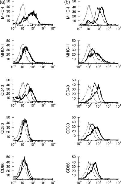
Figure 4.
Expression of surface molecules on MΦ and DC 24 h after exposure to S. typhimurium. MΦ (a) and DC (b) were stained with monoclonal antibodies to bovine MHC-I, MHC-II, CD40, CD80 and CD86 24 hr after exposure to a low dose of Salmonella (thick lines) or non-infected negative control (thin lines). These cells were also stained with isotype-matched control antibody, of which data only for non-infected cells were shown (dotted lines) since infected cells showed the same staining pattern with the isotype-matched control antibody. Data are representative of at least four independent experiments.

