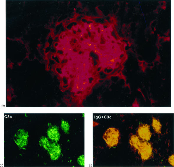
Figure 3.
Direct immunofluorescence of renal tissue from BCG-treated NOD mice at 250 days. (a) IgA deposits in a glomerulus showing mixed mesangial and glomerular capillary wall staining. (b) C3c is deposited both in the mesangium and along the capillary wall. (c) Colocalization of IgG (TRITC) and C3c (FITC) demonstrated by areas of yellow fluorescence.

