Abstract
BACKGROUND: Cardiovascular disease is the main cause of death in patients with type 1 diabetes. Since oxidized low density lipoprotein (LDL) is considered to be a critical factor in the atherosclerotic process, the aim of our study was to assess the influence of different parameters of glycemic control on susceptibility to oxidative stress from low density lipoprotein (LDL) in patients with type 1 diabetes without microvascular or macrovascular complications. METHODS: Forty patients and 33 non-diabetic individuals matched for gender, age and body mass index (BMI) were evaluated. The two groups underwent determination of lipid profile, fasting and postprandial glucose control and measurement of glycated hemoglobin (HbA1c). Spectrophotometric analysis of the LDL oxidation index was performed before and 1, 3, 6 and 24 h after the addition of copper sulfate to purified LDL fractions. RESULTS: The oxidation coefficient for LDL presented similar basal values in the two groups; however, at 3 h, LDL showed a higher degree of oxidation in patients with type 1 diabetes. Correlations with the metabolic control variables were significant only for postprandial glycemia. Stepwise multiple regression showed that post-prandial glycemia and sex were the significant independent variables. CONCLUSION: LDL from patients with type 1 diabetes showed high susceptibility to oxidative stress and this susceptibility was markedly related to the postprandial glucose levels. The influence of our findings on the development of chronic complications in patients with type 1 diabetes must be addressed in prospective studies.
Introduction
Diabetes mellitus is a common metabolic disease with a negative impact on the quality of life of patients as a result of its chronic complications. At present, this disease is the main cause of blindness and is also responsible for a considerable number of cases of terminal renal disease in the USA [1]. Type 1 diabetes is also associated with accelerated atherosclerotic complications. In a multicenter study, cardiovascular disease was ascertained to be the cause of death for 44% of the type 1 diabetic patients, followed by renal disease with a mortality of 21% [2]. A recent study showed a higher risk of mortality from ischemic heart disease and cerebrovascular disease in patients with type 1 diabetes than in the general population [3, 4]. Hyperglycemia is an important determinant factor for chronic microvascular complications, as shown in the Diabetes Complications Control Trial (DCCT) [5]. According to different studies, there was also an association between hyperglycemia and intima-media thickness in type 1 diabetic patients [6] as well as in type 2 diabetes [7]. It has been suggested that the thickness of the arterial intima-media complex is a sensitive marker of coronary and cerebrovascular disease and, as shown by Jarvisalo et al. [8], even children with type 1 diabetes present this modification in their artery walls.
The cited studies evaluated the links between hyperglycemia and chronic complications of diabetes using isolated or individual averages of multiple mea-surements of glycated hemoglobin. This could have masked the impact of acute glycemic excursions, mainly during the postprandial period, on the risk of diabetes complications [9]. Nowadays there is evidence that postprandial hyperglycemia may be an independent risk for cardiovascular disease in patients with type 2 diabetes [10-12]. However, the role of postprandial hyperglycemia in chronic complications of patients with type 1 diabetes has received less attention.
Hyperglycemia is associated with an increase in the generation of superoxide anion and nitric oxide [13]. The increase of these substances is thought to be deleterious since they can interact to produce peroxynitrite, which is a potent oxidant [14]. Hyperglycemia can also be related to the non-enzymatic glycation of proteins, such as low density lipoprotein (LDL). Even mildly glycated LDL affects the microvascular tone in skeletal muscle by decreasing the diameter of both small and terminal arterioles [15]. Interestingly, this was also shown to occur in the postprandial state [16]. Postprandial hyperglycemia is also related to other factors such as the increase of low-density lipoprotein oxidation [17, 18] and other changes that, taken together, predispose patients with type 2 diabetes to a greater cardiovascular risk than the general population.
Oxidized LDL has been implicated as a major factor in the atherosclerotic process in humans and in animal models because of its biological effects on endothelial cells, macrophages and smooth muscle cells [19]. Therefore, the present study was designed firstly to assess the susceptibility of LDL to in vitro oxidation in patients with type 1 diabetes without clinical signs of diabetes complications under routine clinical care and, secondly, to analyze the relationship between parameters of diabetes control and the susceptibility of LDL to in vitro oxidation.
Research design and methods
This is a cross-sectional study and was performed on a group of 40 type 1 diabetic outpatients regularly attendeding at the Diabetes Clinic of the State University of Rio de Janeiro and on 33 non-diabetic subjects (staff members, hospital employees and medical students with fasting blood glucose (FBG) < 100 mg/dl (i.e. 5.56 mmol/l) without first-degree relatives with diabetes mellitus, who were matched for age, gender and body mass index (BMI). The duration of diabetes was less than 5 years in 10 (25%) patients, 5 to 10 years in 20 (50%) and more than 10 years in 10 (25%). The inclusion criteria were patients with diabetes diagnosed before 30 years of age who had been using insulin since the diagnosis and had no symptoms of diabetes decompensation. The exclusion criteria were current smoking, alcoholism, systemic infection, thyroid, hepatic or cardiovascular diseases, diabetic nephropathy, diabetic retinopathy and intake of medicines capable or suspected of interfering with the oxidation process or with susceptibility to oxidation. The insulin dose was 0.79 ± 0.37 U/kg. All subjects underwent a 12-lead resting ECG which was classified according to the Minnesota coding [20], followed by definition of the presence or absence of coronary artery disease [21]. The experimental design was approved by the local ethics committee and all subjects received written instructions and gave informed consent to participate in the study.
Blood pressure was measured by the same observer three times after a 5-min rest in the supine position using a standard mercury sphygmomanometer. Diastolic blood pressure (dBP) was recorded when Korotkoff sounds disappeared (phase 5). The mean of the systolic (sBP) and diastolic blood pressure measurements was used. Weight and height were measured to the nearest 0.1 kg and 0.1 cm, respectively. Body mass index (BMI) (kg/m2) was calculated from these measurements. Blood samples were drawn in the morning between 7:30 and 8:30 am after an overnight fast. After centrifugation at 2500 g for 15 min at room temperature (19°C), aliquots of plasma and sera were stored at -70°C until analysis. Apolipoproteins A1 (apoA1) and B (apoB) were determined by immunoturbidimetry (Behring Turbidimeter, Marburg, Germany). The detection limits and intra-assay CV% were: ApoA1 30 mg/dl (4.2%) and ApoB 40 mg/dl (5.5%). HbA1c was determined by high performance liquid chromatography (L-9100 Merck Hitachi, Frankfurt, Germany) (reference range: 4.5 - 6.2%). Fasting blood glucose (FBG), triglycerides, HDL cholesterol and total cholesterol levels were measured by colorimetric reactions using an auto-analyzer (Cobas-Mira Roche). LDL cholesterol was calculated using the Friedewald equation [22]. After the samples were taken, patients had their usual breakfast, containing a mean of 400 kcal, and another sample was taken two hours later in order to measure postprandial glucose (PPG).
In accordance with the guidelines recommended by the European Diabetes Policy Group, the cutoff point for good metabolic control was set at 6.5% HbA1c [23]. Patients with FBG ≥ 110 mg/dl (i.e. 6.11 mmol/l) and PPG ≥ 135 mg/dl (i.e. 7.49 mmol/l) were considered to be at higher macrovascular risk [23]. Thus, an increase in glucose levels 2 h after ingestion of more than 25 mg/dl (i.e. 1.39 mmol/l) was not considered desirable.
All subjects were asked to provide three accurately timed overnight urine samples over three months. They passed urine immediately before 8:00 pm, discarded this sample and recorded the time. All urine passed until 6:00 am was collected into containers without a preservative to determine the albumin excretion rate (AER). The urine volume was recorded and aliquots were stored in glass tubes at -70°C until analysis. The urinary albumin concentration was estimated by double antibody radioimmunoassay (Diagnostic, Los Angeles, CA, sensitivity of 0.3 μg/ml) with an intra-assay and interassay coefficient of variation of 8.7% and 8.3%, respectively. Based on AER, only subjects with normoalbuminuria (AER < 20 μg/min in two out of three overnight urine specimens) were included. The diabetic type 1 patients underwent fundoscopy through a dilated pupil by ophthalmoscopy performed by the same ophthalmologist in order to evaluate diabetic retinopathy.
LDL oxidation was assessed indirectly as follows: LDL was first isolated from fasting plasma obtained by taking blood with EDTA and centrifuging it at 800 x g for 20 minutes at 4°C. Plasma density was then immediately adjusted to 1.3 g/ml by the addition of KBr and plasma was ultracentrifuged against a saline solution at 150,000 x g for 3 h at 4°C. Fractions with 1.019 to 1.063 g/ml density were then collected [24, 25]. Secondly, LDL samples were dialyzed against PBS 1 X overnight and oxidized by exposure to copper sulfate (CuSO4) during a 24-hour period. One μl of 20 mM CuSO4 for each 1 ml of LDL was added and the material was left in a 37°C bath for 24 h. Thirdly, oxidative modification of LDL was monitored indirectly by calculation of the oxidation coefficient (OxC), which uses spectrophotometric readings at 205, 232 and 280 nm at different time points such as 0 h, 1 h, 3 h, 6 h and 24 h after CuSO4 addition. The OxC was calculated using the formula: OxC = Abs 205 - Abs 280/Abs 232 - Abs 280. The reading at 205 nm reveals phospholipid polyunsaturated fatty acid double connections, at 232 nm the conjugated dienes, and at 280 nm the protein component – apo B [26, 27].
Statistical analysis
Results are expressed as mean ± SD. Variables without Gaussian distribution were log transformed before analysis. Differences in clinical characteristics and laboratory variables between diabetic and non-diabetic subjects were determined by the two-tailed Student t-test. Pearson correlation was used to assess the correlation between independent variables and the oxidation coefficient three hours after the addition of CuSO4 (OxC 3h). Because an interaction was observed between PPG and the postprandial glucose increment, we chose the former as an independent variable. Stepwise multiple regression was fitted to OxC 3h as the dependent variable and to variables with p < 0.1 in the Pearson correlation and to variables that possibly influence the oxidative process as independent variables. These analyses were performed using the SPSS (Statistical Package for the Social Science) for Windows version 10.0.
Results
Seven (17.5%) patients with type 1 diabetes had good diabetes control and 33 (82.5%) had moderate control. We observed that patients had higher apoA1 levels (110.74 ± 31.63 vs. 94.06 ± 24.47 mg/dl, p = 0.0188) and albumin excretion rate (9.51 ± 4.58 vs. 4.17 ± 3.13 μ/min, p = 0.0001) than non-diabetic subjects and lower uric acid levels (3.62 ± 1.03 vs. 4.75 ± 1.34 mg/dl, p = 0.0001). No difference was observed between patients and non-diabetic subjects with respect to the other variables analyzed. These data are shown in Table 1.
Table 1. Demographic and clinical characteristics and metabolic control variables.
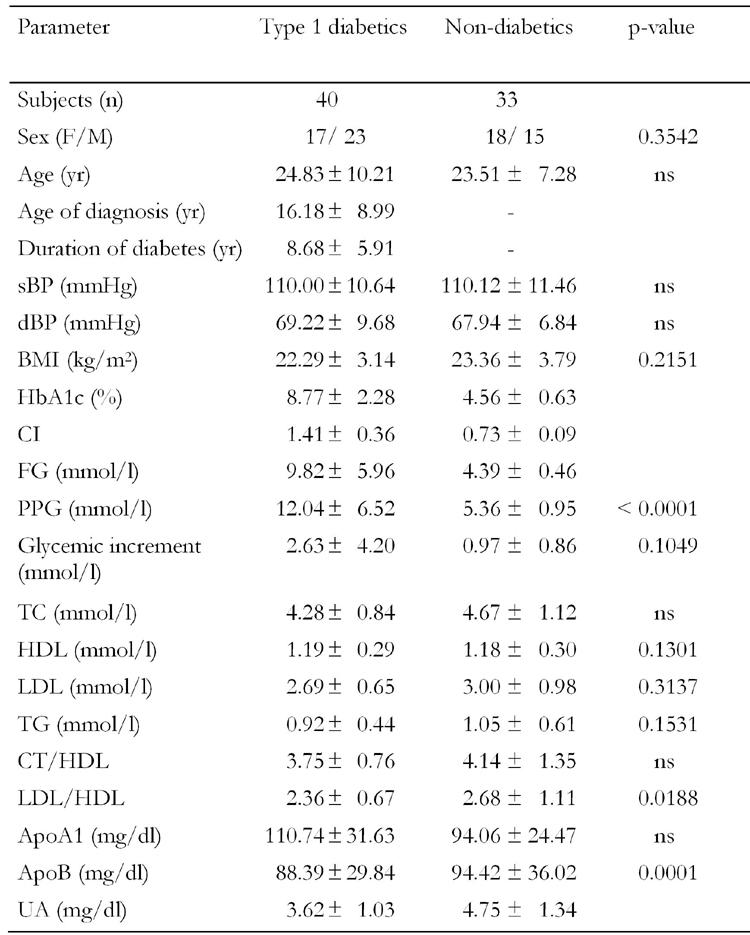
sBP: systolic blood pressure, dBP: diastolic blood pressure, BMI: body mass index, HbA1c: glycated hemoglobin, CI: glucose control index, FG: fasting glucose, PPG: postprandial glucose, TC: total cholesterol, HDL: high density lipoprotein, LDL: low den-sity lipoprotein, TG: triglycerides, ApoA1: apolipoprotein A1, ApoB: apolipoprotein B, UA: uric acid , AER: urinary albumin excretion rate, ns: not significant.
Basal OxC values were similar in the two groups. After the addition of CuSO4, patients with type 1 diabetes had earlier LDL oxidation than non-diabetic subjects (7.02 ± 1.35 vs. 7.90 ± 1.43, p = 0.01) (Figure 1). The difference between diabetic and non-diabetic subjects in the LDL cholesterol reaction was observed 3 h after addition of CuSO4.
Figure 1.
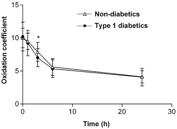
LDL oxidation coefficient (OxC) curve showing a significant difference between patients with type 1 diabe-tes (n = 40) and non-diabetic subjects (n = 33) 3 h after administration of CuSO4. * p = 0.01.
The increment of glycemia between the fasting and postprandial state was greater in patients than in non-diabetic subjects (47.34 ± 75.63 vs. 17.41 ± 15.44 mg/dl, p = 0.0000) (i.e. 2.63 ± 4.20 vs. 0.97 ± 0.86 mmol/l). By grouping all the individuals enrolled in the study (patients with type 1 diabetes and non-diabetic subjects) according to the glycemic increment (> or < 25 mg/dl (i.e. 1.39 mmol/l)), we observed that individuals with the smallest increment had greater OxC 3h (7.79 ± 1.60 vs. 6.95 ± 1.18, p = 0.0117) (Figure 2). The OxC 3h was also greater in the group classified as having a lower macrovascular risk (7.75 ± 1.43 vs. 6.88 ± 1.39, p = 0.0177) (Figure 3).
Figure 2.
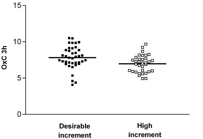
Distribution of OxC 3h according to the postprandial glucose increment in the total study population (n = 73). Desirable increment of glucose: ≤ 1.39 mmol/l (n = 39), high increment of glucose: < 1.39 mmol/l (n = 34). p = 0.0117.
Figure 3.
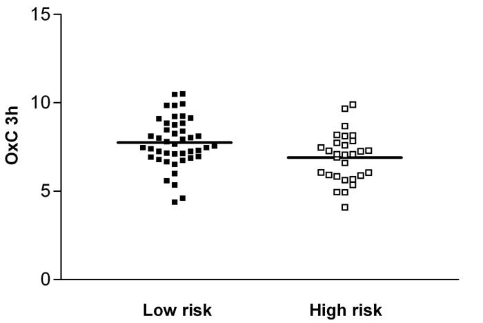
Distribution of OxC 3h according to the risk of arterial disease development in the total study population (n = 73). Low risk: PPG < 7.49 mmol/l (n = 44), high risk: PPG ≥ 7.49 mmol/l (n = 29). p = 0.0177.
In the pooled group (patients with type 1 diabetes and non-diabetic subjects), OxC 3h correlated with PPG (r = -0.3123, p = 0.0101) (Figure 4). Data are presented in Table 2. Stepwise multiple regression applied to the data with age, sex, BMI, FBG, HbA1c and PPG as independent variables and OxC 3h as the dependent variable showed that PPG (r = 0.3088, r2 = 0.0953 (95% CI (-0.0078 to -0.0009), B = -0.0044, p = 0.0138)) and female sex (r = 0.4014, r2 = 0.1611, B = -0.0048, p = 0.0051) were the significant independent variables. In the diabetic group no correlation was found between OxC 3h and any of the demographic, clinical and laboratory variables analyzed, including the insulin dose.
Figure 4.
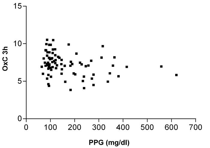
Correlation between LDL oxidation coefficient (OxC 3h) and postprandial hyperglycemia (PPG) 3 h after CuSO4 administration measured in the total study population (n = 73). p = 0.0101.
Table 2. Correlation between OxC 3h and metabolic parameters and apolipoproteins from the pooled group (n = 73).
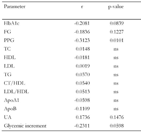
HbA1c: glycated hemoglobin, FG: fasting glucose, PPG: postprandial glucose, TC: total cholesterol, HDL: high density lipoprotein, LDL: low density lipoprotein, TG: triglycerides, ApoA1: apolipoprotein A1, ApoB: apolipoprotein B, UA: uric acid, AER: urinary albumin excretion rate, r: correlation coefficient, ns: not significant.
Discussion
In this cross-sectional study we evaluated the susceptibility of LDL to in vitro oxidation by CuSO4 in patients with type 1 diabetes and in non-diabetic subjects matched for gender, age and BMI. Since the reaction of LDL with CuSO4 is similar to the one determined by the presence of activated macrophages, it is possible that the former reproduces these modifications in vivo [27]. Assuming that this hypothesis is true, the greater susceptibility of LDL to oxidative stress may be one of the pathophysiological mechanisms involved in atherosclerotic disease in type 1 diabetes, helping to explain the higher prevalence of this illness in these patients than in the general population [28].
Oxidation of LDL is thought to play a major role in the initiation and development of atherosclerosis through the generation of inflammatory lipids and the covalent modification of this particle [29, 30]. Some of the pro-atherogenic effects of modified LDL are toxicity to endothelial cells, monocyte, neutrophil and eosinophil attraction, inhibition of macrophage mobility and promotion of foam cell formation [19, 31]. Although several animal studies have shown that antioxidants have been effective (reviewed in [19]), in humans, four clinical trials reported negative results in relation to vitamin E interference with atherogenesis, but they did not prove that the oxidative modification hypothesis is not valid [32-35]. However, a recent study showed different results and suggested that vitamin E protects against the risk of cardiovascular disease by reducing the susceptibility of LDL to oxidative modification in patients with hypertension [36].
There are controversial data regarding LDL oxidation in patients with type 1 diabetes [37-39]. These conflicting results may reflect the differences in the diabetic populations studied by each group in terms of glycemia levels, presence and severity of diabetes complications, vitamin content in the diet and, possibly, race [40]. Our data, which are based on the formation of conjugated dienes and oxidation of apoB, showed a greater susceptibility of LDL to in vitro oxidation in patients with type 1 diabetes without clinical evidence of diabetes complications. Since 82.5% of our patients did not present good metabolic control, it is possible that this fact interfered with the oxidation process. Liguori et al. [38] showed that the improvement of glycemic control led to less susceptibility of LDL to oxidation in patients with type 1 diabetes.
Our samples showed no difference in OxC before CuSO4 addition, meaning that in the basal state there was no difference in this atherogenic component. However, 3 h after exposure of LDL to oxidative stress we observed that LDL isolated from patients with type 1 diabetes became more oxidized than LDL obtained from matched non-diabetic subjects, even though this difference was small.
In our study, as in the literature [40], the greater LDL susceptibility to oxidation in patients with type 1 diabetes cannot be justified by the presence of dyslipidemia. This suggests that differences in lipid metabolism may not be the only and main factor determining this process. Thus, according to our data, hyperglycemia contributes critically to the higher susceptibility of LDL to in vitro oxidation.
As proposed by the European Diabetes Policy Group [23], diabetic patients with PPG ≥ 135 mg/dl (i.e. 7.49 mmol/l) are at higher risk of developing arterial disease. This cutoff was determined for patients with type 2 diabetes. Since there are no data available on the level of postprandial glucose that determines a higher risk of cardiovascular disease in patients with type 1 diabetes, we adopted the same cutoff. We observed that the OxC 3h was higher in the group with a low risk for cardiovascular disease and, as the patients with type 1 diabetes had higher PPG levels than non-diabetic subjects, these data suggest that the postprandial hyperglycemia is indeed related to the greater LDL susceptibility to oxidation. Since oxidized LDL is considered to be a relevant factor in the pathogenesis of atherosclerosis, the observation that the group at higher cardiovascular risk had lower OxC 3h corroborates our hypothesis.
In the pooled group studied as a whole, we found a negative correlation between OxC 3h and PPG, showing that the higher the levels of this variable, the lower the OxC 3h and therefore the greater the oxidation of LDL particles. However, in contrast to type 2 diabetes where postprandial hyperglycemia is recognized as an important risk factor for the development of cardiovascular disease [41, 42], to the best of our knowledge there are no data as yet that prove an association of postprandial hyperglycemia with risk factors for atherosclerotic disease in type 1 diabetes.
We may infer that the association of hyperglycemia and LDL oxidation may be related to the elevated glycation of LDL during the hyperglycemic state, which turns it into a form more susceptible to the oxidation process [43]. However, we cannot exclude a direct effect of hyperglycemia on the overproduction of reactive oxygen species. This effect can cause a greater consumption of the endogenous antioxidant substances, resulting in an imbalance between free radical production and antioxidant defense, and it may contribute to LDL oxidation. In contrast to the latter statement is the observation that normal or even higher levels of vitamin E, a potent endogenous antioxidant substance, were found in patients with type 1 diabetes [44]. In the present study, we showed diminished levels of uric acid, which is another potent endogenous antioxidant, in patients with type 1 diabetes compared to non-diabetic subjects. The same observation was made in the studies by Hoeldtke et al. [45] and Tsai et al. [37].
Interestingly, the stepwise multiple regression model showed that female sex and postprandial hyperglycemia were the only variables related to the greater susceptibility of LDL to in vitro oxidation in patients with type 1 diabetes. As shown by other authors, diabetes reduces gender protection against vascular disease in premenopausal women and also increases cardiovascular risk to a greater extent in women than in men [46-48]. Since the loss of relative protection in women with type 1 diabetes cannot be explained by traditional risk factors for coronary artery disease [49] or by differences in lipoprotein particle size [50], the increased oxidative stress and reduced antioxidant defense in these patients, as observed by Marra et al. [51], could be involved in the pathophysiology of atherosclerotic disease. A decrease in the endothelium-dependent vasodilatation induced by estrogen has been demonstrated [52]. The protective effect of estrogen on lipoprotein oxidizability [53] and on endothelium-dependent vasodilatation [54] might be impaired by the increased oxidative stress in diabetic women.
Some limitations of our study concerning the microvascular and macrovascular complications of diabetes should be discussed. Our measurement of retinopathy was less sensitive in detecting the initial stages of non-proliferative retinopathy than fundus photographs and/or fluorescein angiography. However, according to Willems et al. [55], there are no differences in titers of circulating autoantibodies against oxidized LDL between patients with type 1 diabetes with and without subclinical retinopathy. With regard to the macrovascular complications, we excluded patients with clinical cardiovascular disease using the Minnesota coding of 12-lead ECGs. Thus patients with preclinical atherosclerosis should have been included.
In conclusion, the present study showed that even LDL isolated from patients with type 1 diabetes without clinical microvascular complications or cardiovascular disease is more susceptible to in vitro oxidation than LDL from non-diabetic subjects. Since there are few studies using this approach in this group of patients, additional prospective studies are needed in order to establish the predictive factors for LDL susceptibility to oxidation during exposure to pro-oxidative agents and its influences on chronic complications of diabetes.
References
- 1.Ruderman NB, Williamson JR, Brownlee M. Glucose and diabetic vascular disease. FASEB. 1992;6:2905–2914. doi: 10.1096/fasebj.6.11.1644256. [DOI] [PubMed] [Google Scholar]
- 2.Morrish NJ, Wang SL, Stevens LK, Fuller JH, Keen H. Mortality and causes of death in the WHO multinational study of vascular disease in diabetes. Diabetologia. 2001;44(Suppl 2):S14–S21. doi: 10.1007/pl00002934. [DOI] [PubMed] [Google Scholar]
- 3.Laing SP, Swerdlow AJ, Slater SD, Burden AC, Morris A, Waugh NR, Gatling W, Bingley PJ, Patterson CC. Mortality from heart disease in a cohort of 23,000 patients with insulin-treated diabetes. Diabetologia. 2003;46:760–765. doi: 10.1007/s00125-003-1116-6. [DOI] [PubMed] [Google Scholar]
- 4.Laing SP, Swerdlow AJ, Carpenter LM, Slater SD, Burden AC, Botha JL, Morris AD, Waugh NR, Gatling W, Gale EA, Patterson CC, Qiao Z, Keen H. Mortality from cerebrovascular disease in a cohort of 23 000 patients with insulin-treated diabetes. Stroke. 2003;34:418–421. doi: 10.1161/01.str.0000053843.03997.35. [DOI] [PubMed] [Google Scholar]
- 5.The DCCT Research Group. The effect of intensive treatment of diabetes on the development and progression of long-term complications in insulin-dependent diabetes mellitus. N Engl J Med. 1993;329:977–986. doi: 10.1056/NEJM199309303291401. [DOI] [PubMed] [Google Scholar]
- 6.Nathan DM, Lachin J, Cleary P, Orchard T, Brillon DJ, Backlund JY, O'Leary DH, Genuth S. Intensive diabetes therapy and carotid intima-media thickness in type 1 diabetes mellitus. Diabetes Control and Complications Trial. Epidemiology of Diabetes Interventions and Complications Research Group. N Engl J Med. 2003;348:2294–2303. doi: 10.1056/NEJMoa022314. [DOI] [PMC free article] [PubMed] [Google Scholar]
- 7.Yamasaki Y, Kawamori R, Matsushima H, Nishizawa H, Kodama M, Kubota M, Kajimoto Y, Kamada T. Asymptomatic hyperglycaemia is associated with increased intimal plus medial thickness of the carotid artery. Diabetologia. 1995;38:585–591. doi: 10.1007/BF00400728. [DOI] [PubMed] [Google Scholar]
- 8.Jarvisalo MJ, Putto-Laurila A, Jartti L, Lehtimaki T, Solakivi T, Ronnemaa T, Raitakari OT. Carotid artery intima-media thickness in children with type 1 diabetes. Diabetes. 2002;51:493–498. doi: 10.2337/diabetes.51.2.493. [DOI] [PubMed] [Google Scholar]
- 9.Hirsch IB, Brownlee M. Should minimal blood glucose variability become the gold standard of glycemic control? J Diabetes Complications. 2005;19:178–181. doi: 10.1016/j.jdiacomp.2004.10.001. [DOI] [PubMed] [Google Scholar]
- 10.Laakso M. Hyperglycemia and cardiovascular disease in type 2 diabetes. Diabetes. 1999;44:937–942. doi: 10.2337/diabetes.48.5.937. [DOI] [PubMed] [Google Scholar]
- 11.Stratton IM, Adler AI, Neil HA, Matthews DR, Manley SE, Cull CA, Hadden D, Turner RC, Holman RR. Association of glycaemia with macrovascular and microvascular complications of type 2 diabetes (UKPDS 35) BMJ. 2000;321:405–412. doi: 10.1136/bmj.321.7258.405. [DOI] [PMC free article] [PubMed] [Google Scholar]
- 12.Ceriello A. The emerging role of post-prandial hyperglycaemic spikes in the pathogenesis of diabetic complications. Diabet Med. 1998;15:188–193. doi: 10.1002/(SICI)1096-9136(199803)15:3<188::AID-DIA545>3.0.CO;2-V. [DOI] [PubMed] [Google Scholar]
- 13.Cosentino F, Hishikawa K, Katusic ZS, Luscher TF. High glucose increases nitric oxide synthase expression and superoxide anion generation in human aortic endothelial cells. Circulation. 1997;96:25–28. doi: 10.1161/01.cir.96.1.25. [DOI] [PubMed] [Google Scholar]
- 14.Beckman JS, Koppenol WH. Nitric oxide, superoxide, and peroxynitrite: the good, the bad, and ugly. Am J Physiol. 1996;271:C1424–C1437. doi: 10.1152/ajpcell.1996.271.5.C1424. [DOI] [PubMed] [Google Scholar]
- 15.Nivoit P, Wiernsperger N, Moulin P, Lagarde M, Renaudin C. Effect of glycated LDL on microvascular tone in mice: a comparative study with LDL modified in vitro or isolated from diabetic patients. Diabetologia. 2003;46:1550–1558. doi: 10.1007/s00125-003-1225-2. [DOI] [PubMed] [Google Scholar]
- 16.Ceriello A, Quagliaro L, Catone B, Pascon R, Piazzola M, Bais B, Marra G, Tonutti L, Taboga C, Motz E. Role of hyperglycemia in nitrotyrosine postprandial generation. Diabetes Care. 2002;25:1439–1443. doi: 10.2337/diacare.25.8.1439. [DOI] [PubMed] [Google Scholar]
- 17.Diwadkar VA, Anderson JW, Bridges SR, Gowri MS, Oelgten PR. Postprandial low density lipoproteins in type 2 diabetes are oxidized more extensively than fasting diabetes and control samples. Proc Soc Exp Biol Med. 1999;222:178–184. doi: 10.1046/j.1525-1373.1999.d01-129.x. [DOI] [PubMed] [Google Scholar]
- 18.Ceriello A, Bortolotti N, Motz E, Pieri C, Marra M, Tonutti L, Lizzio S, Feletto F, Catone B, Taboga C. Meal induced oxidative stress and low-density lipoprotein (LDL) oxidation in diabetes: the possible role of hyperglycemia. Metabolism. 1999;48:1503–1508. doi: 10.1016/s0026-0495(99)90237-8. [DOI] [PubMed] [Google Scholar]
- 19.Chisolm GM, Steinberg D. The oxidative modification hypothesis of atherogenesis: an overview. Free Radical Biol Med. 2000;28:1815–1826. doi: 10.1016/s0891-5849(00)00344-0. [DOI] [PubMed] [Google Scholar]
- 20.Rose GA, Blackburn H, Gillum RF, Prineas RJ. World Health Organization; 1982. Cardiovascular survey methods. 2nd edition. Anex 1. [PubMed] [Google Scholar]
- 21.Howard BV, Lee ET, Cowan L, Fabsitz R, Oopik AJ, Robbins DC, Savage PJ, Yeh JL, Welty TK. Coronary heart disease prevalence and its relation to heart disease in American Indians: The Strong Heart Study. Am J Epidemiol. 1995;142:254–268. doi: 10.1093/oxfordjournals.aje.a117632. [DOI] [PubMed] [Google Scholar]
- 22.Friedwald WT, Levy R, Fredrickson DS. Estimations of serum low density lipoprotein cholesterol without use of preparative ultracentrifuge. Clin Chem. 1972;18:499–502. [PubMed] [Google Scholar]
- 23.European Diabetes Policy Group. A desktop guide to type 2 diabetes mellitus. Diabet Med. 1999;16:716–730. [PubMed] [Google Scholar]
- 24.Chung BH, Wilkinson T, Geer JC, Segrest JP. Preparative and quantitative isolation of plasma lipoproteins: rapid, single discontinuous density gradient ultracentrifugation in a vertical rotor. J Lipid Res. 1980;21:284–291. [PubMed] [Google Scholar]
- 25.Heery JM, Kozak M, Stafforini DM, Jones DA, Zimmerman GA, McIntyre TM. Oxidatively modified LDL contains phospholipids with platelet-activating factor-like activity and stimulates the growth of smooth muscle cells. J Clin Invest. 1995;96:2322–2330. doi: 10.1172/JCI118288. [DOI] [PMC free article] [PubMed] [Google Scholar]
- 26.Kim BS, LaBella FS. Comparison of analytical methods for monitoring autoxidation profiles of authentic lipids. J Lipid Res. 1987;28:1110–1117. [PubMed] [Google Scholar]
- 27.Esterbauer H, Gebicki J, Puhl H, Jürgens G. The role of lipid peroxidation and antioxidants in oxidative modification of LDL. Free Radical Biol Med. 1992;13:341–390. doi: 10.1016/0891-5849(92)90181-f. [DOI] [PubMed] [Google Scholar]
- 28.Krolewski AS, Warram JH, Valsania P, Martin BC, Laffel LMB, Christlieb AR. Evolving natural history of coronary artery disease in diabetes mellitus. Am J Med. 1991;90:S56–S61. doi: 10.1016/0002-9343(91)90040-5. [DOI] [PubMed] [Google Scholar]
- 29.Steinberg D. Low density lipoprotein oxidation and its pathobiological significance. J Biol Chem. 1997;272:20963–20966. doi: 10.1074/jbc.272.34.20963. [DOI] [PubMed] [Google Scholar]
- 30.Berliner JA, Heinecke JW. The role of oxidized lipoproteins in atherogenesis. Free Radical Biol Med. 1996;20:707–727. doi: 10.1016/0891-5849(95)02173-6. [DOI] [PubMed] [Google Scholar]
- 31.Marathe GK, Davies SS, Harrison KA, Silva AR, Murphy RC, Castro-Faria-Neto H, Prescott SM, Zimmerman GA, McIntyre TM. Inflammatory platelet-activating factor-like phospholipids in oxidized low density lipoproteins are fragmented alkyl phosphatidylcholines. J Biol Chem. 1999;274:28395–28404. doi: 10.1074/jbc.274.40.28395. [DOI] [PubMed] [Google Scholar]
- 32.Stephens NG, Parsons A, Schofield PM, Kelly F, Cheeseman K, Mitchinson MJ. Randomised controlled trial of vitamin E in patients with coronary disease: Cambridge Heart Antioxidant Study (CHAOS) Lancet. 1996;347:781–786. doi: 10.1016/s0140-6736(96)90866-1. [DOI] [PubMed] [Google Scholar]
- 33.Gruppo Italiano per lo Studio della Sopravvivenza nell’Infarto miocardico. Dietary supplementation with n-3 polyunsaturated fatty acids and vitamin E after myocardical infarction: results of the GISSI-Prevenzione trial. Lancet. 1999;354:447–455. [PubMed] [Google Scholar]
- 34.Leppala JM, Virtamo J, Fogelholm R, Huttunen JK, Albanes D, Taylor PR, Heinonen OP. Controlled trial of alpha-tocopherol and beta-carotene supplements on stroke incidence and mortality in male smokers. Arterioscler Thromb Vasc Biol. 2000;20:230–235. doi: 10.1161/01.atv.20.1.230. [DOI] [PubMed] [Google Scholar]
- 35.Yusuf S, Dagenais G, Pogue J, Bosch J, Sleight P. Vitamin E supplementation and cardiovascular events in high-risk patients. The Heart Outcomes Prevention Evaluation Study investigators. N Engl J Med. 2000;342:154–160. doi: 10.1056/NEJM200001203420302. [DOI] [PubMed] [Google Scholar]
- 36.Brockes C, Buchili C, Locher R, Koch J, Velter W. Vitamin E prevents extensive lipid peroxidation in patients with hypertension. Br J Biomed Sci. 2003;60:5–8. doi: 10.1080/09674845.2003.11783669. [DOI] [PubMed] [Google Scholar]
- 37.Tsai EC, Hirsch IB, Brunzell JD, Chait A. Reduced plasma peroxyl radical trapping capacity and increased susceptibility of LDL to oxidation in poorly controlled IDDM. Diabetes. 1994;43:1010–1014. doi: 10.2337/diab.43.8.1010. [DOI] [PubMed] [Google Scholar]
- 38.Liguori A, Abete P, Hayden JM, Cacciatore F, Rengo F, Ambrosio G, Bonaduce D, Condorelli M, Reaven PD, Napoli C. Effect of glycaemic control and age on low-density lipoprotein susceptibility to oxidation in diabetes mellitus type 1. Eur Heart J. 2001;22:2075–2084. doi: 10.1053/euhj.2001.2655. [DOI] [PubMed] [Google Scholar]
- 39.Jenkins AJ, Klein RL, Chassereau CN, Hermayer KL, Lopes-Virella MF. LDL from patients with well-controlled IDDM is not more susceptible to in vitro oxidation. Diabetes. 1996;45:762–767. doi: 10.2337/diab.45.6.762. [DOI] [PubMed] [Google Scholar]
- 40.Kuyvenhoven JP, Meinders AE. Oxidative stress and diabetes mellitus – Pathogenesis of long-term complications. Eur J Intern Med. 1999;10:9–19. [Google Scholar]
- 41.Hanefeld M, Fischer S, Julius U, Schulze J, Schwanebeck U, Schmechel H, Ziegelasch HJ, Lindner J. Risk factors for myocardial infarction and death in newly detected NIDDM: the Diabetes Intervention Study, 11-year follow-up. Diabetologia. 1996;39:1577–1583. doi: 10.1007/s001250050617. [DOI] [PubMed] [Google Scholar]
- 42.Fujishima M, Kiyohara Y, Kato I, Ohmura T, Iwamoto H, Nakayama K, Ohmori S, Yoshitake T. Diabetes and cardiovascular disease in a prospective population survey in Japan: The Hisayama Study. Diabetes. 1996;45:S14–S16. doi: 10.2337/diab.45.3.s14. [DOI] [PubMed] [Google Scholar]
- 43.Kobayashi K, Watanabe J, Umeda F, Nawata H. Glycation accelerates the oxidation of low density lipoprotein by copper ions. Endocrinol J. 1995;42:461–465. doi: 10.1507/endocrj.42.461. [DOI] [PubMed] [Google Scholar]
- 44.Nhadimana J, Dorchy H, Vertongen F. Activité anti-oxydante erythrocytaire et plasmatique dans le diabète de type I. Presse Méd. 1996;25:188–192. [PubMed] [Google Scholar]
- 45.Hoeldtke RD, Bryner KD, McNeill DR, Hobbs GR, Riggs JE, Warehime SS, Christie I, Ganser G, Van Dyke K. Nitrosative stress, uric acid and peripheral nerve function in early type 1 diabetes. Diabetes. 2002;51:2817–2825. doi: 10.2337/diabetes.51.9.2817. [DOI] [PubMed] [Google Scholar]
- 46.Heyden S, Heiss G, Bartel AG, Hames CG. Sex differences in coronary mortality among diabetics in Evans County, Georgia. J Chron Dis. 1980;33:265–273. doi: 10.1016/0021-9681(80)90021-1. [DOI] [PubMed] [Google Scholar]
- 47.Gu K, Cowie CC, Harris MI. Diabetes and decline in heart disease mortality in US adults. JAMA. 1999;281:1291–1297. doi: 10.1001/jama.281.14.1291. [DOI] [PubMed] [Google Scholar]
- 48.Dabelea D, Kinney G, Snell-Bergeon JK, Hokanson JE, Eckel RH, Ehrlich J, Garg S, Hamman RF, Rewers M; The Coronary Artery Calcification in Type 1 Diabetes Study. Effect of type 1 diabetes on the gender difference in coronary artery calcification: a role for insulin resistance? The Coronary Artery Calcification in Type 1 Diabetes (CACTI) Study. Diabetes. 2003;52:2833–2839. doi: 10.2337/diabetes.52.11.2833. [DOI] [PubMed] [Google Scholar]
- 49.Colhoum HM, Rubens MB, Underwood R, Fuller JH. The effect of type 1 diabetes mellitus on the gender difference in coronary artery calcification. J Am Coll Cardiol. 2000;36:2160–2167. doi: 10.1016/s0735-1097(00)00986-4. [DOI] [PubMed] [Google Scholar]
- 50.Colhoum HM, Otvos JD, Rubens MB, Taskinen MR, Underwood SR, Fuller JH. Lipoprotein subclasses and particle size and their relation with coronary artery calcification in men and women with and without type 1 diabetes. Diabetes. 2002;51:1949–1956. doi: 10.2337/diabetes.51.6.1949. [DOI] [PubMed] [Google Scholar]
- 51.Marra G, Cotroneo P, Pitocco D, Manto A, Di Leo MA, Ruotolo V, Caputo S, Giardina B, Ghirlanda G, Santini SA. Early increase of oxidative stress and reduced antioxidant defenses in patients with uncomplicated type 1 diabetes. Diabetes Care. 2002;25:370–375. doi: 10.2337/diacare.25.2.370. [DOI] [PubMed] [Google Scholar]
- 52.Steinberg HO, Paradisi G, Cronin J, Crowde K, Hempfling A, Hook G, Baron AD. Type II diabetes abrogates gender differences in endothelial function in premenopausal women. Circulation. 2000;101:2040–2046. doi: 10.1161/01.cir.101.17.2040. [DOI] [PubMed] [Google Scholar]
- 53.Grady D, Rubin SM, Petitti DB, Fox CS, Black D, Ettinger B, Ernster VL, Cummings SR. Hormone therapy to prevent disease and prolong life in postmenopausal women. Ann Intern Med. 1992;117:1016–1037. doi: 10.7326/0003-4819-117-12-1016. [DOI] [PubMed] [Google Scholar]
- 54.Collins P, Rosano GM, Sarrel PM, Ulrich L, Adamopoulos S, Beale CM, McNeill JG, Poole-Wilson PA. 17 beta-estradiol attenuates acetylcholine-induced coronary artery disease. Circulation. 1995;92:24–30. doi: 10.1161/01.cir.92.1.24. [DOI] [PubMed] [Google Scholar]
- 55.Willems D, Dorchy H, Dufrasne D. Serum antioxidant status and oxidized LDL in well-controlled young type 1 diabetic patients with and without subclinical complications. Atherosclerosis. 1998;137:S61–S64. doi: 10.1016/s0021-9150(97)00320-1. [DOI] [PubMed] [Google Scholar]


