Scientific documentation on the optimal injection procedure for knee joint injection is sparse.1 In the absence of a knee effusion, placing the needle precisely into the intraarticular space presents a challenge to clinicians.2 One study showed that about one third of knee injections were extraarticular; another showed that about 90% were extraarticular.1
Osteoarthritis is one of the most common and costly chronic medical conditions. Most current therapies are directed toward minimizing pain and swelling, maintaining joint mobility, and reducing associated disability.2 To achieve the maximum potential benefit, hyaluronan-based preparations should be delivered directly into the intraarticular space, not into the anterior fat pad or the subsynovial tissue layers.2
The technique I describe was discovered by doing 5 to 10 of these injections a month for 2 to 3 years, by getting feedback from patients and colleagues, and by drawing on my own personal clinical experience.
To verify that the procedure had never been done, I searched MEDLINE, EMBASE, and PubMed from 1966 to 2004 using the MeSH words “knee injection,” “aspiration,” “lateral approach,” “mid-patella,” “knee flexion,” and “triangle.” No studies on the technique were found. Combining “injections” and “mid-patella,” however, did reveal 1 article mentioning a lateral midpatella approach with the knee extended.2
Indications for knee aspiration include unexplained effusion, possible septic arthritis, and relief of discomfort caused by an effusion.3 Indications for injection include delivery of corticosteroids for advanced osteoarthritis and other noninfectious inflammatory arthritides, such as gout, calcium pyrophosphate deposition disease, or delivery of viscosupplementation. Viscosupplementation and corticosteroid therapies are not used concomitantly.3
Contraindications to injections are superimposed septic arthritis in rheumatoid arthritis patients, acutely inflamed joints, effusion not detected by clinical examination but detected by aspiration before injections,4 skin lesions, and risk of infection.
Materials
The size of syringe or needle depends on its application:
30-mL to 60-mL syringe for aspiration;
3-mL syringe for injection;
1.5-inch or 1-inch 25-gauge needle for injection; and
22-gauge needle for aspiration.
You will also require 2 mL of 2% lidocaine for local anesthetic, 40 mg/mL or 80 mg/mL of methylprednisolone, a marker, and alcohol swabs or iodine.
Technique
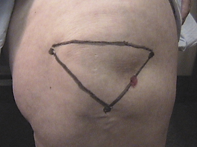
The patient should be sitting with the knee flexed at 90˚. Locate the apex of the patella by palpation. This is also the apex of the triangle. Draw a line from the apex to the lateral upper pole of the patella and another line from the apex to the medial upper pole of the patella. Join these lines, with the base of the triangle forming the upper border of the patella. This position with the knee flexed is used for injecting or aspirating the knee.
On the lateral side of the isosceles triangle find the midpoint. Mark the midpoint with ink. This is where the needle entry for injection will be (approximately midpatella).
Mix 1 mL of either 40 mg/mL or 80 mg/mL of methylprednisolone (depending on required dosage) with 2 mL of lidocaine. Draw up the 2 mL of lidocaine first and then the methylprednisolone as it mixes better that way.
Clean the area with alcohol or iodine. Insert the needle into the space between the patella and femur parallel to the middle facet of the patella using the ink spot as the point of entry. Angle the needle to the centre of the patella and inject the mixture into the space between the patella and the femur (Figure 1).
Figure 1.
Insert the needle into the space between the patella and femur parallel to the middle facet of the patella.
In obese patients landmarks are sometimes difficult to palpate. When this is the case, locate the apex of the patella by palpation. Draw a line from this apex to the outermost medial and lateral aspects of the knee. Join all these lines to form an upside down isosceles triangle and proceed.
For obese patients, use a 1.5-inch 25-gauge needle, and for thin patients, use a 1-inch 25-gauge needle. The distance measured from the edge of the skin to the articular surface of the femoral condyle can range from 4.5 to 5.5 cm.2 The additional 1 cm will help the needle to clear the intraarticular fat pad and reach the intraarticular space.2
The lidocaine-methylprednisolone mixture passes into the joint capsule and anesthetizes the articular surfaces of the knee joint; this effect gives patients immediate relief and they feel like moving the joint instantaneously. Most current methods of assessing the accuracy of needle placement, such as imaging or injecting air or contrast material into the knee joint, are too invasive for routine clinical application. My accuracy rate of 85 out of 95 placements was determined by questioning patients and assessing the following symptoms and signs: pain, range of movement, tenderness, willingness to repeat or repeating the procedure in 4 to 6 months, and improvement in social and occupational functioning. These parameters were assessed at the time of the procedure, 2 to 4 weeks later, and 4 to 6 months after that and noted in patients’ charts.
I still see some side effects of the procedure, such as infection, flushing, and hypotension, as described in the literature. Aspirating and injecting knee joints provides family physicians with a wealth of knowledge about knee pathology as well as about therapeutic measures to relieve pain and suffering. One of the main reasons these techniques are not widely used in family practice is that some physicians are anxious about injecting needles into joint spaces, and some patients think that because needles are involved, the procedure must be painful.
When inserting the needle, try not to poke around too much as it might be uncomfortable for patients. Entry should be deliberate and smooth at an angle of between 15˚ and 20˚. If the needle meets an obstruction, pull back slightly and aim anteriorly. I use only the lateral midpatellar knee-flexed position for knee injections. Some physicians infiltrate the skin with local anesthetic before injecting into the intraarticular space. I do not use local anesthetic in this way. The only cost to patients is the vial of methylprednisolone and pharmacy fees.
Other techniques
Many rheumatologists prefer the medial approach with knee extended and patient lying down because the lateral patellofemoral cleft is narrower, and the joint capsule is tougher laterally than medially. These conditions did not hinder my lateral approach. The anterior approach preferred by some physicians might involve greater risk of meniscal injury caused by the needle.3
Some physicians use anterolateral, anteromedial, and upper-lateral approaches. Some use a lateral midpatella approach with the knee extended. In this technique, the physician uses his or her free hand to manually evert and move the patella laterally so the needle can enter the knee space. I find this method cumbersome and time-consuming.
Conclusion
My experience with the triangle technique has been rewarding. My accuracy rate is about 90% and in keeping with an accuracy rate of 93% reported by some authors using other techniques.2
Biography
Dr Lockman practises family medicine at the St Vital Family Medical Clinic in Winnipeg, Man.
References
- 1.Weitoft T, Uddenfeldt P. Importance of synovial fluid aspiration when injecting intra-articular corticosteroids. Ann Rheum Dis. 2000;59:233–235. doi: 10.1136/ard.59.3.233. [DOI] [PMC free article] [PubMed] [Google Scholar]
- 2.Jackson DW, Evans NA, Thomas BM. Accuracy of needle placement into the intra-articular space of the knee. J Bone Joint Surg Am. 2002;84-A(9):1522–1527. doi: 10.2106/00004623-200209000-00003. [DOI] [PubMed] [Google Scholar]
- 3.Cardone DA, Tallia AF. Diagnostic and therapeutic injection of the hip and knee. Am Fam Physician. 2003;67(10):2147–2152. [PubMed] [Google Scholar]
- 4.Bliddal H. Placement of intra-articular injections verified by mini air-arthrography. Ann Rheum Dis. 1999;58:641–643. doi: 10.1136/ard.58.10.641. [DOI] [PMC free article] [PubMed] [Google Scholar]


