

Abstract
Vaginal smears from seven cats were examined at two-day intervals for 32 days in order to describe the cyclical pattern of epithelial cells exfoliated throughout the stages of the estrus cycle. Vaginal epithelial cells were classified as parabasal, intermediate and superficial (nucleate and anucleate) cells, and their dimensions were measured for the purpose of definition. The percentages of the epithelial cell populations (i.e. Maturation Index) from Wright's stained smears, were determined at all stages of the estrus cycle. The Eosinophilic Index was estimated on Papanicolaou stained smears.
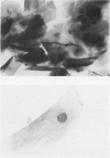
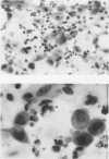
Smears of cats in estrus were populated almost entirely with nucleate and anucleate superficial epithelial cells. Proestrus was characterized by intermediate epithelial cells with increasing eosinophilia, and rare neutrophils. Metestrus was associated with desquamation of intermediate and parabasal epithelial cells, neutrophils and debris. In the anestrus period, groups of intermediate cells and some parabasal epithelial cells were exfoliated.
Two cats in the study did not cycle and exhibited anestrus. Of the five cats cycling, eight estrus periods were observed of two to five days duration. The cycles were of 15 to 17 days interval in three normal cats. Two cats did not show a second estrus within 30 days, and were subsequently found to have bacterial growth on the culture of vaginal swabs, however the presence of an initial ovular estrus cannot be ruled out. The rare presence of erythrocytes was associated with vaginal bacterial infections and discharge in two cats.
Full text
PDF


Images in this article


Selected References
These references are in PubMed. This may not be the complete list of references from this article.
- MICHAEL R. P., SCOTT P. P. THE ACTIVATION OF SEXUAL BEHAVIOUR IN CATS BY THE SUBCUTANEOUS ADMINISTRATION OF OESTROGEN. J Physiol. 1964 Jun;171:254–274. doi: 10.1113/jphysiol.1964.sp007376. [DOI] [PMC free article] [PubMed] [Google Scholar]
- Mowrer R. T., Conti P. A., Rossow C. F. Vaginal cytology an approach of improvement of cat breeding. Vet Med Small Anim Clin. 1975 Jun;70(6):691–696. [PubMed] [Google Scholar]
- Schutte A. P. Canine vaginal cytology. 3. Compilation and evaluation of cellular indices. J Small Anim Pract. 1967 Jun;8(6):313–317. doi: 10.1111/j.1748-5827.1967.tb04556.x. [DOI] [PubMed] [Google Scholar]
- Strasser H., Brunk R., Baeder C. Untersuchungen zum Sexualzyklus der Katze. Berl Munch Tierarztl Wochenschr. 1971 Jul 1;84(13):253–254. [PubMed] [Google Scholar]