Abstract
We show here that a cell-wall invertase encoded by the Incw1 gene is regulated at both the transcriptional and posttranscriptional levels by sugars in a heterotrophic cell suspension culture of maize. The Incw1 gene encoded two transcripts: Incw1-S (small) and Incw1-L (large); the size variation was attributable to different lengths in the 3′ untranslated region. Both metabolizable and nonmetabolizable sugars induced Incw1-L RNA apparently by default. However, only the metabolizable sugars, sucrose and d-glucose, were associated with the increased steady-state abundance of Incw1-S RNA, the concomitant increased levels of INCW1 protein and enzyme activity, and the downstream metabolic repression of the sucrose synthase gene, Sh1. Conversely, nonmetabolizable sugars, including the two glucose analogs 3-O-methylglucose and 2-deoxyglucose, induced greater steady-state levels of the Incw1-L RNA, but this increase did not lead to either an increase in the levels of the INCW1 protein/enzyme activity or the repression of the Sh1 gene. We conclude that sugar sensing and the induction of the Incw1 gene is independent of the hexokinase pathway. More importantly, our results also suggest that the 3′ untranslated region of the Incw1 gene acts as a regulatory sensor of carbon starvation and may constitute a link between sink metabolism and cellular translation in plants.
The enzyme invertase (EC 3.2.1.26), also known as β-fructofuranosidase, is known to catalyze sucrose (suc) cleavage to its monosaccharide constituents, glucose (glc) and fructose, in an irreversible manner. At least two forms of the enzyme, soluble and particulate, are features common to all invertases, and recent molecular analyses indicate that each belongs to two distinct class of small gene families (ref. 1 and references therein). The physiological role of the invertases is believed to be in suc partitioning between source and sink regions of the plant. In general, the vacuolar invertases are believed to be important in the regulation of hexose levels in certain specific tissues and in the use of stored suc in vacuoles. The cell-wall forms (extracellular or secreted) are associated with rapidly growing tissues and have been implicated in phloem unloading and source/sink regulation (2).
Although the experimental evidence is largely correlative in nature, a lot of insight regarding the roles of invertases is now emerging from studies of transgenic or mutant plants. Among several studies on transgenic plants, the most detailed analyses are reported in potato where yeast invertase has been expressed in both the cytosol and apoplast (3). Tissue-specific elevated expression of invertase in both cellular locations leads to substantial reductions in tuber suc content and a corresponding large increase in glc content. More importantly, the apoplastic and cytosolic increases in enzyme activities are associated with different phenotypic responses: an increased tuber size but reduced tuber number in the former and a reversed pattern in the latter. In contrast, when antisense technology was used to reduce the levels of the two invertases in transgenic carrot plants, there were significant alterations in early seedling morphology and much altered leaf-to-root ratios (4). In maize, the miniature1 (mn1) seed mutation is a seed-specific invertase-deficient trait caused by the loss of the Mn1 (Incw2)-encoded cell-wall invertase 2 protein INCW2 (1, 5). The mn1 seed shows a loss of ≈70–80% of mature seed weight because of early cessation of kernel growth. Overall, these data from diverse plant species show that invertases play a crucial role in the control of metabolic fluxes, downstream suc partitioning, and ultimately, plant development and crop productivity.
Despite their central role, very little is known on the mode of regulation of invertases in plants. Suc is now widely recognized as a signal molecule in gene expression in plants (see refs. 6 and 7 for recent reviews). Because invertase provides an entry point for suc into cellular metabolism, it is not surprising that it is among the group of sugar-modulated genes (2, 8). In a photoautotrophic cell culture of Chenopodium rubrum, cell-wall invertase enzyme activity is up-regulated by suc or glc (2, 9). Another mode of control is through an invertase inhibitor protein by protein–protein interactions (10, 11). In contrast to our lack of understanding on the regulatory controls of plant invertase genes, there is a wealth of knowledge on a single invertase gene, sucrase2 (suc2), in yeast. Interestingly, much of the knowledge about yeast is from general studies on how glc is used as a preferred source of carbon (catabolite or glc repression). These studies have discovered a plethora of regulatory genes that control the expression of the suc2 gene. Transcriptional repression of the suc2 gene is mediated by a zinc-finger protein, MIG1, that binds to the suc2 promoter and other glc-repressible genes when glc is available; and, under glc starvation, the protein moves to the cytoplasm causing derepression of suc2 gene. A protein kinase, Snf1, is also regulated in response to the glc signal and is required in differential phosphorylation and dephosphorylation of MIG1. A phosphorylated MIG1 protein remains in the nucleus, but a dephosphorylated form moves to the cytoplasm. There is, in fact, a multitude of coinducers and corepressors that form multisubunit complexes with Snf1 and/or MIG1 proteins leading to a large network of controls in energy metabolism. Indeed, glc metabolism in yeast cells determines the expression of ≈11% of its total genomic content (12, 13).
In plants, suc is the predominant sugar of transport between a source tissue (autotrophic) and heterotrophic sink organs, such as developing seeds and tubers. A cell-wall invertase in the apoplast is the first enzyme to commit suc carbon into downstream metabolism inside a cell. Here, we have used a heterotrophic cell suspension culture of maize (14), which is readily amenable to exogenous metabolic manipulations, to investigate the molecular nature and the controls in the regulation of a cell-wall invertase gene, Incw1. The usefulness of cell cultures in such studies is well documented (2, 9, 15, 16). Grotewald et al. (17) have in fact observed that molecular regulatory controls in maize are similar in cell culture and whole plants for the flavonoid pathway. We report that both metabolizable and nonmetabolizable sugars control the induction of the Incw1 gene. However, of the two transcripts encoded by the gene, only one accumulated specifically in response to metabolizable sugars. Induction of Incw1-L RNA by sugar-deficiency is not reflected in an increase in enzyme activity, whereas sugar sufficiency was associated with Incw1-S and a concomitant increase in the levels of both INCW protein and enzyme activity. Finally, based on specific differences in the 3′ untranslated region (UTR) of the two RNAs, we suggest that sugar sensing may be linked to the translational control of the Incw1 RNA.
MATERIALS AND METHODS
Maintenance of Maize-Suspension-Cultured Cells.
The Black Mexican Sweet suspension cell line was cultured as described (14). It was subcultured once a week by transferring ≈0.4 g fresh weight of cells in 5 ml of medium into 125-ml flasks with 50 ml of fresh medium. All sugar treatments were preceded by a 7-day preculture in a normal suc+ medium, thereafter pooled together, washed thrice with sugar-free medium, and dispensed in 5-ml aliquots (≈0.8 g fresh weight) to filter-sterilized fresh medium with or without sugar. Sugar concentration in the medium was 58.4 mM, except for 2-deoxyglucose (2-dGlc), which had a concentration of 29.2 mM. Cells were harvested at desired time points and stored at −80°C until used.
Enzyme Activity and Protein Assays.
Crude protein extracts were prepared from frozen-suspension-cultured cells in a 1:10 (wt/vol) ratio of extraction buffer by using a chilled mortar and pestle. The extraction buffer contained 50 mM Tris-maleate (pH 7.0) and 1 mM DTT (18). The homogenate was centrifuged at 14,000 × g for 10 min, and the supernatant was used in the assay of suc synthase (SS) proteins. The pellet was washed thrice, resuspended in extraction buffer containing 1 M NaCl in a 1:2 (wt/vol) ratio, shaken on a rotating shaker for 2–3 hr at 3°C, and centrifuged as described above. The dialyzed supernatant was assayed for cell-wall invertase enzyme activity as described (5). SDS immunoblot analyses for the SS and INCW proteins were performed as described (1, 19). Immunodetection of specific proteins was done via a chemiluminescence detection kit (Pierce). Primary antibodies were the polyclonal carrot cell-wall invertase (ref. 20; a kind gift from A. Sturm, Friedrich Miescher Institute, Basel) and the monoclonal antibodies against maize SSs, SS1 and SS2 (19).
RNA Blot Analysis.
Total RNA was isolated from frozen-suspension-cultured cells as described (21). RNA was glyoxylated and size-fractionated by 1.2% agarose gel electrophoresis, followed by transfer to a Nytran membrane (Schleicher & Schuell). Prehybridization and hybridization conditions have been described (1). Hybridization probes were cDNA inserts from clones corresponding to Incw1 and Incw2 (22, 23), Sh1, and Sus1 (24) genes. After hybridization, the blots were exposed to x-ray film at −80°C.
Screening and Isolation of Incw1 cDNA Clones.
Two cDNA libraries were made from poly(A)+ RNA isolated from either 48-hr-starved or from 5-day-old suc+ cell cultures by using the ZAP-cDNA synthesis kit (Stratagene) following the manufacturer’s instructions. Screening for the Incw1 clones was done by using the 5′ rapid amplification of Incw1 cDNA ends (5′ RACE) product as a probe (see below for the primer sequence). A total of eight nearly full-length clones, seven from suc-deficient and one from the suc+ library, were isolated and sequenced after in vivo excision. All sequence reactions were performed at the DNA Sequence Core Unit of the University of Florida. Sequences were analyzed with the program seqaid.
5′ and 3′ RACE Analyses and Cloning.
All RACE analyses were performed on RNAs from the cells cultured in suc+ or suc-deficient medium for 48 hr. The first strand of the 5′ RACE was synthesized from poly(A)+ RNA by using an Incw1-specific primer (5′-GCGGAGCAGCGGGTCG-3′), 546 bp from the 5′ end of the Incw1 RNA (GenBank accession no. AF050129), and a poly(dC) tail added according to the manufacturer’s instructions (GIBCO/BRL). The RACE product was amplified in a PCR by using a nested primer (5′-GGGTCGGACGCGTCCTTGGG-3′), 536 bp from the 5′ end, and an anchor primer to the poly(dC) tail. The first strand of the 3′ RACE was synthesized by using total RNA with the oligo(dT) primer, according to the manufacturer’s instructions (GIBCO/BRL). Two successive PCRs amplified the 3′ RACE products; the first amplification was based on the anchor primer to the poly(A)+ tail and Incw1-specific primer 1 (5′-AGCTTGATCGATCGGTCCGTGG-3′), 189 bp 5′ of the stop codon. The second amplification used the poly(A)+ anchor primer and Incw1-specific primer 2 (5′-GCTTCGGCGCGGGAGGCAAGAC-3′), 161 bp 5′ to the stop codon. The 3′ RACE products were excised from a gel and sequenced directly.
RESULTS
Sugar-Dependent Loss of Cell-Wall Invertase Protein and Enzyme Activity.
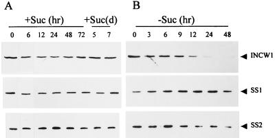
Table 1 shows cell-wall invertase enzyme activities in cells cultured in medium free of suc or glc for 12–72 hr. The most striking effect of sugar-free medium, after preculture in suc+ medium for a 7-day period, was the time-dependent loss of enzyme activity; the highest and the lowest levels were seen at 0 and 72 hr, respectively. Although a low level of the enzyme activity was still present at 72 hr (≈29.7% of the control), cells at this time point were associated with secondary effects, including reduction in the levels of soluble protein and cellular lysis (data not shown). Hence, nearly all gene expression analyses described here are limited to a 48-hr starvation that retained 44.2% of the control enzyme activity. Sugar-dependent loss of enzyme activity was reversible if these cells were transferred to the normal sugar+ medium. A transfer to suc+ or glc+ medium for 12 hr led to a 69% or 49% increase in enzyme activity, respectively, as compared with the levels seen after the 48-hr starvation (greater increases were seen with longer duration on suc+ medium; data not shown). However, such increases were specific to media with metabolizable sugars; sugar analogs that are not metabolized, 3-O-methylglucose (3-O-mGlc) and 2-dGlc, showed only reductions in activity compared with the activity at the 48-hr starvation time point. The time-dependent loss in cell-wall invertase activity was also seen in the levels of the INCW1 polypeptide in SDS Western blots (Fig. 1). Whereas the INCW1 levels remained relatively unchanged in a normal suc+ medium during a week-long culture, transfer to a suc-deficient medium led to its gradual reduction, especially in cells cultured for 24–48 hr (Fig. 1). The two SS isozymes, SS1 and SS2, are included here as an internal control and have shown no major detectable differences under these treatments (also see below).
Table 1.
Sugar-modulated control of cell-wall invertase in suspension cells
| Culture | Invertase specific activity (%) |
|---|---|
| Suc-deficient | |
| 0 hr | 0.770 ± 0.0054 (100) |
| 12 hr | 0.519 ± 0.042 (67.4) |
| 24 hr | 0.415 ± 0.058 (53.9) |
| 48 hr | 0.340 ± 0.035 (44.2) |
| 72 hr | 0.229 ± 0.011 (29.7) |
| Suc-deficient for 48 hr followed by 12 hr with sugar | |
| +suc | 0.576 ± 0.014 (74.8) |
| +D-Glc | 0.507 ± 0.036 (65.8) |
| +3-O-mGlc | 0.209 ± 0.041 (27.1) |
| +2-dGlc | 0.292 ± 0.022 (37.9) |
Values are means ± SD of three independent experiments (except for +2DG, for which the value represents the mean ± SD of two experiments); 0 hr represents a 7-day-old culture washed three times with suc-free medium. D-Glc, d-glucose.
Micromoles of reducing sugars per milligram of protein per minute.
Figure 1.
SDS Western blot showing INCW1, SS1, and SS2 proteins in crude extracts of cell cultures at various time points shown in hours (hr) or days (d) in suc+ (A) and suc-deficient media (B). Each lane contains 50, 10, and 5 μg of protein for INCW1, SS1, and SS2 proteins, respectively.
Control of Expression at the RNA Level Is Complex. RNA blots prepared from the above cell cultures were hybridized to a full-length cDNA clone of the Incw1 gene (Fig. 2). No hybridization was seen with the Incw2 probe (data not shown). The time course for steady-state levels of Incw1 RNAs in cells from suc+ medium indicated little or no change until 72 hr; thereafter, there was a gradual reduction (data not shown) until day 7, when the levels were extremely low (Fig. 2A). The lack of concordance between INCW protein (Fig. 1A) and the corresponding RNA (Fig. 2A) suggests that the protein may have a longer half-life than the RNA during the 7-day period of culture. After the 7-day culture in suc+ medium, cells were transferred to a fresh suc-deficient medium to test whether the time-dependent losses at the protein/enzyme level correlated with the RNA level of expression. In contrast with the protein data, steady-state levels of the Incw1 RNAs did not show major reductions in the 6- to 24-hr duration (Fig. 2B). In fact, there was an induction of two RNAs, Incw1-L (large) and Incw1-S (small), which were readily seen at the 6-hr time point. Thereafter, a gradual change occurred that led to an increased abundance of the Incw1-L relative to the Incw1-S by the 48-hr time point, and, by 72 hr, only the Incw1-L form was seen, albeit at greatly reduced levels. Interestingly, the same two RNAs were also detectable in the first 6 hr of culture in a fresh suc+ medium, subsequent to the 48-hr culture in suc-deficient medium (Fig. 2C). However, unlike the suc-deficient medium, the suc+ medium led to a gradual increase in the levels of Incw1-S at 12 hr, and by the 24-hr time point, Incw1-S became a major RNA in these cells (Fig. 2C). It is worth noting here that both forms of the Incw1 RNA were polyadenylated (see Fig. 5A).
Figure 2.
RNA blot showing Incw1, Sh1, and Sus1 transcripts in 20 μg of total RNA at various time points (hours). (A) Suc+ medium. (B) Fresh suc-deficient medium. (C) Fresh suc-deficient medium at 0 and 48 hr, thereafter in the suc+ medium as shown. (D) Mannitol (M) or suc (S) is added after a 48-hr preculture in suc-deficient medium for the duration as shown. For all treatments, 0 hr represents 7-day cultured cells that were washed three times in suc-deficient medium.
Figure 5.
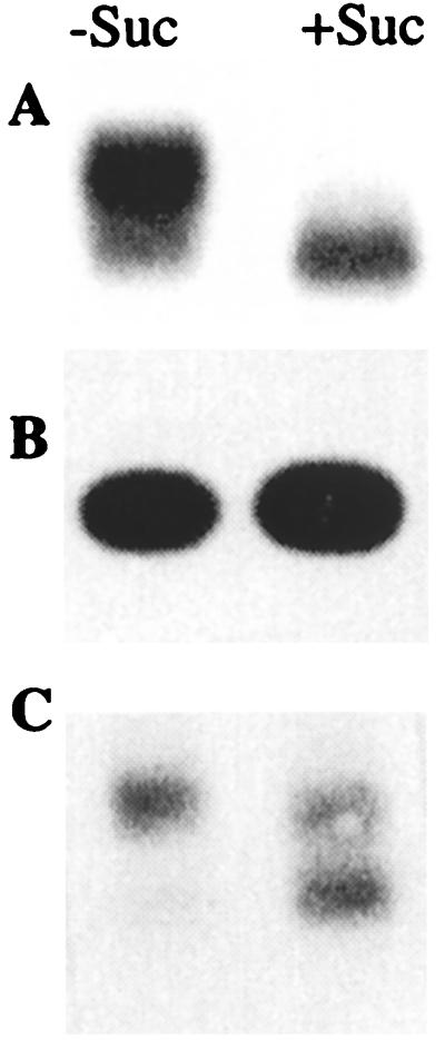
(A) Poly(A)+ RNA samples; (B) 5′ and (C) 3′ RACE products of RNAs from cells in suc+ (+S) or suc-deficient (−S) medium. An identically sized fragment is seen in A, and two fragments, “small” and “large,” are seen in B on hybridization with the Incw1 cDNA probe.
Because both Incw RNAs were rapidly induced by transfer to new medium with or without suc (Fig. 2 C and B, respectively), it was important to determine whether suc played any role in the induction process. Thus, a 10× solution of suc or mannitol (an osmoticum) was added directly to the suc-deficient medium with cells precultured for 48 hr to attain the original 2% level of sugar concentration. A time-course analysis of the RNA profile is shown in Fig. 2D. Suc, but not mannitol, led to a gradual induction of the two Incw1 RNAs during the 3- to 6-hr period, and only the Incw1-S RNA was seen by the 24-hr time point. The mannitol-supplemented medium, like the fresh suc-deficient medium (Fig. 2B), has shown only the Incw1-L RNA.
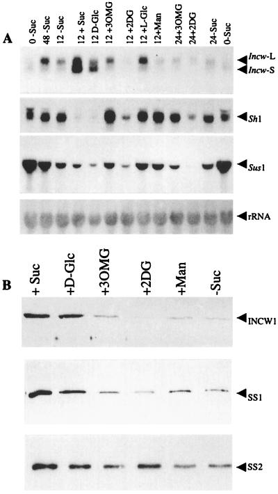
RNA blot analyses were also done on samples from cells cultured on nonmetabolizable sugars, including l-Glc and glc analogs, 3-O-mGlc and 2-dGlc (Fig. 3A). The control samples from suc+ or glc+ (d-Glc) medium showed both Incw1-L and Incw1-S transcripts at the 12-hr time point. However, media with 3-O-mGlc and l-Glc showed only the Incw1-L form, and greatly reduced levels of the same RNA were also seen in media with 2-dGlc and mannitol. The significance of the reduced levels of the Incw1-L message in the last two samples is not obvious, although physiological toxicity at the levels tested is possible. At 24 hr, media with nonmetabolizable sugars or no suc showed greatly reduced levels of the Incw1 RNAs. At the protein level, the INCW protein was detectably reduced in all samples except those with suc or glc (Fig. 3B). As with the enzyme activity (Table 1), media with 3-O-mGlc or 2-dGlc showed reduced levels of the INCW protein. Thus, the collective data suggest that, unlike the Incw1-S, samples with the Incw1-L RNA (Figs. 2 and 3) were not correlated with the INCW protein or the enzyme activity.
Figure 3.
(A) RNA blot showing Incw1, Sh1, and Sus1 transcripts in 20 μg of total RNA after incubation for 0 or 48 hr in suc-deficient medium (−Suc; first two lanes and last lane) or after incubation in suc-deficient medium for 48 hr followed by addition of water (−Suc), +Suc, +d-Glc, 3-O-mGlc (+3OMG), +l-Glc, or mannitol (+Man) for 12 or 24 hr. (B) SDS Western blot showing INCW1, SS1, and SS2 proteins in crude extracts of cells in the media as above. Each lane contains 50, 10, and 5 μg of protein for INCW1, SS1, and SS2 proteins, respectively.
Sugar-Modulated Control of SS Genes.
Although the predominant focus of this study is on the cell-wall invertase genes, products of the SS genes, Sh1 and Sus1, were included initially as an internal loading control for comparisons against the Incw1 expression. However, both SS genes were also responsive to certain metabolic manipulations, as has been described in maize protoplasts (25) and in excised root tips (26). Figs. 2 and 3 show hybridization patterns with the Sh1 and the Sus1 cDNAs to either the parallel Northern blots (i.e., the same RNA samples) or to the same membranes that were first hybridized with the Incw1 probe. During the 7-day growth in a suc+ medium, there was no change in the Sh1 transcripts and an increase in the levels of the Sus1 RNA in only the later phase of growth (Fig. 2A). In a suc-deficient medium (Fig. 2B), there was an increase in the steady-state levels of the Sh1 RNA in the first 12-hr period and no change thereafter during the 24- to 72-hr period. The Sus1 transcripts were at the highest levels at 0 hr and remained unchanged throughout the 6- to 72-hr period. Interestingly, the most dramatic changes in the Sh1 expression were seen on transfer to suc+ medium (Fig. 2C). Steady-state levels of the Sh1 RNA were reduced significantly by 6 hr and were undetectable during the 12- to 24-hr period. A similar pattern was seen with the Sus1 gene, except that the magnitude of change was less drastic (Fig. 2 C and D). Obviously, the metabolizable sugars suc and glc led to the Sh1 repression, whereas sugar deficiency or the nonmetabolizable sugars 3-O-mGlc, mannitol, l-Glc, and 2-dGlc did not elicit such repression during the 24-hr duration (Fig. 3A).
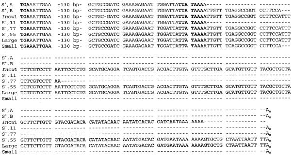
On the Molecular Nature of the Incw1-L and Incw1-S RNAs.Incw clones, four from suc+ and five from suc-deficient libraries, were isolated and compared against each other and with the previously isolated clone, Incw1-1 (22), from an independently isolated library of the same cell line (with poorly defined growth and medium conditions). cDNA inserts were Incw1-specific (data not shown; also see below) and showed no hybridization to the paralogous endosperm-specific gene Incw2, reported previously (23). Full-length sequencing of the five clones from the suc-deficient library and one clone from the suc+ library has shown perfect sequence identity with the Incw1-1 gene (data not shown), except for a high level of heterogeneity in the 3′ UTR. Fig. 4 depicts the relevant sequences of the three unique 3′ UTRs represented among the five suc-deficient clones; the other two were identical to the representative clone S−,55 (see Fig. 4). The 3′ UTRs of the clones from the suc+ library were also sequenced by using the primers as shown in Materials and Methods. Only two types of 3′ UTRs were seen in the four clones from the suc+ library; the most common sequence, shared by three clones, is shown in the S+,B clone. Most importantly, all four suc+ clones were ≈150 bp shorter in length than four of the five clones from the suc-deficient library and the reference clone Incw1-1 (Fig. 4). It is significant that the size variation in the 3′ UTRs of the suc+ and suc-depleted clones was in general agreement with the size differences in the Incw1-S and Incw1-L RNAs, respectively (Figs. 2 and 3).
Figure 4.
Alignment of the 3′ ends of Incw1 cDNAs and RACE products from cell cultures grown in the presence (S+) or absence (S−) of suc. The series of bold letters TGA and TTATAAA are the presumed translation termination codon and poly(A)+ signal site, respectively.
To establish further the possible relationships between the 3′ UTR size differences of the cDNA clones and the two different sized transcripts Incw1-S and Incw1-L, we examined suc+ and suc-deficient cellular RNAs directly by using the 5′ and 3′ RACE approach. Results of the 5′ RACE analyses are shown in Fig. 5 (see Materials and Methods for the details on primers). An identically sized ≈500-bp 5′ RACE fragment was seen in both RNA samples (Fig. 5B), indicating that there was no detectable difference in the positions of their transcription initiation sites. The 3′ RACE products using a universal amplification primer annealed to poly(A)+ tail and primer 1 (first amplification cycle); universal amplification primer and primer 2 (second amplification cycle) led to two major products that were detectable in both ethidium-bromide-stained gels (not shown) and by hybridization to the Incw1 probe (Fig. 5C). Two amplified fragments, identified as large and small, with a size variation of ≈150 bp (see below), were seen regardless of whether the RNA template was from suc+ or the suc-deficient cells. Direct sequencing of the small RACE fragment revealed that it was identical to the 3′ end sequences of the cDNA clone S+,B. Similarly, the large RACE fragment was similar to the most frequently seen sequence of an additional ≈150 bp, represented by the clone S−,55 from the suc-deficient library. Database searches and sequence alignments show that the ≈150 bp of the large fragment is similar to the genomic sequence (GenBank accession no. AF050129) of the Incw1 gene (23). It is worth noting that we observed perfect sequence identity between the large and small RACE fragments and the 3′ cDNA ends of clones S−,55 and S+,B, which were isolated from suc-depleted and suc+ libraries, respectively. We infer from the collective data that 3′ UTR of the Incw1-L RNA carried ≈150 bp of genomic sequence that was otherwise lacking from the Incw1-S transcript.
DISCUSSION
We report here three important observations relating to sugar-modulated control of gene expression in plant cells. First, both suc and glc control the expression of Incw1 gene at the levels of RNA, protein, and subsequent enzyme activity. Second, transcriptional induction of the Incw1 gene is associated with two RNAs, Incw1-S and Incw1-L, and their relative abundance depends on the metabolic status of apoplastic and/or intracellular sugars. Metabolizable sugars lead to Incw1-S RNA and a concomitant increase in the levels of INCW protein and enzyme activity; conversely, nonmetabolizable sugars are associated with the Incw1-L RNA and no increase in the levels of the protein and/or enzyme activity. Finally, the two RNAs are identical in the coding region and in the positions of their transcription initiation sites but divergent in the 3′ UTR.
We have shown that endosperm-specific cell-wall invertase (INCW2) encoded by the Mn1 locus is spatially and temporally the first gene to metabolize incoming suc in maize endosperm (1). In the heterotrophic cell culture used in this study, the very rapid induction of the Incw1 gene in response to suc or glc (Table 1 and Fig. 2 B and C) is consistent with a view that suc hydrolysis is the first step in the use of suc. On uptake into the apoplast, suc hydrolysis by a cell-wall invertase must provide an entry point for the carbon into the downstream reactions. Interestingly glc, an end product of this enzyme, also acts as an inducer, albeit the level of enzyme activity is lower. It is unclear how glc acts as an inducer of this enzyme in maize cell culture. In a typical model of catabolite repression, glc is expected to repress an invertase gene. However, a similar pattern of induction by the hexose sugars is also seen in C. rubrum cell culture (2) and for the Incw2 gene in developing endosperm in vitro kernel cultures (27). A “futile cycle” of suc synthesis and cleavage, similar to the previously proposed suc turn-over reactions in several plant systems (2, 28, 29), is postulated as a possible basis for the hexose induction of the two Incw genes. In sycamore suspension cells, an SS pathway is proposed to be relatively more important than an invertase pathway when suc is limiting (15). Because both SS isozymes are present throughout a 7-day culture period in maize cells, suc synthesis by either or both of the isozymes, in particular the plasma membrane-associated forms (19), may yield suc for the Incw1 induction in glc-supplemented cells.
Hexoses and hexokinase play a central role in sugar sensing in yeast, bacteria, and mammals (see ref. 6 for a review), and a similar control is also reported in plant cells (16, 30, 31). In this regard, glc analogs that are taken up and phosphorylated (2-dGlc) or not phosphorylated (3-O-mGlc and 6-deoxyglucose) have yielded significant insight as to whether the sensing is mediated by hexokinases. Several observations here suggest that the Incw1 induction is independent of the hexokinase pathway. First, 3-O-mGlc induced only the Incw1-L form of RNA, a response that is similar to the nonmetabolizable sugars. Second, 2-dGlc did not induce any increases in the levels of INCW protein and enzyme activity, and there were greatly reduced steady-state levels of the Incw1-L RNA relative to suc or glc. Finally, there was no Sh1 repression at the RNA level in 3-O-mGlc cells, as is the case with media lacking metabolizable sugars (Fig. 3). The time lag in the Sh1 repression as compared with the rapid induction of the Incw1 gene (Fig. 3) indicated that such repression may depend on the INCW-mediated suc hydrolysis and the subsequent metabolic reactions. The Sh1 response may serve as an excellent metabolic marker of sugar use or nonuse in heterotrophic sinks.
Remarkably, a hexokinase-independent pathway of sugar sensing in the INCW (Cin) induction in C. rubrum autotrophic suspension cells has also been described (9). The Cin mRNA in these cells is at nearly the same steady-state levels in a medium with the nonmetabolizable glc analog 6-deoxyglucose as it is in glc medium (2, 9). Because cell-wall invertase is a secretory glycoprotein that depends intimately on cotranslational translocation in its expression and function, we suggest that the endoplasmic reticulum may serve as an ideal vehicle in signal transduction to the nucleus. It is noteworthy that maize SS is partly localized in the plasma membrane (19); thus, suc turn-over reactions in the apoplast may be an important link in this pathway. A suc-dependent, but hexokinase-independent, proton-suc symporter in sugar beets has also been described (32).
Sequence identities in the coding region of several Incw1 cDNA clones, presumably representative of the Incw1-S and Incw1-L transcripts, indicate that they are encoded by the Incw1 gene. A similar mode of control is also described for the yeast cells where the Suc2 gene encodes two forms of invertase transcripts that differ by 100 bp, and intriguingly, as in maize, one form is glc-repressed and another is glc-derepressed (33). However, unlike yeast where the two RNAs correspond to extracellular and intracellular invertases, in maize, only one of the two forms, the Incw1-S, corresponded with protein and enzyme activity. It is noteworthy that the same two Incw1 transcripts have also been observed in several whole-plant tissues both by RNA blot and by 3′ RACE analyses (W.-H.C., E.W.T., and P.S.C., unpublished data). Whether this well conserved mode of the two mRNAs from a single invertase gene in response to sugars will also be seen in other plant species is currently unknown.
Two well established posttranslational mechanisms of controlling gene expression are mRNA stability and mRNA translation efficiency. In maize cells, although both Incw1 RNAs were induced, their relative steady-state levels depend on whether a given culture medium contained a metabolizable sugar or not. Obviously, the inductional signals are different from those that stabilize the two RNAs. The presence of metabolizable sugars leads to greater steady-state levels of the Incw1-S transcript and, ultimately, the increased protein and enzyme activity. Thus, the translatability of the message seems to play a major role in its stability. A similar situation is described for pea ferredoxin1 (Fed1) mRNA (34) where the translatable message is associated with an increased half-life. Conversely, if not translated, the same sized Fed1 mRNA has a shorter half-life and is preferentially degraded. The relationship between the two Incw1 transcripts and whether the polyadenylated Incw1-L RNA is ever translated are not known. It is conceivable that the two RNAs have a pre-mRNA–mRNA relationship, and, soon after induction, their relative steady-state abundance is sensed by the metabolic status of apoplastic and/or intracellular sugars.
In this regard, the differences in the 3′ UTR, but no detectable differences in the transcription initiation site, of the two Incw1 RNAs are of special significance. The 3′ UTR of a gene is generally a sequence between the translation termination codon and the poly(A) tail. Based on cDNA and 3′ RACE sequences, we show that the increased length of Incw1-L relative to the Incw1-S form is contributed by ≈150 bp of the Incw1 genomic sequences beyond the putative poly(A) signal. All Incw1 clones have shown a well conserved hexanucleotide motif (ATAAAA; Fig. 4) that is similar to a consensus poly(A) signal for plants (35). Significantly, three of the four clones from the suc+ library and the 3′ RACE products identified as the small fragment (Fig. 5), presumably representative of the Incw1-S transcript, show only 21 bp in between the poly(A) signal and the poly(A) tail. In general, the poly(A) site is located 10–30 nt from a poly(A) signal (35); thus, most likely these clones represent a normal 3′ UTR of the gene. Clearly, these changes in the 3′ UTR, in particular the region in between the poly(A) signal and the poly(A) tail, are of crucial importance in the metabolism of the two Incw1 RNAs. Consistent with these interpretations are data from yeast and other eukaryotic systems where the entire 3′ UTR is known to regulate both translational control and mRNA stability. Such controls are mediated through interactions of the 3′ UTR with various factors that lead to sequential endonucleolytic cleavage and polyadenylation of the mRNA (36). Also of significant interest are the recent data from rice cell cultures that implicate the 3′ UTR as a major determinant of mRNA stability of the α-amylase3 gene (37). In maize, the increased length of the Incw1-L RNA in the 3′ UTR may contribute to an alternative RNA metabolism and translational fate in suc-deficient cells. Collectively, our results provide an important link between sink metabolism and cellular translational processes in plants.
Acknowledgments
We thank Drs. Susan J. Carlson and Daryl R. Pring for critical reading of the manuscript. We gratefully acknowledge the services of the DNA Sequencing Core of the Interdisciplinary Center for Biotechnology Research, University of Florida. This work was supported in part by U.S. Department of Agriculture/National Research Initiative Competitive Grants Program Grant 98-35301-6135. This study was a cooperative investigation of the U.S. Department of Agriculture/Agricultural Research Service and the Institute of Food and Agricultural Science, University of Florida. This paper is Agricultural Experimental Journal Series no. R-06760.
ABBREVIATIONS
- suc
sucrose
- glc
glucose
- 2-dGlc
2-deoxyglucose
- 3-O-mGlc
3-O-methylglucose
- SS
sucrose synthase
- UTR
untranslated region
- RACE
rapid amplification of cDNA ends
References
- 1.Cheng W-H, Taliercio E W, Chourey P S. Plant Cell. 1996;8:971–983. doi: 10.1105/tpc.8.6.971. [DOI] [PMC free article] [PubMed] [Google Scholar]
- 2.Roitsch T, Bittner M, Godt D E. Plant Physiol. 1995;108:285–294. doi: 10.1104/pp.108.1.285. [DOI] [PMC free article] [PubMed] [Google Scholar]
- 3.Sonnewald U, Hajirezaei M-R, Kossmann J, Heyer A, Trethewey R N, Willmitzer L. Nat Biotechnol. 1997;15:794–797. doi: 10.1038/nbt0897-794. [DOI] [PubMed] [Google Scholar]
- 4.Tang G-Q, Luscher M, Sturm A. Plant Cell. 1999;11:177–189. doi: 10.1105/tpc.11.2.177. [DOI] [PMC free article] [PubMed] [Google Scholar]
- 5.Miller M E, Chourey P S. Plant Cell. 1992;4:297–305. doi: 10.1105/tpc.4.3.297. [DOI] [PMC free article] [PubMed] [Google Scholar]
- 6.Jang J-C, Sheen J. Trends Plant Sci. 1997;2:208–214. [Google Scholar]
- 7.Smeekens S, Rook F. Plant Physiol. 1997;115:7–13. doi: 10.1104/pp.115.1.7. [DOI] [PMC free article] [PubMed] [Google Scholar]
- 8.Xu J, Avigne W T, McCarty D R, Koch K E. Plant Cell. 1996;8:1209–1220. doi: 10.1105/tpc.8.7.1209. [DOI] [PMC free article] [PubMed] [Google Scholar]
- 9.Ehness R, Ecker M, Godt D E, Roitsch T. Plant Cell. 1997;9:1825–1841. doi: 10.1105/tpc.9.10.1825. [DOI] [PMC free article] [PubMed] [Google Scholar]
- 10.Krausgrill S, Greiner S, Koster U, Vogel R, Rausch T. Plant J. 1998;13:275–280. [Google Scholar]
- 11.Greiner S, Krausgrill S, Rausch T. Plant Physiol. 1998;116:733–742. doi: 10.1104/pp.116.2.733. [DOI] [PMC free article] [PubMed] [Google Scholar]
- 12.Lutfiyya L L, Iyer V R, DeRisi J, DeVit M J, Brown P O, Johnston M. Genetics. 1998;150:1377–1391. doi: 10.1093/genetics/150.4.1377. [DOI] [PMC free article] [PubMed] [Google Scholar]
- 13.Ludin K, Jiang R, Carlson M. Proc Natl Acad Sci USA. 1998;95:6245–6250. doi: 10.1073/pnas.95.11.6245. [DOI] [PMC free article] [PubMed] [Google Scholar]
- 14.Chourey P S, Zurawski D B. Theor Appl Genet. 1981;59:341–344. doi: 10.1007/BF00276446. [DOI] [PubMed] [Google Scholar]
- 15.Huber S C, Akazawa T. Plant Physiol. 1986;81:1008–1013. doi: 10.1104/pp.81.4.1008. [DOI] [PMC free article] [PubMed] [Google Scholar]
- 16.Graham I A, Denby K J, Leaver C J. Plant Cell. 1994;6:761–772. doi: 10.1105/tpc.6.5.761. [DOI] [PMC free article] [PubMed] [Google Scholar]
- 17.Grotewold E, Chamberlin M, Snook M, Siame B, Butler L, Swenson J, Maddock S, St. Clair G, Bowen B. Plant Cell. 1998;10:721–740. [PMC free article] [PubMed] [Google Scholar]
- 18.Doehlert C D, Felker F C. Physiol Plant. 1987;70:1–57. [Google Scholar]
- 19.Carlson S J, Chourey P S. Mol Gen Genet. 1996;252:303–310. doi: 10.1007/BF02173776. [DOI] [PubMed] [Google Scholar]
- 20.Ramloch-Lorenz K, Knudsen S, Sturm A. Plant J. 1993;4:545–554. doi: 10.1046/j.1365-313x.1993.04030545.x. [DOI] [PubMed] [Google Scholar]
- 21.Wadsworth G J, Redinbaugh M G, Scandalios J G. Anal Biochem. 1988;172:279–283. doi: 10.1016/0003-2697(88)90443-5. [DOI] [PubMed] [Google Scholar]
- 22.Shanker S, Salazar R W, Taliercio E W, Chourey P S. Plant Physiol. 1995;108:873–874. doi: 10.1104/pp.108.2.873. [DOI] [PMC free article] [PubMed] [Google Scholar]
- 23.Taliercio, E. W., Kim, J.-Y., Mahe, A., Shanker, S., Choi, J., Cheng, W.-H., Prioul, J.-L. & Chourey, P. S. (1999) J. Plant Physiol., in press.
- 24.Gupta M, Chourey P S, Burr B, Still P E. Plant Mol Biol. 1988;10:215–224. doi: 10.1007/BF00027398. [DOI] [PubMed] [Google Scholar]
- 25.Maas C, Schall S, Werr W. EMBO J. 1990;9:3447–3452. doi: 10.1002/j.1460-2075.1990.tb07552.x. [DOI] [PMC free article] [PubMed] [Google Scholar]
- 26.Koch K E, Nolte K D, Duke E R, McCarty D R, Avigne W T. Plant Cell. 1992;4:59–69. doi: 10.1105/tpc.4.1.59. [DOI] [PMC free article] [PubMed] [Google Scholar]
- 27.Cheng W-H, Chourey P S. Theor Appl Genet. 1999;98:485–495. [Google Scholar]
- 28.Wendler R, Veith R, Dancer J, Stitt M, Komor E. Planta. 1990;183:183–189. doi: 10.1007/BF00197564. [DOI] [PubMed] [Google Scholar]
- 29.Dancer J, Hatzfeld W D, Stitt M. Planta. 1990;182:223–231. doi: 10.1007/BF00197115. [DOI] [PubMed] [Google Scholar]
- 30.Jang J-C, Sheen J. Plant Cell. 1994;6:1665–1679. doi: 10.1105/tpc.6.11.1665. [DOI] [PMC free article] [PubMed] [Google Scholar]
- 31.Zhou L, Jang J-C, Sheen J. Proc Natl Acad Sci USA. 1998;95:10294–10299. doi: 10.1073/pnas.95.17.10294. [DOI] [PMC free article] [PubMed] [Google Scholar]
- 32.Chiou T J, Bush D R. Proc Natl Acad Sci USA. 1998;95:4784–4788. doi: 10.1073/pnas.95.8.4784. [DOI] [PMC free article] [PubMed] [Google Scholar]
- 33.Carlson M, Botstein D. Cell. 1982;28:145–154. doi: 10.1016/0092-8674(82)90384-1. [DOI] [PubMed] [Google Scholar]
- 34.Petracek M E, Dickey L F, Nguyen T T, Gatz C, Sowinski D A, Allen G C, Thompson W F. Proc Natl Acad Sci USA. 1998;95:9009–9013. doi: 10.1073/pnas.95.15.9009. [DOI] [PMC free article] [PubMed] [Google Scholar]
- 35.Wu L, Ueda T, Messing J. Plant J. 1995;8:323–329. doi: 10.1046/j.1365-313x.1995.08030323.x. [DOI] [PubMed] [Google Scholar]
- 36.Manley J L, Takagaki Y. Science. 1996;274:1481–1482. doi: 10.1126/science.274.5292.1481. [DOI] [PubMed] [Google Scholar]
- 37.Chan M-T, Yu S-M. Proc Natl Acad Sci USA. 1998;95:6543–6547. doi: 10.1073/pnas.95.11.6543. [DOI] [PMC free article] [PubMed] [Google Scholar]


