Abstract
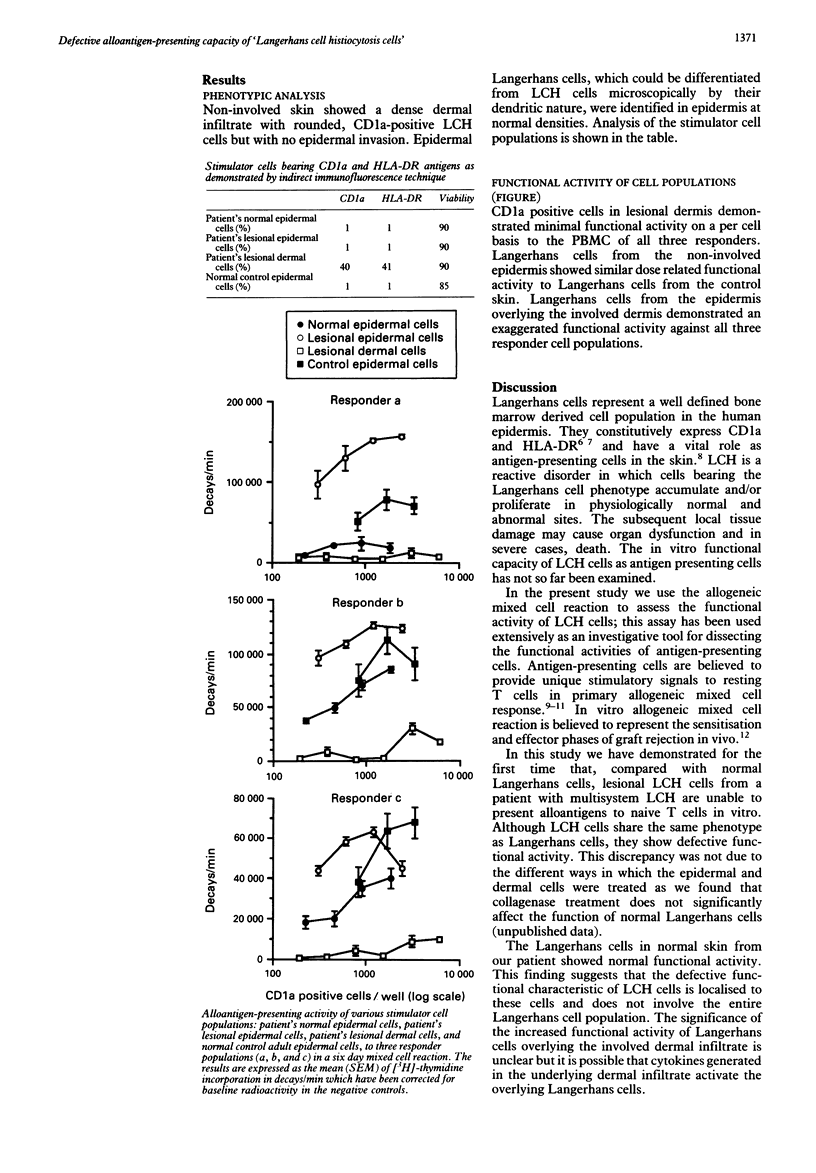
The functional activity of skin cells derived from an infant who died of multisystem Langerhans cell histiocytosis (LCH) was examined. Involved and non-involved skin was obtained at postmortem examination within three hours of death; normal epidermal Langerhans cells and 'LCH cells' were separated by means of dispase digestion. The functional activity of different populations of CD1a positive cells was assessed using the conventional six day allogeneic mixed cell reaction. Compared with Langerhans cells from a healthy control, LCH cells showed minimal functional activity. However, Langerhans cells from non-involved skin showed normal and Langerhans cells overlying involved skin showed augmented functional activity. These findings suggest that LCH is a disease in which abnormal Langerhans cells accumulate and/or proliferate in various tissues but it does not affect the entire Langerhans cell population.
Full text
PDF


Selected References
These references are in PubMed. This may not be the complete list of references from this article.
- Arenzana-Seisdedos F., Barbey S., Virelizier J. L., Kornprobst M., Nezelof C. Histiocytosis X. Purified (T6+) cells from bone granuloma produce interleukin 1 and prostaglandin E2 in culture. J Clin Invest. 1986 Jan;77(1):326–329. doi: 10.1172/JCI112296. [DOI] [PMC free article] [PubMed] [Google Scholar]
- Austyn J. M. Lymphoid dendritic cells. Immunology. 1987 Oct;62(2):161–170. [PMC free article] [PubMed] [Google Scholar]
- Bjercke S., Elgø J., Braathen L., Thorsby E. Enriched epidermal Langerhans cells are potent antigen-presenting cells for T cells. J Invest Dermatol. 1984 Oct;83(4):286–289. doi: 10.1111/1523-1747.ep12340417. [DOI] [PubMed] [Google Scholar]
- Chu T., D'Angio G. J., Favara B. E., Ladisch S., Nesbit M., Pritchard J. Histiocytosis syndromes in children. Lancet. 1987 Jul 4;2(8549):41–42. doi: 10.1016/s0140-6736(87)93074-1. [DOI] [PubMed] [Google Scholar]
- Groh V., Gadner H., Radaszkiewicz T., Rappersberger K., Konrad K., Wolff K., Stingl G. The phenotypic spectrum of histiocytosis X cells. J Invest Dermatol. 1988 Apr;90(4):441–447. doi: 10.1111/1523-1747.ep12460878. [DOI] [PubMed] [Google Scholar]
- Inaba K., Schuler G., Witmer M. D., Valinksy J., Atassi B., Steinman R. M. Immunologic properties of purified epidermal Langerhans cells. Distinct requirements for stimulation of unprimed and sensitized T lymphocytes. J Exp Med. 1986 Aug 1;164(2):605–613. doi: 10.1084/jem.164.2.605. [DOI] [PMC free article] [PubMed] [Google Scholar]
- Inaba K., Steinman R. M. Resting and sensitized T lymphocytes exhibit distinct stimulatory (antigen-presenting cell) requirements for growth and lymphokine release. J Exp Med. 1984 Dec 1;160(6):1717–1735. doi: 10.1084/jem.160.6.1717. [DOI] [PMC free article] [PubMed] [Google Scholar]
- McLelland J., Newton J., Malone M., Camplejohn R. S., Chu A. C. A flow cytometric study of Langerhans cell histiocytosis. Br J Dermatol. 1989 Apr;120(4):485–491. doi: 10.1111/j.1365-2133.1989.tb01321.x. [DOI] [PubMed] [Google Scholar]
- Murphy G. F., Bhan A. K., Sato S., Mihm M. C., Jr, Harrist T. J. A new immunologic marker for human Langerhans cells. N Engl J Med. 1981 Mar 26;304(13):791–792. doi: 10.1056/NEJM198103263041320. [DOI] [PubMed] [Google Scholar]
- Rowden G., Lewis M. G., Sullivan A. K. Ia antigen expression on human epidermal Langerhans cells. Nature. 1977 Jul 21;268(5617):247–248. doi: 10.1038/268247a0. [DOI] [PubMed] [Google Scholar]


