Abstract
The production of the vegetative mosquitocidal toxin Mtx1 from Bacillus sphaericus was redirected to the sporulation phase by replacement of its weak, native promoter with the strong sporulation promoter of the bin genes. Recombinant bacilli developed toxicity during early sporulation, but this declined rapidly in later stages, indicating the proteolytic instability of the toxin. Inhibition studies indicated the action of a serine proteinase, and similar degradation was also seen with the purified B. sphaericus enzyme sphericase. Following the identification of the initial cleavage site involved in this degradation, mutant Mtx1 proteins were expressed in an attempt to overcome destructive cleavage while remaining capable of proteolytic activation. However, the apparently broad specificity of sphericase seems to make this impossible. The stability of a further vegetative toxin, Mtx2, was also found to be low when it was exposed to sphericase or conditioned medium. Random mutation of the receptor binding loops of the Bacillus thuringiensis Cry1Aa toxin did, in contrast, allow production of significant levels of spore-associated protein in the form of parasporal crystals. The exploitation of vegetative toxins may, therefore, be greatly limited by their susceptibility to proteinases produced by the host bacteria, whereas the sequestration of sporulation-associated toxins into crystals may make them more amenable to use in strain improvement.
Strains of Bacillus sphaericus and Bacillus thuringiensis are used extensively in the field for the control of insect pests and vectors of disease. While some strains, notably B. thuringiensis subsp. israelensis, have never produced resistant insects in the field, resistance has proved a problem with some B. thuringiensis strains (26), with B. sphaericus (22, 34; G. Sinegre, M. Babinot, J. M. Quermel, and B. Gavan, presented at the 8th European Meeting of the Society of Vector Ecology, Barcelona, Spain, 1994), and with transgenic plants transformed with B. thuringiensis toxins under laboratory and greenhouse conditions (25). Biotechnology offers the potential of supplementing existing toxins within Bacillus species, either with naturally occurring toxins or with engineered toxin variants with new properties.
B. sphaericus produces binary toxin (Bin) during its sporulation phase and Mtx toxins (Mtx1, Mtx2, and Mtx3) (14, 28, 29) during vegetative growth. Due to their low level of expression and their susceptibility to a subtilisin-like serine proteinase (32) also known as sphericase (2), Mtx1 and Mtx2 are not able to contribute to the toxicity of the insecticidal spore formulations used in mosquito control programs. However, these toxins have mechanisms of action distinct from that of the Bin toxin and have significant activity against Aedes aegypti larvae (27) that show low sensitivity to Bin toxins (7). Recent studies have shown that a vegetative insecticidal protein from B. thuringiensis can be redirected in its time of production (6), and a similar retargeting of Mtx1 might enhance B. sphaericus strain activity. In this study, we have examined the possibility of supplementing the binary toxins of B. sphaericus with Mtx toxins from this species and attempted to direct the production of Mtx1 during sporulation and to produce mutants that are proteolytically stable. In addition, the stability of Mtx2 to proteolytic degradation was also examined.
With the importance of proteolytic stability in toxin production and increasing attempts to modify other insecticidal proteins for the selection of novel activities, for instance by phage display (12, 16, 17, 31), we also considered it to be of interest to analyze mutants of a B. thuringiensis Cry toxin to determine whether specific mutation of receptor binding regions might also introduce proteolytic sensitivity. B. thuringiensis strains produce a variety of Cry toxins with a wide variety of insecticidal specificities. X-ray crystallographic studies of these toxins have revealed structures with three domains (13). Domain I appears to be involved in pore formation in target cell membranes, and domain III may influence receptor binding, but it is domain II that plays the major part in this process (18). The toxin-receptor interactions of domain II are believed to be mediated through three loops (1, 24), and specific substitutions can be introduced into these features to retarget the Cry1Aa toxin (15). The similarity of the members of the Cry family of toxins suggests that genetic recombination between family members may be a factor in producing new toxin variants in nature (9). In some instances, however, there is evidence that a significant proportion of the variants produced will suffer from proteolytic instability in the producing organism (3). In this work, we have constructed a library of precisely targeted loop variants derived from the Cry1Aa protein and have assessed the stability of these proteins upon production in B. thuringiensis.
MATERIALS AND METHODS
Bacterial strains.
B. thuringiensis subsp. israelensis strain 4Q7 (also known as 4Q2-81) from the Bacillus Genetic Stock Center, is a crystal-minus strain used for the transformation and expression of recombinant proteins. B. thuringiensis subsp. kurstaki HD1, which carries the cry1Aa gene, was kindly provided by David Ellar, University of Cambridge, United Kingdom. B. sphaericus 2362, a commonly used strain in the production of commercial formulations, was obtained from the Institut Pasteur, and B. sphaericus 1693, a proteinase-negative strain that has been used previously for the production of Mtx1 protein (30), was from the Bacillus Genetic Stock Center.
DNA manipulations and vector construction were carried out with Escherichia coli strain DH5α. B. sphaericus strains were grown in MBS medium (10 g/liter tryptone, 2 g/liter yeast extract, 0.02 g/liter CaCl2, 0.3 g/liter MgSO4 · 7H2O, 6.8 g/liter KH2SO4, 0.2 g/liter MnSO4, 0.02 g/liter FeSO4, 0.02 g/liter ZnSO4; pH 7.0). B. thuringiensis strains were grown in G-Tris medium [0.08 g/liter CaCl2, 2 g/liter (NH4)2SO4, 0.5 g/liter K2HPO4, 50 mM Tris-HCl (pH 7.5), 1.5 g/liter yeast extract, and 1% G-Tris stock salt solution, which comprises 0.25 g/liter FeSO4 · H2O, 0.5 g/liter CuSO4, 0.5 g/liter ZnSO4 · 7H2O, 5 g/liter MnSO4 · H2O, and 20 g/liter MgSO4]. Sterile glucose solution was added before use to a final concentration of 2 g/liter). E. coli was grown in LB medium.
Fusion of the bin promoter and mtx1 gene.
In an attempt to drive the production of the Mtx1 toxin during the sporulation phase of growth, the coding sequence of mtx1 was joined to the sporulation promoter from the bin operon (Pbin), as follows. A new NcoI site was introduced at the ATG initiation codon of the binB gene by site-directed mutagenesis of the plasmid pCW-2, which contains the bin operon from B. sphaericus strain C3-41 (33). For mutagenesis, we employed the QuikChange kit (Stratagene) according to the manufacturer's instructions, using the primers BinmutF and BinmutR (Table 1). The Pbin promoter was then isolated by digestion with SalI and NcoI and ligated into the pGEMT-Easy plasmid (Promega), which had been prepared with the same enzymes, to produce plasmid pTBP.
TABLE 1.
Primers used in this study
| Category | Primer | Sequencea | Cut site(s) |
|---|---|---|---|
| PBin-mtx1 fusion | BinmutF | GGAGATGAAGAAACCATGGGCGATTCAAAAGAC | NcoI |
| BinmutR | GTCTTTTGAATCGCCCATGGTTTCTTCATCTCC | NcoI | |
| mtx1F | CCATGGCTTCACCTAATTCTCCAAAAG | NcoI | |
| mtx1R | CCATGGTCATTACTATCTAGGTTCTACAC | NcoI | |
| mtx1 mutants | MtxF | CCGGAATTCCCGGATCCTCACCTAATTCTCCAAAAGAT | EcoRI, BamHI |
| MtxR | GGCCCGGGATCCTATCTAGGTTCTACACCTAATG | SmaI, BamHI | |
| MutLeuF | GGATTCTAAAGGTTTGATACTAGATTTAG | ||
| MutGlyF | GGATTCTAAAGGTGGTATACTAGATTTAG | ||
| MutArgF | GGATTCTAAAGGTCGTATACTAGATTTAG | ||
| MutKKF | GGAAATAATATGGATAAGAAAGGTAAGATACTAGATTTAG | ||
| MutLeuR | CTAAATCTAGTATCAAACCTTTAGAATCC | ||
| MutGlyR | CTAAATCTAGTATACCACCTTTAGAATCC | ||
| MutArgR | CTAAATCTAGTATACGACCTTTAGAATCC | ||
| MutKKR | CTAAATCTAGTATCTTACCTTTCTTATCCATATTATTTCC | ||
| Mtx2 production | mtx2N15TF | GAAGGATCCATGGAATTGTTTGTAAACGGAAGTATATATACG | BamHI, NcoI |
| mtx2N15TR | GGGCTCGAGTTATTTAAAAGAAATTTCTTTAACATCTATTA | XhoI | |
| Domain II variants | A | GGATCCTTTTGATGGTAGTTTTCGTGGA | BamHI |
| B | GACGTCAGTATAAATGGTTATACTATTAAG | AatII | |
| C | AATTATTGGTCAGGGCATCAAA | ||
| D | ATATAAAGGTGAAGATAATGTTCT | ||
| E | CTGTTTGTCCTTGATGGAACG | ||
| F | CAGCATTGTAACATGACTCAATC | ||
| G | TTGAGAGCTCCAACGTTTTCTTGGCAGCATCGCAGTGCGGCCG | SacI, NotI | |
| H | CGGCCGCACTGCGATGCTGCCAAGAAAACGTTGGAGCTCTCAA | NotI, SacI | |
| 1.1 | CTTAATAGTATAACCATTTATACTGACGTC(NNS)4AATTTATTGGTCAGGGCATCAAA | AatII | |
| 1.2 | CTTAATAGTATAACCATTTATACTGACGTC(NNS)3AATTTATTGGTCAGGGCATCAAA | AatII | |
| 1.3 | CTTAATAGTATAACCATTTATACTGACGTC(NNS)5AATTTATTGGTCAGGGCATCAAA | AatII | |
| 2.1 | AGAACATTATCTTCACCTTTATAT(NNS)13CTGTTTGTCCTTGATGGAACG | ||
| 2.2 | AGAACATTATCTTCACCTTTATAT(NNS)12CTGTTTGTCCTTGATGGAACG | ||
| 2.3 | AGAACATTATCTTCACCTTTATAT(NNS)14CTGTTTGTCCTTGATGGAACG | ||
| 3.1 | GATTGAGTCATGTTACAATGCTG(NNS)9TTGAGAGCTCCAACGTTTTCT | ||
| 3.2 | GATTGAGTCATGTTACAATGCTG(NNS)8TTGAGAGCTCCAACGTTTTCT | ||
| 3.3 | GATTGAGTCATGTTACAATGCTG(NNS)10TTGAGAGCTCCAACGTTTTCT | ||
| cry1Aa construction | N-termF | GGATCCTAAGTAGATTGTTAACACCCTG | BamHI |
| C-term F | GACGTCCATAGAGGCTTTAATTATTGGTC | AatII | |
| C-termR | GGATCCCTATTCCTCCATAAGGAGTAATTC | BamHI |
The sequences of the restriction enzymes noted in the fourth column are underlined.
The region of mtx1 encoding the 97-kDa truncated protein (lacking the N-terminal signal peptide) was amplified from plasmid pTH21 (27) by PCR using the primers mtx1F and mtx1R (Table 1), which contain NcoI sites. The resulting PCR product was gel purified; digested with NcoI; ligated into NcoI-cut, phosphatase-treated pTBP; and transformed into E. coli DH5α. A recombinant plasmid which contained Pbin and mtx1 in the correct orientation was designated pTBPM. For expression of Mtx1 in bacilli, the Pbin-mtx1 cassette was isolated by digestion of pTBPM with SphI and ligation into the SphI-digested shuttle vector pBU4 (8), resulting in the recombinant plasmid pBM97, in which the transcriptional orientation of the inserted mtx1 gene was the same as that of the vector's lacZ gene. The plasmid pBM97 was transferred into B. thuringiensis subsp. israelensis 4Q7 by electroporation, and a recombinant was identified for further investigation.
Production of Mtx1 mutants.
Mutations in mtx1 were created by an overlapping PCR protocol. Plasmid pTH21, encoding the 97-kDa truncation of Mtx1 (27), was used as the template for two separate PCRs for each mutation. To amplify the 5′ end of the gene, primer MtxF was used in conjunction with a reverse primer that incorporated the desired mutation (MutLeuR, MutGlyR, MutArgR, or MutKKR [Table 1]), using EasyA polymerase (Stratagene) in 30 cycles of 95°C for 1 min, 55°C for 1 min, and 72°C for 1 min. The 3′ end of the gene was amplified under the same conditions, using a reverse primer, MtxR, in combination with one of the forward primers containing the same mutations (MutLeuF, MutGlyF, MutArgF, or MutKKF [Table 1]). Finally, the two parts of each mutant construct were combined by amplification as described above using the primers MtxF and MtxR. Most of the mtx1 gene in the original pTH21 vector was replaced by the above-described mutated mtx1 amplicon at the BamHI and SnaBI sites, giving recombinant plasmid pTH21-M. Production and purification of the mutant fusion proteins and of wild-type Mtx1 were carried out as previously described (27).
Production of Mtx2.
The plasmid pTH84 (29) was a kind gift from Alan Porter (Institute of Molecular and Cell Biology, National University of Singapore) and encodes a glutathione S-transferase (GST)-Mtx2 fusion protein. In order to produce and purify significant amounts of the fusion protein, the mtx2 gene was amplified from pTH84 using primers mtx2N15TF and mtx2N15TR (Table 1) in 30 cycles of 95°C for 1 min, 55°C for 1 min, and 72°C for 3 min with EasyA polymerase (Stratagene). The amplicon encoding an approximately 30-kDa portion of Mtx2 was digested with BamHI and XhoI and ligated into the pGEX4T2 vector (Amersham Biosciences), which had been prepared with the same enzymes. The expression and purification of the GST-Mtx2 fusion protein then proceeded as previously described for GST-Mtx1 fusion proteins (27).
Construction of Cry1Aa domain II variants.
Variants of Cry1Aa were constructed in a modular fashion using a “gene assembly” approach. The region encoding domain II was first produced with sequences that varied in the three loop regions and was then linked to sequences encoding domains I and III in order to reconstitute the toxin sequence.
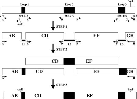
The region encoding Cry1Aa domain II (amino acids Phe276 to Ser458) was amplified from B. thuringiensis subsp. kurstaki HD1 by PCR using primers A and H (Table 1) and Taq polymerase (Promega) under the following conditions: 30 cycles of 95°C for 1 min, 60°C for 1 min, and 72°C for 1 min. The resulting amplicon was gel purified and ligated into the vector pGEM-T for transformation into E. coli DH5α. One colony was selected and the plasmid prepared for DNA sequencing, which confirmed that the expected region had been amplified without mutation. This plasmid was used as the template for further domain II manipulation. Synthesis of the region encoding domain II with hypervariable binding loops occurred stepwise as illustrated in Fig. 1. First, the regions surrounding the loops to be mutated were produced by PCR from pairs of flanking primers (primers A to F) (Table 1; Fig. 1, step 1). Primer B includes an AatII site unique to the cry1Aa gene, produced through silent mutations just upstream of loop 1. Primer A includes at its 5′ end a BamHI site, and primer H includes at its 5′ end a NotI site to facilitate later stages of gene construction (see below). Synthesis of the fragments AB, CD, and EF (Fig. 1) used Kod Hot Start DNA polymerase (Novagen) in a touch-down PCR protocol in which denaturation was at 96°C for 1 min, extension was at 72°C for 1 min, and the annealing temperature decreased from 70°C to 56°C (five cycles each in 2°C steps with a 1-min annealing time). To produce the 35-bp region downstream of loop 3 (GH in Fig. 1), the complementary oligonucleotides G and H were annealed by heating to 100°C for 10 min, followed by slow cooling to room temperature over 30 min. The sequences encoding the loops were synthesized as oligonucleotides (L1, L2, and L3 in Fig. 1) with sufficient overlap with the flanking regions to allow annealing to the interloop regions in subsequent steps. The sequences encoding the amino acids of the loops were of sufficient degeneracy to encode any amino acid at any position. To limit unnecessary degeneracy and reduce the occurrence of in-frame stop codons, these regions were encoded by oligonucleotide sequences of the form [N-N-S]n (where N is A, G, C, or T; S is G or C; and n is the number of amino acids to be encoded). In the original Cry1Aa toxin, loops 1, 2, and 3 consist of 4, 13, and 9 amino acids, respectively. In other toxins, these loops may be of different lengths; therefore, for each loop we produced oligonucleotides encoding the original length loop (1.1, 2.1, and 3.1 [Table 1]) as well as loops that were shorter and longer by one amino acid (1.2, 2.2, and 3.2 and 1.3, 2.3, and 3.3, respectively). Amplicons containing two interloop regions and one hypervariable loop were then produced by PCR using the Kod Hot Start enzyme (Fig. 1, step 2). The AB-CD fragment was produced using primers A, D, 1.1, 1.2, and 1.3 in addition to fragments AB and CD in a touch-down PCR protocol as described above. CD-EF was produced using primers C, F, 2.1, 2.2, and 2.3 and fragments CD and EF (30 cycles of 96°C for 1 min, 56°C for 1 min, and 72°C for 1 min). EF-GH was synthesized using primers E, H, 3.1, 3.2, and 3.3 and fragments EF and GH (30 cycles of 96°C for 1 min, 60°C for 1 min, and 72°C for 1 min). Finally, the entire domain II with hypervariable loops was amplified using primers A and H in the presence of fragments AB-CD, CD-EF, and EF-GH under the touch-down conditions described above (Fig. 1, step 3).
FIG. 1.
Construction of Cry1Aa domain II variants. Schematic diagram of stages of the assembly process. Right-pointing arrows represent forward primers; left-pointing arrows represent reverse primers. Loop regions are shown as dark boxes.
The above protocol produced a range of domain II-derived sequences, conserved in the interloop areas but hypervariable in the loops. These were then incorporated with Cry1Aa domain I, domain III, and the C terminus to reconstitute protoxin sequences as follows.
The nucleotide sequence containing the cry1Aa promoter and encoding the N-terminal region of Cry1Aa encompassing domain I and a part of domain II was amplified using primers N-termF and B, which contain BamHI and AatII sites, respectively. PCR was carried out using vegetative cells of B. thuringiensis subsp. kurstaki to provide the template under the following conditions: 96°C for 5 min and then 30 cycles of 96°C for 1 min, 55°C for 1 min, and 72°C for 1 min. The sequence encoding part of domain II, domain III, and the C terminus of the Cry1Aa protoxin was amplified using primers C-termF and C-termR, which contain AatII and BamHI sites, respectively, in a PCR under conditions identical to those employed to amplify the N-terminal region, as described above. Both amplicons were cloned, separately, into the pGEM-T vector (Promega), and the entire DNA sequences were verified to ensure that they were free of unintended mutations. To allow cloning in B. thuringiensis, the shuttle vector pHT304 (5) was chosen. This vector, however, contains restriction enzyme recognition sites for AatII and SacI, which our strategy required us to remove. Plasmid pHT304 was, therefore, digested with AatII and SmaI before blunt ending of the products using T4 polymerase in the presence of a 2 mM concentration of the deoxynucleoside triphosphates. The larger fragment was gel purified (the smaller fragment that includes the SacI site was discarded) and self-ligated to produce plasmid pHT304mod. To produce a clone able to express full-length Cry1Aa, the clones encoding N- and C-terminal portions of the gene were digested with AatII and BamHI; the gene fragments were gel purified and mixed in a three-way ligation with BamHI-cut, phosphatase-treated pHT304mod. After the transformation of E. coli, the resulting plasmid was sequenced to check the cry1Aa gene and was designated pHT304-cry1Aa.
In the final stage, domain II loop variants were introduced into this plasmid by digestion of the loop variant PCR mix (Fig. 1, final product) with AatII and SacI and ligation of the resulting fragments into pHT304-cry1Aa, which had been cut with the same enzymes and gel purified. The resulting library of domain II-variant cry1Aa genes was transformed into B. thuringiensis subsp. israelensis strain 4Q7, and individual erythromycin-resistant colonies were picked and grown to sporulation for preparation of crystal protein extracts. At the same time, plasmid was isolated from the same clones and was subjected to sequencing to confirm that loop variations had been created.
Western blot analysis.
The polyclonal antiserum against Mtx1 and Western blot analysis of Mtx1 production were performed as described by Thanabalu and Porter (30).
Bioassays.
Bacilli were prepared for bioassay by the resuspension of an inoculating loop of cells and spores from a plate, in 2 ml sterile water, followed by a heat shock at 72°C for 20 min to eliminate vegetative cells. This preparation was then inoculated into 50 ml G-Tris medium and grown at 30°C and 300 rpm, with samples taken every 4 h to monitor growth phases, and bioassays of representative samples were carried out so that a time course of toxin production could be assessed. E. coli cultures were grown in LB medium overnight at 37°C and 220 rpm for use in assays. For each assay, 25 second-instar larvae of a susceptible Culex quinquefasciatus colony were placed in 100 ml dechlorinated water and different bacterial dilutions were added. At least five concentrations giving a mortality between 2 and 98% were tested, and mortality was recorded after incubation at 26°C for 48 h. Bioassays were performed in duplicate, and the tests were replicated on at least three different days. Fifty and 90% lethal concentrations (LC50 and LC90, respectively) were determined by Probit analysis (10) with a program indicating means and standard errors.
Preparation of proteolytic extracts.
To assess the proteolytic potential of host bacilli, samples were prepared from the lysis of vegetative cells of B. thuringiensis subsp. israelensis 4Q7 and medium conditioned by the growth of this strain. Cultures were grown in MBS (B. sphaericus) or G-Tris (B. thuringiensis) medium until approximately 50% sporulation was obtained. The culture was then harvested by centrifugation, and the conditioned medium was removed and further clarified by sterile filtration through a 0.22-μm filter (Millipore). The cell pellet was washed twice with phosphate-buffered saline and then resuspended in the same buffer prior to disruption using a Soniprobe sonicator (Lucas Dawe Ultrasonics Ltd., London, United Kingdom) for 1 min on 60% power. The lysed cell suspension was then clarified by centrifugation at 12,000 × g, and the soluble cell extract was used in further proteolysis assays. An equivalent conditioned medium was produced by the growth of B. sphaericus strains 2362 and 1693. Purified sphericase was a kind gift from Daniela Klein (Hebrew University of Jerusalem, Jerusalem, Israel). The protein content of all preparations was estimated by a Bradford assay using a Bio-Rad protein assay kit (Bio-Rad; Hertfordshire, United Kingdom) and bovine serum albumin standards.
The ability of 1 μg of protein from conditioned medium or from cell extracts to degrade 5 μg of Mtx1 toxin proteins was assessed by sodium dodecyl sulfate-polyacrylamide gel electrophoresis following incubations at 30°C in phosphate-buffered saline buffer, while sphericase activity was assessed in 50 mM Tris-HCl, 0.1 mM CaCl2, pH 7.7. N-terminal sequencing of digestion products was performed by Alta Biosciences (Birmingham, United Kingdom) following transfer of proteins to a polyvinylidene difluoride membrane from a Tricine-containing gel.
To assess Cry toxin stability, crystals were purified from sporulated cultures (23) and solubilized in 50 mM sodium carbonate buffer, pH 10.5, and the solution was neutralized before estimation of their concentrations (by measuring absorbance at 280 nm) and use in proteolysis assays.
RESULTS AND DISCUSSION
Expression of mtx1 clones.
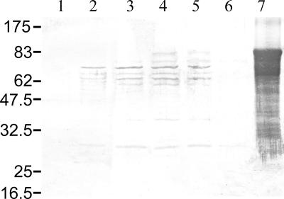
The E. coli strain transformed with plasmid pBM97 resulted in an LC50 of 2.4 × 10⁻⁵ g/ml, compared to 3.3 × 10⁻⁴ g/ml for the strain transformed with pTH21 (27). This demonstrates that the pBM97 clone is able to produce active toxin. This construct was then transformed into recombinant B. thuringiensis 4Q7, and its toxicity was assessed at different phases of growth (Table 2). The strain shows no toxicity when all cells are in the vegetative phase, but activity increases and peaks in early sporulation, consistent with the regulation of Mtx1 synthesis from the bin sporulation promoter. Toxicity then declines in later stages and is absent from the final 52-h preparation. These results were consistent with those of Western blot analysis of Mtx1 (Fig. 2) and indicate successful redirection of Mtx1 synthesis to sporulation stages, but they appear to indicate the degradation of the resulting protein at the end of the sporulation process.
TABLE 2.
Toxicity of recombinant B. thuringiensis 4Q7-pBM97 cultures against C. quinquefasciatus
| Incubation time (h) | Growth phase | Toxicity against Culex quinquefasciatusa
|
|
|---|---|---|---|
| LC50 at 95% confidence limits | LC90 at 95% confidence limits | ||
| 12 | Vegetative | >10−2 | ND |
| 16 | Early sporulation | >10−2 | ND |
| 20 | 0.91 (0.59 to ∼1.46) × 10−5 | 27.8 (12.3 to ∼97.8) × 10−5 | |
| 24 | 0.71 (0.48 to ∼1.05) × 10−5 | 15.4 (7.89 to ∼41.2) × 10−5 | |
| 28 | 0.29 (0.18 to ∼0.43) × 10−5 | 8.72 (4.41 to ∼24.21) × 10−5 | |
| 32 | Spore shedding | 1.02 (0.68 to ∼1.54) × 10−5 | 20.3 (9.92 to ∼64.3) × 10−5 |
| 36 | 2.62 (1.72 to ∼3.86) × 10−5 | 38.4 (21.2 to ∼95.5) × 10−5 | |
| 40 | All spores shed | 8.81 (6.25 to ∼12.34) × 10−4 | 96.9 (57.2 to ∼211) × 10−4 |
| 44 | 5.30 (4.35 to ∼6.60) × 10−3 | 21.3 (14.8 to ∼39.0) × 10−3 | |
| 48 | >10−2 | >10−2 | |
| 52 | >10−2 | >10−2 | |
LC50 and LC90 values are expressed as dilutions of final whole-cell cultures. ND, not determined.
FIG. 2.
Western blot analysis of Mtx1 expression. Lane 1, untransformed B. thuringiensis subsp. israelensis 4Q7; lanes 2 to 6, B. thuringiensis subsp. israelensis 4Q7 transformed with pBM97 at different growth times (for lane 2, 16 h; for lane 3, 20 h; for lane 4, 24 h; for lane 5, 28 h; and for lane 6, 43 h); lane 7, purified Mtx1 (97 kDa). Numbers at the left are molecular masses (in kilodaltons).
Proteinase activity.
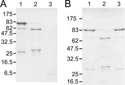
Previous work (30) suggested that proteinase sensitivity might account for the degradation of the Mtx1 toxin and, thus, the loss of toxicity of the strains. To assess this, we exposed Mtx1 toxin, purified as previously described (27), to conditioned growth medium and cell extracts from B. thuringiensis cultures. Over an extended incubation, both preparations caused the degradation of the Mtx1 protein, with greater activity occurring in the conditioned medium than in the cell extract (Fig. 3A). Identical results were seen when conditioned medium from B. sphaericus 2362 was used in place of that from B. thuringiensis and on incubation with purified sphericase (not shown). In shorter (∼1-h) incubations (Fig. 3B, lane 2), two primary degradation products were observed, with the approximately 62-kDa band being slightly larger in the 1-h incubations than in the 16-h experiments (compare Fig. 3A and B), perhaps suggesting further processing. N-terminal sequencing of bands from the 1-h incubation yielded the residues G-S-M-A-S∼ (for the smaller peptide, corresponding to the N terminus of the thrombin-cleaved Mtx1 product (27), and I-L-D-L-D∼, indicating cleavage after residue Phe264 in the region known to be involved in the processing of Mtx1 into its two functional toxin derivatives (27). The recently solved crystal structure of a catalytically inactive and truncated form of Mtx1 clearly shows that this part of Mtx1 forms a surface-exposed region that is an obvious target for proteolytic activity (19). The degradation by the above-described extracts was completely inhibited by preincubation of the proteinase source with phenylmethylsulfonyl fluoride (PMSF) (Fig. 3B, lane 3), thus confirming the nature of the degrading enzyme as a serine proteinase.
FIG. 3.
Degradation of Mtx1 protein. (A) Degradation over 16 h. Lane 1, Mtx1 protein; lane 2, Mtx1 incubated with cell extract; lane 3, Mtx1 incubated with conditioned medium. (B) Inhibition of degradation with PMSF. Lane 1, Mtx1 protein; lane 2, Mtx1 incubated for 1 h with conditioned medium; lane 3, Mtx1 incubated for 1 h with conditioned medium that had been preincubated with PMSF. Numbers at the left of the gels are molecular masses (in kilodaltons).
Mtx1 mutants.
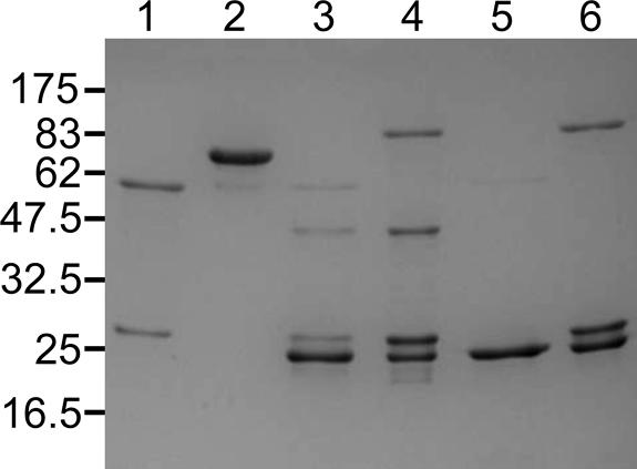
In an attempt to stabilize Mtx1 against host proteolytic attack, the challenge is to eliminate degradation without preventing the proteolytic activation of Mtx1 into its two components in the insect gut. Previous experiments have shown the cleavage of Mtx1 by trypsin after residue Lys262 and a product 2 residues shorter after treatment with mosquito gut extracts, representing a cleavage after Phe264 (27). The latter cleavage is presumably due to chymotrypsin-like enzymes that have been shown to carry out this processing (20) and is at the same site as that of the initial stages of degradation in B. sphaericus (described above), assumed to be by sphericase. Therefore, we attempted to produce mutants of Mtx1 that would resist sphericase treatment while remaining sensitive to trypsin activation, since trypsin-like enzymes are known to be present in mosquito guts (11). Sphericase is a subtilisin-like serine proteinase (32), and as such, its substrate preferences are likely to be determined by residues in the P4 to P1 positions, upstream of the cut site with the greatest influence in the P1 and P4 positions, in which aromatic or large nonpolar residues may be preferred (21). In order to preserve a trypsin cut site, the Lys262 residue (the P3 residue with respect to sphericase cleavage) was maintained, while we hoped to eliminate sphericase degradation by replacement of the P1 residue Phe264 with Leu (MutLeu), Gly (MutGly), or Arg (MutArg) or to replace both Phe264 at P1 and Ser261 at P4 with Lys residues (MutKK). Recombinant GST fusion proteins for each of these mutants were prepared and subjected to incubation with both the B. thuringiensis 4Q7 culture supernatant and purified sphericase, as described above. Unfortunately, in each case, cleavage of the mutant proteins was also observed (sample results for MutGly and MutLeu are shown in Fig. 4, where the lower bands in lanes 3 to 6 correspond to free GST, derived from the fusion proteins used in these cases). Interestingly, degradation was also seen when culture supernatants from B. sphaericus strain 1693 were used (results not shown). This strain belongs to a DNA homology group different from that of the toxin-producing B. sphaericus strains and was previously reported to be suitable for Mtx1 production owing to a lack of degrading proteinase activity (30). The reason for the discrepancy between our results and those of the previous studies is not clear, although in our experiments, we expressed Mtx1 from a sporulation promoter whereas Thanabalu and Porter (30) used its native vegetative promoter. Perhaps vegetative toxin production occurs before the expression of the degrading proteinase. The above results indicate that sphericase has a broad substrate specificity and that mutagenesis of the cleavage site in Mtx1 may not be feasible to protect it from this enzyme, which, like other subtilisins, may have a wide, general-purpose substrate specificity (21). It also appears that B. thuringiensis may have equivalent enzymes able to carry out a similar degradation of Mtx1. Indeed, a BLAST search (4) of the B. thuringiensis subsp. konkukian genome (performed at SIB using the BLAST network service at http://www.expasy.org/tools/blast) reveals several similar proteinases in this organism.
FIG. 4.
Degradation of Mtx1 mutants. Samples were incubated for 1 h. Lane 1, Mtx1 incubated with conditioned medium; lane 2, Mtx1; lane 3, MutGly-GST fusion protein incubated with conditioned medium; lane 4, MutGly-GST fusion protein; lane 5, MutLeu-GST fusion protein incubated with conditioned medium; lane 6, MutLeu-GST fusion protein. Numbers at the left are molecular masses (in kilodaltons).
Proteinase sensitivity of Mtx2.
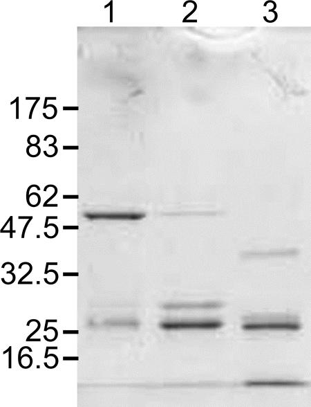
Given the harsh proteolytic environment that may be experienced by proteins produced by Bacillus species, we investigated the susceptibility of a further B. sphaericus toxin, Mtx2, to degradation. A GST fusion protein with Mtx2 (approximately 56 kDa) was incubated with both culture supernatants and purified sphericase, and in both cases degradation of the protein was observed (Fig. 5), indicating that this toxin, like Mtx1, is susceptible to proteolytic degradation that is likely to limit its use in bacilli to supplement the mosquitocidal activities of spores.
FIG. 5.
Degradation of Mtx2. Lane 1, Mtx2-GST fusion protein; lane 2, Mtx2-GST fusion protein incubated with conditioned medium; lane 3, Mtx2-GST fusion protein incubated with sphericase. Numbers at the left are molecular masses (in kilodaltons).
Cry1Aa variants.
In contrast to Mtx1 and Mtx2, Cry toxins are normally produced in bacilli during sporulation and are resistant to their host proteinases. We analyzed the stability of four domain II loop variants in order to determine whether general mutations in these regions, designed to aid retargeting of the toxins, would be likely to render mutants susceptible to proteolysis. The four mutants were chosen at random from a pool of variant sequences, and the residues comprising their loop regions were determined by DNA sequencing (Table 3). All mutants produced crystals, indicating some degree of stability and an ability to produce these proteins under normal sporulation conditions in B. thuringiensis. The formation of crystals may also imply a normal folding of the mutant proteins, which would indicate that loop randomization alters the potential ligand binding loops without the disruption of proper protoxin folding, thus suggesting that this is a viable strategy for mutant production that might be used in screening strategies such as phage display.
TABLE 3.
Cry1Aa loop variantsa
| Clone | Loop | Loop sequence |
|---|---|---|
| WT | 1 | CAT AGA GGC TTT |
| H R G F | ||
| 2 | AGA AGA ATT ATA CTT GGT TCA GGC CCA AAT AAT CAG GAA | |
| R R I I L G S G P N N Q E | ||
| 3 | AGC CAA GCA GCT GGA GCA GTT TAC ACC | |
| S Q A A G A V Y T | ||
| 1 | 1 | GAG CGG GGG GTG |
| E R G V | ||
| 2 | GGG GCC GCG GCC CAG CGG GGG TAC GGG TTC GCC TGC ACG | |
| G A A A Q R G Y G F A C T | ||
| 3 | CCG TTG ACG GGG AAG GGG GCG TCG | |
| P L T G K G A S | ||
| 2 | 1 | ACG ACC GGC CTC AAC |
| T T G L N | ||
| 2 | TTG GAC AAC AGC TGG CGG GAG GAG AAG GGG GTG CGG GGC | |
| L D N S W R E E K G V R G | ||
| 3 | TTC CAG GCG AGC CTC AGC TGG CAC AAG GCG | |
| F Q A S L S W H K A | ||
| 4 | 1 | AAG GTG GAG CTG CAG |
| K V E L Q | ||
| 2 | AAG TTG ATG TGG GGC TCC CTG GGC GAG GTG CAG CGG TGC | |
| K L M W G S L G E V Q R C | ||
| 3 | CGG GGG GCC GCG CGG TAC TTG GTG | |
| R G A A R Y L V | ||
| 5 | 1 | GAG CGC CTC |
| E R L | ||
| 2 | GCC TGC CCG TCG GTG CCG CAG ATC TCG ATG ATC TCG TAC GGC | |
| A C P S V P Q I S M I S Y G | ||
| 3 | CTG GCG GGC GCC GTA AAA CAG | |
| L A G A V K Q |
The sequences of the wild-type (WT) loops are shown for comparison. Mutant loops show variations in sequence and length from the original sequences.
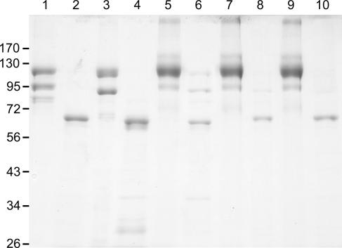
When solubilized, both wild-type and mutant Cry1Aa proteins showed susceptibility to the proteolytic activities of both conditioned medium and B. thuringiensis cell extracts, although mutants may show a somewhat higher level of degradation (Fig. 6). This suggests that for wild-type protoxin during normal production in a harsh proteolytic environment, crystallization is important to sequester the protein into crystalline inclusions that are inaccessible to proteinases, and this may be an important evolutionary force behind the acquisition of crystal-forming properties.
FIG. 6.
Incubation of Cry1Aa and mutants with conditioned medium. Lane 1, Cry1Aa; lane 2, Cry1Aa incubated with conditioned medium; lane 3, Cry variant 1; lane 4, Cry variant 1 incubated with conditioned medium; lane 5, Cry variant 2; lane 6, Cry variant 2 incubated with conditioned medium; lane 7, Cry variant 4; lane 8, Cry variant 4 incubated with conditioned medium; lane 9, Cry variant 5; lane 10, Cry variant 5 incubated with conditioned medium. Numbers at the left are molecular masses (in kilodaltons).
All the sporulation-associated toxins known from both B. sphaericus and B. thuringiensis are deposited as crystal proteins, probably for this reason. It appears from this study that retargeting vegetative toxins to spores is likely to be a major challenge and may require a degree of serendipity in choosing toxins that may be able to form other inclusion types (6). An understanding of the process(es) of crystallization and the motifs that might promote this phenomenon may, in the future, allow us to link the redirection of Mtx toxin synthesis to the sporulation phase with sequences to drive the proteins into a protected, insoluble form. In the meantime, the successful production in crystal form of loop variant Cry protoxins may indicate a more feasible means of adding extra toxins to existing arsenals.
Acknowledgments
This work received financial support from the Biotechnology and Biological Sciences Research Council (L.W. and A.G.; grant number 72/G16021), a National 973 grant (2003CB114201), and an NFSC grant (30470037) from China and the British Council UK-China Science and Technology Fund (C.B., Y.Y., and Z.Y.).
Footnotes
Published ahead of print on 10 November 2006.
REFERENCES
- 1.Abdul-Rauf, M., and D. J. Ellar. 1999. Mutations of loop 2 and loop 3 residues in domain II of Bacillus thuringiensis Cry1C delta-endotoxin affect insecticidal specificity and initial binding to Spodoptera littoralis and Aedes aegypti midgut membranes. Curr. Microbiol. 39:94-98. [DOI] [PubMed] [Google Scholar]
- 2.Almog, O., A. Gonzalez, D. Klein, H. M. Greenblatt, S. Braun, and G. Shoham. 2003. The 0.93Å crystal structure of sphericase: a calcium-loaded serine protease from Bacillus sphaericus. J. Mol. Biol. 332:1071-1082. [DOI] [PubMed] [Google Scholar]
- 3.Almond, B. D., and D. H. Dean. 1994. Intracellular proteolysis and limited diversity of the Bacillus thuringiensis CryIA family of the insecticidal crystal proteins. Biochem. Biophys. Res. Commun. 201:788-794. [DOI] [PubMed] [Google Scholar]
- 4.Altschul, S. F., W. Gish, W. Miller, E. W. Myers, and D. J. Lipman. 1990. Basic local alignment search tool. J. Mol. Biol. 215:403-410. [DOI] [PubMed] [Google Scholar]
- 5.Arantes, O., and D. Lereclus. 1991. Construction of cloning vectors for Bacillus thuringiensis. Gene 108:115-119. [DOI] [PubMed] [Google Scholar]
- 6.Arora, N., A. Selvapandiyan, N. Agrawal, and R. K. Bhatnagar. 2003. Relocation of vegetative insecticidal protein into mother cell of Bacillus thuringiensis. Biochem. Biophys. Res. Commun. 310:158-162. [DOI] [PubMed] [Google Scholar]
- 7.Berry, C., J. Hindley, A. F. Ehrhardt, T. Grounds, I. de Souza, and E. W. Davidson. 1993. Genetic determinants of the host range of the Bacillus sphaericus mosquito larvicidal toxins. J. Bacteriol. 175:510-518. [DOI] [PMC free article] [PubMed] [Google Scholar]
- 8.Bourgouin, C., A. Delécluse, F. de la Torre, and J. Szulmajster. 1990. Transfer of the toxin protein genes of Bacillus sphaericus into Bacillus thuringiensis subsp. israelensis and their expression. Appl. Environ. Microbiol. 56:340-344. [DOI] [PMC free article] [PubMed] [Google Scholar]
- 9.de Maagd, R. A., A. Bravo, C. Berry, N. Crickmore, and H. E. Schnepf. 2003. Structure, diversity, and evolution of protein toxins from spore-forming entomopathogenic bacteria. Annu. Rev. Genet. 37:409-433. [DOI] [PubMed] [Google Scholar]
- 10.Finney, D. J. 1971. Probit analysis—a statistical treatment of the sigmoid response curve, 3rd ed. Cambridge University Press, Cambridge, United Kingdom.
- 11.Graf, R., and H. Briegel. 1985. Isolation of trypsin isozymes from the mosquito Aedes aegypti (L.). Insect Biochem. 15:611-618. [Google Scholar]
- 12.Kasman, L. M., A. A. Lukowiak, S. F. Garczynski, R. J. McNall, P. Youngman, and M. J. Adang. 1998. Phage display of a biologically active Bacillus thuringiensis toxin. Appl. Environ. Microbiol. 64:2995-3003. [DOI] [PMC free article] [PubMed] [Google Scholar]
- 13.Li, J. D., J. Carroll, and D. J. Ellar. 1991. Crystal structure of insecticidal delta-endotoxin from Bacillus thuringiensis at 2.5 Å resolution. Nature 353:815-821. [DOI] [PubMed] [Google Scholar]
- 14.Liu, J.-W., A. G. Porter, B. Y. Wee, and T. Thanabalu. 1996. New gene from nine Bacillus sphaericus strains encoding highly conserved 35.8-kilodalton mosquitocidal toxins. Appl. Environ. Microbiol. 62:2174-2176. [DOI] [PMC free article] [PubMed] [Google Scholar]
- 15.Liu, X. S., and D. H. Dean. 2006. Redesigning Bacillus thuringiensis Cry1Aa toxin into a mosquitocidal toxin. Protein Eng. Design Select. 19:107-111. [DOI] [PubMed] [Google Scholar]
- 16.Marzari, R., P. Edomi, R. K. Bhatnagar, S. Ahmad, A. Selvapandiyan, and A. Bradbury. 1997. Phage display of Bacillus thuringiensis CryIA(a) insecticidal toxin. FEBS Lett. 411:27-31. [DOI] [PubMed] [Google Scholar]
- 17.Pacheco, S., I. Gomez, R. Sato, A. Bravo, and M. Soberon. 2006. Functional display of Bacillus thuringiensis Cry1Ac toxin on T7 phage. J. Invertebr. Pathol. 92:45-49. [DOI] [PubMed] [Google Scholar]
- 18.Rajamohan, F., M. K. Lee, and D. H. Dean. 1998. Bacillus thuringiensis insecticidal proteins: molecular mode of action. Prog. Nucleic Acid Res. Mol. Biol. 60:1-27. [DOI] [PubMed] [Google Scholar]
- 19.Reinert, D. J., I. Carpusca, K. Aktories, and G. E. Schulz. 2006. Structure of the mosquitocidal toxins from Bacillus sphaericus. J. Mol. Biol. 357:1226-1236. [DOI] [PubMed] [Google Scholar]
- 20.Schirmer, J., I. Just, and K. Aktories. 2002. The ADP-ribosylating mosquitocidal toxin from Bacillus sphaericus. J. Biol. Chem. 277:11941-11948. [DOI] [PubMed] [Google Scholar]
- 21.Siezen, R. J., and J. A. Leunissen. 1997. Subtilases: the superfamily of subtilisin-like serine proteases. Protein Sci. 6:501-523. [DOI] [PMC free article] [PubMed] [Google Scholar]
- 22.Silva-Filha, M.-H., L. Regis, C. Nielsen-LeRoux, and J.-F. Charles. 1995. Low-level resistance to Bacillus sphaericus in a field-treated population of Culex quinquefasciatus (Diptera: Culicidae). J. Econ. Entomol. 88:525-530. [Google Scholar]
- 23.Silva-Filha, M. H., C. Nielsen-LeRoux, and J.-F. Charles. 1997. Binding kinetics of Bacillus sphaericus binary toxin to midgut brush-border membranes of Anopheles and Culex sp. mosquito larvae. Eur. J. Biochem. 247:754-761. [DOI] [PubMed] [Google Scholar]
- 24.Smith, G. P., and D. J. Ellar. 1994. Mutagenesis of two surface-exposed loops of the Bacillus thuringiensis CryIC delta-endotoxin affects insecticidal specificity. Biochem. J. 302:611-616. [DOI] [PMC free article] [PubMed] [Google Scholar]
- 25.Tabashnik, B. E., Y. Carriere, T. J. Dennehy, S. Morin, M. S. Sisterson, R. T. Roush, A. M. Shelton, and J. Z. Zhao. 2003. Insect resistance to transgenic Bt crops: lessons from the laboratory and field. J. Econ. Entomol. 96:1031-1038. [DOI] [PubMed] [Google Scholar]
- 26.Tabashnik, B. E., N. L. Cushing, N. Finson, and M. W. Johnson. 1990. Field development of resistance to Bacillus thuringiensis in diamondback moth (Lepidoptera: Plutellidae). J. Econ. Entomol. 83:1671-1676. [Google Scholar]
- 27.Thanabalu, T., J. Hindley, and C. Berry. 1992. Proteolytic processing of the mosquitocidal toxin from Bacillus sphaericus SSII-1. J. Bacteriol. 174:5051-5056. [DOI] [PMC free article] [PubMed] [Google Scholar]
- 28.Thanabalu, T., J. Hindley, J. Jackson-Yap, and C. Berry. 1991. Cloning, sequencing and expression of a gene encoding a 100-kilodalton mosquitocidal toxin from Bacillus sphaericus SSII-1. J. Bacteriol. 173:2776-2785. [DOI] [PMC free article] [PubMed] [Google Scholar]
- 29.Thanabalu, T., and A. G. Porter. 1996. A Bacillus sphaericus gene encoding a novel type of mosquitocidal toxin of 31.8kDa. Gene 170:85-89. [DOI] [PubMed] [Google Scholar]
- 30.Thanabalu, T., and A. G. Porter. 1995. Efficient expression of a 100-kilodalton mosquitocidal toxin in protease-deficient recombinant Bacillus sphaericus. Appl. Environ. Microbiol. 61:4031-4036. [DOI] [PMC free article] [PubMed] [Google Scholar]
- 31.Vilchez, S., J. Jacoby, and D. J. Ellar. 2004. Display of biologically functional insecticidal toxin on the surface of lambda phage. Appl. Environ. Microbiol. 70:6587-6594. [DOI] [PMC free article] [PubMed] [Google Scholar]
- 32.Wati, M., T. Thanabalu, and A. Porter. 1997. Gene from tropical Bacillus sphaericus encoding a protease closely related to subtilisins from Antarctic bacilli. Biochim. Biophys. Acta 1352:56-62. [DOI] [PubMed] [Google Scholar]
- 33.Yuan, Z., C. Nielsen-LeRoux, N. Pasteur, A. Delécluse, J. F. Charles, and R. Frutos. 1999. Cloning and expression of the binary toxin genes of Bacillus sphaericus C3-41 in a crystal minus B. thuringiensis subsp. israelensis. Wei Sheng Wu Xue Bao 39:29-35. [PubMed] [Google Scholar]
- 34.Yuan, Z., Y. M. Zhang, Q. X. Cai, and E. Y. Liu. 2000. High-level field resistance to Bacillus sphaericus C3-41 in Culex quinquefasciatus from Southern China. Biocontrol Sci. Technol. 10:41-49. [Google Scholar]


