Abstract
E2 is one of the three envelope glycoproteins of classical swine fever virus (CSFV). Previous studies indicate that E2 is involved in several functions, including virus attachment and entry to target cells, production of antibodies, induction of protective immune response in swine, and virulence. Here, we have investigated the role of E2 glycosylation of the highly virulent CSFV strain Brescia in infection of the natural host. Seven putative glycosylation sites in E2 were modified by site-directed mutagenesis of a CSFV Brescia infectious clone (BICv). A panel of virus mutants was obtained and used to investigate whether the removal of putative glycosylation sites in the E2 glycoprotein would affect viral virulence/pathogenesis in swine. We observed that rescue of viable virus was completely impaired by removal of all putative glycosylation sites in E2 but restored when mutation N185A reverted to wild-type asparagine produced viable virus that was attenuated in swine. Single mutations of each of the E2 glycosylation sites showed that amino acid N116 (N1v virus) was responsible for BICv attenuation. N1v efficiently protected swine from challenge with virulent BICv at 3 and 28 days postinfection, suggesting that glycosylation of E2 could be modified for development of classical swine fever live attenuated vaccines.
Classical swine fever (CSF) is a highly contagious disease of swine. The etiological agent, CSF virus (CSFV), is a small, enveloped virus with a positive, single-stranded RNA genome and, along with bovine viral diarrhea virus (BVDV) and border disease virus, is classified as a member of the genus Pestivirus within the family Flaviridae (3). The 12.5-kb CSFV genome contains a single open reading frame that encodes a 3,898-amino-acid polyprotein and ultimately yields 11 to 12 final cleavage products (NH2-Npro-C-Erns-E1-E2-p7-NS2-NS3-NS4A-NS4B-NS5A-NS5B-COOH) through co- and posttranslational processing of the polyprotein by cellular and viral proteases (28). Structural components of the CSFV virion include the capsid (C) protein and glycoproteins Erns, E1, and E2. E1 and E2 are anchored to the envelope at their carboxyl termini, and Erns loosely associates with the viral envelope (38, 44, 45). E1 and E2 are type I transmembrane proteins with an N-terminal ectodomain and a C-terminal hydrophobic anchor (38). E2 is considered essential for CSFV replication, as virus mutants containing partial or complete deletions of the E2 gene have proven nonviable (39). E2 is the most immunogenic of the CSFV glycoproteins (15, 40, 44), inducing neutralizing antibodies and protection against lethal CSFV challenge. E2 has been implicated, along with Erns (14) and E1 (43), in viral adsorption to host cells; indeed, chimeric pestiviruses exhibit infectivity and cell tropism phenotypes consistent with those of the E2 gene donor (18, 40). Modifications introduced into these glycoproteins appear to have an important effect on CSFV virulence (21, 29, 31, 41).
Glycosylation is one of the most common types of protein modifications. N-linked oligosaccharides are added to specific asparagine residues in the context of the consensus sequence Asn-X-Ser/Thr (16). Intracellular O glycosylation is characterized by the addition of N-acetylglucosamine to serine and threonine residues in a protein, although the acceptor site does not display a definite consensus sequence (8). Not all predicted sites in a protein sequence are used for carbohydrates, since many of them are inefficiently glycosylated (33) or remain unglycosylated (7).
Putative N-glycosylation sites within CSFV E2 have been predicted previously (23, 42). According to a glycosylation analysis algorithm (http://www.cbs.dtu.dk/services/), E2 of the CSFV strain Brescia has five putative N-linked sites and one putative O-linked glycosylation site, although this is not confirmed by experimental evidence. A sixth N-linked glycosylation site is present in several CSFV strains, with the Brescia sequence differing in one amino acid from the consensus (Asn-X-Ser/Thr). Predicted E2 glycosylation sites are highly conserved among CSFV isolates. Even though glycosylation of Erns, E1, or E2 protein may play a significant role in the CSFV viral replication cycle, the function of added oligosaccharides is not known. In general, glycosylation of enveloped virus structural proteins has been shown to be important for receptor binding, membrane fusion, penetration, virus budding, and infectivity as analyzed in cultured cells (1, 5, 9, 34, 35). However, the significance of viral envelope protein glycosylation in virus replication, pathogenesis, and virulence in the natural host is unknown. Recently it has been shown that glycosylation of porcine respiratory and reproductive syndrome virus GP5 affects virus infectivity, antigenicity, and ability to induce neutralizing antibodies in swine (2). Loss of N-linked glycosylation from the hemagglutinin-neuraminidase (HN) protein from Newcastle disease virus (NDV), a bird pathogen, results in attenuation of the virus in chickens (25). Similarly, degrees of virulence in chickens have been associated with glycosylation patterns of surface proteins hemagglutinin and neuraminidase of the highly pathogenic avian influenza virus H5N1 (13). Pathogenic phenotypes observed upon infection of natural hosts with modified viruses link glycosylation of virus surface proteins with mechanisms such as evasion of the immune system in porcine respiratory and reproductive syndrome virus (2), attenuation in NDV (25), and determinants of virulence in avian influenza (13).
In this study, we used oligonucleotide site-directed mutagenesis of the highly virulent CSFV strain Brescia E2 gene to construct a panel of glycosylation mutants. These mutants were applied to investigate whether the removal of each of these glycosylation sites in the E2 glycoprotein could affect viral infectivity and virulence in swine. We found that rescue of viable virus was completely impaired by removal of all putative glycosylation sites in E2 but was restored when E2 amino acid residue A185 in the mutant virus was reverted to wild-type N. Interestingly, one of the seven single mutations introduced in E2, N116A, renders an attenuated virus (N1v) with decreased virus replication and shedding in infected swine.
MATERIALS AND METHODS
Viruses and cells.
Swine kidney cells (SK6) (37), free of BVDV, were cultured in Dulbecco's minimal essential medium (Gibco, Grand Island, NY) with 10% fetal calf serum (Atlas Biologicals, Fort Collins, CO). CSFV strain Brescia was propagated in SK6 cells and used for the construction of an infectious cDNA clone (29). Growth kinetics was assessed on primary swine macrophage cultures prepared as described by Zsak et al. (46). Titration of CSFV from clinical samples was performed using SK6 cells in 96-well plates (Costar, Cambridge, MA). Viral infectivity was detected, after 4 days in culture, by an immunoperoxidase assay using the CSFV monoclonal antibody (MAb) WH303 (6) and a Vectastain ABC kit (Vector Laboratories, Burlingame, CA). Titers were calculated using the method of Reed and Muench (27) and are expressed as 50% tissue culture infective dose (TCID50)/ml. As performed, test sensitivity was ≥log10 1.8 TCID50/ml. Plaque assays were performed using SK6 cells in six-well plates (Costar). SK6 monolayers were infected, overlaid with 0.5% agarose, and incubated at 37°C for 3 days. Plates were fixed with 50% (vol/vol) ethanol-acetone and stained by immunohistochemistry with MAb WH303 (30).
Construction of CSFV glycosylation mutants.
A full-length infectious clone of the virulent Brescia isolate (pBIC) (29) was used as a template in which putative O- and N-linked glycosylation sites in the E2 glycoprotein were mutated. Glycosylation sites were predicted using analysis tools from the Center for Biological Sequence Analysis (http://www.cbs.dtu.dk/services/). Mutations were introduced by site-directed mutagenesis using a QuickChange XL site-directed mutagenesis kit (Stratagene, Cedar Creek, TX) performed per the manufacturer's instructions and using the following primers (only forward primer sequences are shown): for O1v, CATCATTACATAAGGACGCTTTAGCCACTTCCGTGACATTCGAGC; for N1v, CCCTGTAGTCAAGGGAAAGTACGCCACAACCTTGTTGAATGGTAG; for N2v, AAAGTACAACACAACCTTGTTGGCTGGTAGTGCATTCTACCTAGT; for N3v, ATTCTACTGTAAATGGGGGGGCGCTTGGACATGTGTGAAAGGTGA; for N4v, ATAGGTAAGTGCATTTTGGCAGCTGAGACAGGTTACAGAATAGTG; for N5v, GAGTCATGAGTGCTTGATTGGTGCCACAACTGTCAAGGTGCATGC; and for N6v, AAGGAAAACTTCCTGTACATTCGCCTACGCAAAAACTCTGAGGAA.
In vitro rescue of CSFV Brescia and glycosylation mutants.
Full-length genomic clones were linearized with SrfI and in vitro transcribed using a T7 Megascript system (Ambion, Austin, TX). RNA was precipitated with LiCl and transfected into SK6 cells by electroporation at 500 V, 720 Ω, and 100 W with a BTX 630 electroporator (BTX, San Diego, CA). Cells were seeded in 12-well plates and incubated for 4 days at 37°C and 5% CO2. Virus was detected by immunoperoxidase staining as described above, and stocks of rescued viruses were stored at −70°C.
DNA sequencing and analysis.
Full-length clones and in vitro-rescued viruses were completely sequenced with CSFV-specific primers by a dideoxynucleotide chain termination method (32). Viruses recovered from infected animals were sequenced in the mutated area. Sequencing reaction mixtures were prepared with a Dye Terminator cycle sequencing kit (Applied Biosystems, Foster City, CA). Reaction products were sequenced on a PRISM 3730xl automated DNA sequencer (Applied Biosystems). Sequence data were assembled with the Phrap software program (http://www.phrap.org), with confirmatory assemblies performed using CAP3 (12). The final DNA consensus sequence represented an average fivefold redundancy at each base position. Sequence comparisons were conducted using BioEdit software (http://www.mbio.ncsu.edu/BioEdit/bioedit.html).
Western blot analysis.
Glycosylation status of the E2 glycoprotein of CSFV Brescia infectious clone (BICv) and mutant viruses in lysates of SK6 infected cells was analyzed by Western immunoblotting. CSFV E2 was detected with MAb WH303. SK6 monolayers were infected (multiplicity of infection [MOI] of 1) with BICv or glycosylation mutants, harvested at 48 h postinoculation using a NuPAGE LDS sample buffer system (Invitrogen, Carlsbad, CA), and incubated at 80°C for 20 min. Samples were run under reducing or nonreducing conditions in precast NuPAGE 12% bis-Tris acrylamide gels (Invitrogen). Western immunoblot analyses were performed using a WesternBreeze chemiluminescent immunodetection system (Invitrogen).
Treatment of infected cell extracts with peptide N-glycosidase F (PNGase F) (New England BioLabs, Ipswich, MA) was performed following the manufacturer's directions. Briefly, infected cell extracts were denatured at 100°C for 10 min in glycoprotein denaturing buffer (New England BioLabs). The reaction mixture was then put on ice for 5 min, and PNGase F digestion was performed for 20 h in the presence of 1% NP-40.
Animal infections.
Each of the glycosylation mutants was initially screened for its virulence phenotype in swine relative to that of virulent Brescia virus. Swine used in all animal studies were 10- to 12-week-old, 40-lb commercial-breed pigs inoculated intranasally with 105 TCID50 of either mutant or wild-type virus. For screening, 18 pigs were randomly allocated into nine groups of two animals each, and pigs in each group were inoculated with one of the single glycosylation mutants, ΔO1N1-N6/N3v or BICv. Clinical signs (anorexia, depression, fever, purple skin discoloration, staggering gait, diarrhea, and cough) and changes in body temperature were recorded daily throughout the experiment and scored as previously described (22), with modifications.
To assess the effect of the N1v mutation on virus shedding and distribution in different organs during infection, pigs were randomly allocated into two groups of nine animals each and intranasally inoculated (see above) with N1v or BICv. One pig per group was sacrificed at 2, 4, 6, 8, and 12 days postinfection (dpi). Blood, nasal swabs, and tonsil scraping samples were obtained from pigs at necropsy. Tissue samples (tonsil, mandibular lymph node, spleen, and kidney) were snap-frozen in liquid nitrogen for subsequent virus titration. The remaining four pigs in each room were monitored to check for appearance of clinical signs during a 21-day period.
For protection studies, 12 pigs were randomly allocated into three groups of four animals each. Pigs in groups 1 and 2 were inoculated with N1v, and pigs in group 3 were mock infected. At 3 dpi (group 1) or 28 dpi (group 2), animals were challenged with BICv along with animals in group 3. Clinical signs and body temperature were recorded daily throughout the experiment as described above. Blood, serum, nasal swabs, and tonsil scrapings were collected at various times postchallenge, with blood obtained from the anterior vena cava in EDTA-containing tubes (Vacutainer) for total and differential white blood cell counts. Total and differential white blood cell and platelet counts were obtained using a Beckman Coulter ACT (Beckman Coulter, Fullerton, CA).
RESULTS
Construction of CSFV glycosylation mutant viruses.
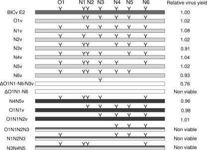
Infectious RNA was in vitro transcribed from full-length infectious clones of the CSFV Brescia strain or a set of glycosylation mutants (Table 1; Fig. 1) and used to transfect SK6 cells. Mutants referred to as O1, N1, N2, N3, N4, N5, and N6 represent each of seven putative glycosylation sites starting from the N terminus of E2 (Table 1), whereas multiple mutants are represented by combinations of indicated sites (Fig. 1). Viruses were rescued from transfected cells by day 4 posttransfection. Nucleotide sequences of the rescued virus genomes were identical to parental DNA plasmids, confirming that only mutations at predicted glycosylation sites were reflected in rescued viruses.
TABLE 1.
Set of CSFV E2 glycosylation mutant viruses constructed in this study
| E2 position(s) | Wild-type sequence(s) | Mutant sequence(s) | Codon change(s) | Mutant |
|---|---|---|---|---|
| 75 | P | A | CCC → GCC | O1v |
| 116 | NTTL | ATTL | AAC → GCC | N1v |
| 121 | NGSA | AGSA | AAT → GCT | N2v |
| 185 | NWTC | AWTC | AAC → GCC | N3v |
| 229 | NETG | AETG | AAT → GCT | N4v |
| 260 | NTTV | ATTV | AAC → GCC | N5v |
| 297 | NYAK | AYAK | AAC → GCC | N6v |
| 75, 116, 121, 229, 260, 297 | P, NTTL, NGSA, NETG, NTTV, NYAK | A, ATTL, AGSA, AETG, ATTV, AYAK | CCC → GCC, AAC → GCC, AAT → GCT, AAT → GCT, AAC → GCC, AAC → GCC | ΔO1N1-N6/N3v |
| 75, 116 | P, NTTL | A, ATTL | CCC → GCC, AAC → GCC | O1N1v |
| 75, 116, 121 | P, NTTL, NGSA | A, ATTL, AGSA | CCC → GCC, AAC → GCC, AAT → GCT | O1N1N2v |
| 229, 260 | NETG, NTTV | AETG, ATTV | AAT → GCT, AAC → GCC | N4N5v |
FIG. 1.
Schematic representation of glycosylation mutants of CSFV E2 protein, generated by site-directed mutagenesis of a cDNA full-length clone, pBIC. Wild-type E2 glycoprotein is shown at the top. Y, putative glycosylation site. Mutants were named with an O (O-linked glycosylation) or an N (N-linked glycosylation) followed by a number(s) that represents the relative position(s) of putative glycosylation sites within the E2 amino acid sequence (76, 116, 121, 185, 229, 260, or 297). The relative virus yield is the final-point virus yield as a proportion of the final-endpoint (72 h postinfection) virus yield of parental BICv.
Replication of glycosylation mutants in vitro.
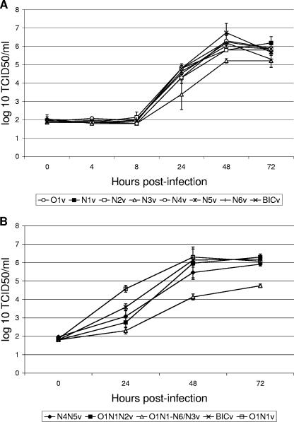
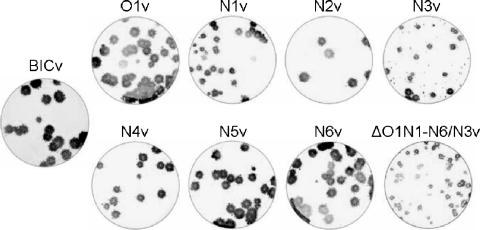
In vitro growth characteristics of mutant viruses O1v, N1v, N2v, N3v, N4v, N5v, and N6v and multiple site mutants ΔO1N1-N6/N3v, O1N1v, O1N1N2v, and N4N5v were evaluated relative to those of parental BICv in a multistep growth curve (Fig. 2A and B). Primary swine macrophage cultures were infected at an MOI of 0.01 TCID50 per cell. Virus was adsorbed for 1 h (time zero), and samples were collected at various times postinfection through 72 h. All single glycosylation site mutants, except N3v, exhibited growth characteristics practically indistinguishable from those of BICv. N3v exhibited a lower rate of growth and a 10-fold decrease in the final virus yield (Fig. 2A). Multiple glycosylation site mutant ΔO1N1-N6/N3v also exhibited delayed growth and reduced viral yield compared with multiple site mutants N4N5v, O1N1v, and O1N1N2v and parental BICv (Fig. 2B). Additionally, when viruses were tested for their plaque size in SK6 cells, N1v, N3v, and ΔO1N1-N6/N3v exhibited a noticeable reduction in plaque size relative to that of BICv (Fig. 3). Interestingly, viruses were not rescued from SK6 cells transfected with RNA transcribed from full-length cDNA clones carrying multiple glycosylation site mutations (ΔO1N1-N6, O1N1N2N3, N1N2N3, and N3N4N5) that included substitutions at the N3 position (N185).
FIG. 2.
In vitro growth characteristics of E2 individual (A) and multiple (B) glycosylation mutants and parental BICv. Primary swine macrophage cultures were infected (MOI of 0.01) with each of the mutants or BICv and virus yield titers determined at various times postinfection in SK6 cells. Data represent means and standard deviations from two independent experiments. Sensitivity of virus detection was ≥log10 1.8 TCID50/ml.
FIG. 3.
Plaque formation of E2 glycosylation mutants and BICv. SK6 monolayers were infected, overlaid with 0.5% agarose, and incubated at 37°C for 3 days. Plates were fixed with 50% (vol/vol) ethanol-acetone and stained by immunohistochemistry with MAb WH303 (34).
Analysis of relative electrophoretic mobilities of E2 in virus mutants.
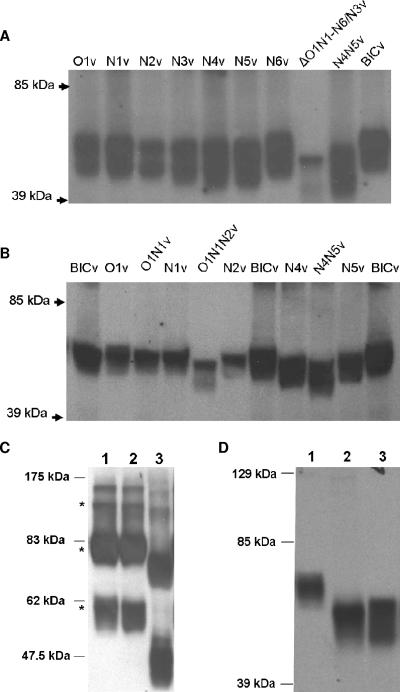
Relative electrophoretic mobilities of E2 were analyzed by Western blotting in lysates of SK6 cells infected with different mutant and parental BICv viruses by use of MAb WH303. Assuming that differences in E2 mobility among mutants and parental viruses are likely due to the number of carbohydrate moieties attached to the protein, we observed that E2 from single mutants N1v, N2v, N3v, N4v, and N5v migrated further than E2 from mutants O1v and N6v or parental BICv (Fig. 4A and B). The data suggested that, while N1, N2, N3, N4, and N5 sites are targeted for the addition of glycans in swine cells, O1 and N6 sites (Table 1) are not used for glycosylation of BICv E2. Those observations were further confirmed by analyzing the mobility of E2 in lysates from cells infected with mutant viruses missing two or more putative glycosylation sites (Fig. 1 and 4A and B). For example, double mutant O1N1v E2 migrated faster than O1v and BICv E2 but paralleled N1v E2 mobility (Fig. 4B). Triple mutant O1N1N2v E2 protein migrated faster than N1v or N2v E2 protein (Fig. 4B), probably due to the additive effect of the mobility shift caused by lack of glycans at both the N1 and N2 sites. N3v E2, like N2v E2, migrated more quickly than parental BICv E2 (Fig. 4A). As observed with O1N1N2v, double mutant N4N5v E2 migrated further than N4v and N5v E2 proteins (Fig. 4B), suggesting that the disparity in mobility is due to the absence of both glycosylation sites. The largest change in E2 mobility was observed with multiple site mutant ΔO1N1-N6/N3v. This virus is deficient in four of five N-linked glycosylation sites that showed different E2 migration patterns (Fig. 4A). Overall, our data firmly suggest that CSFV E2 N1, N2, N3, N4, and N5 sites (Fig. 1) are targeted for glycosylation in swine SK6 cells.
FIG. 4.
Analysis of E2 glycosylation mutants was done by Western immunoblotting. SK6 monolayers were infected (MOI of 1) with each of the mutants or parental BICv or mock infected and harvested 48 h postinfection. Cell lysates were run under reducing (A, B, and D) or nonreducing (C) conditions in 12% sodium dodecyl sulfate-polyacrylamide gels. CSFV E2 was detected with CSFV E2 monoclonal antibody WH303. (C) Lane 1, BICv; lane 2, N1v; lane 3, ΔO1N1-N6/N3v. Asterisks indicate, from top to bottom, E2 homodimers, E1-E2 heterodimers, and monomeric E2, as described by Weiland et al. (44). (D) Lane 1, untreated BICv; lane 2, PNGase F-treated BICv; lane 3, untreated ΔO1N1-N6/N3v.
Absence of glycosylation in N1 and ΔO1N1-N6/N3 viruses does not affect the formation of E2 homo- and heterodimers (Fig. 4C). Western blot analysis performed with extracts of SK6 cells infected with either of the viruses and run under nonreducing conditions demonstrated the presence of bands with apparent molecular masses of 50, 66, and 90 to 100 kDa corresponding to E2 monomers, E2-E1 heterodimers, and E2 homodimers, respectively, as previously described by Weiland et al. (44). The pattern of bands was maintained in N1 and ΔO1N1-N6/N3 viruses with the expected shift in their electrophoretic mobility. Therefore, mutations of CSFV N-linked glycosylation sites would not affect E2 dimerization in infected cells.
Treatment of SK6 BICv-infected cell extracts with PNGase F effectively removed N-linked glycosylation, resulting in a protein that migrated with a slightly faster electrophoretic mobility than the E2 ΔO1N1-N6/N3v protein (Fig. 4D). This is expected, since digestion with PNGase F would generate completely unglycosylated proteins, whereas ΔO1N1-N6/N3v E2 protein still conserves the N3-linked glycosylation site.
Mutants N1v and ΔO1N1-N6/N3v lack determinants necessary for CSFV virulence in swine.
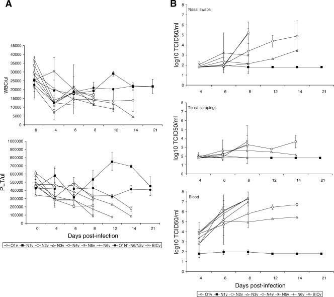
To examine the effect of E2 glycosylation on CSFV virulence, pigs were intranasally inoculated with 105 TCID50 of the ΔO1N1-N6/N3v mutant (Table 1) and monitored for clinical disease. This mutant, lacking all putative glycosylation sites except the N3 site, exhibited an attenuated phenotype (Table 2), with no considerable hematological changes in infected animals (Fig. 5), suggesting a significant role of E2-added oligosaccharides in viral virulence in swine.
TABLE 2.
Swine survival and fever response following infection with CSFV E2 glycosylation mutants and parental BICv
| Virus | No. of survivors/total no. of infected animals | Mean time to death (no. of days ± SD) | Time to onset of fever (no. of days ± SD) | Duration of fever (no. of days ± SD) |
|---|---|---|---|---|
| BICv | 0/2 | 12 (0.00) | 5 (1.41) | 6.5 (0.70) |
| O1v | 0/2 | 9.5 (2.12) | 5 (0.00) | 4.5 (2.12) |
| N1v | 6/6 | |||
| N2v | 0/2 | 11 (0.00) | 4.5 (0.70) | 5.5 (2.12) |
| N3v | 0/2 | 12 (5.65) | 6 (2.82) | 6 (2.82) |
| N4v | 0/2 | 16 (0.00) | 6 (1.41) | 8 (1.41) |
| N5v | 0/2 | 8.5 (0.70) | 5 (0.00) | 3.5 (0.70) |
| N6v | 0/2 | 8 (0.00) | 5 (0.00) | 2.5 (0.70) |
| ΔO1N1- N6/N3v | 2/2 |
FIG. 5.
Hematological data and virus titers of clinical samples from animals infected with CSFV E2 glycosylation mutants and parental BICv. (A) Peripheral white blood cell (WBC) and platelet (PLT) counts in pigs infected with E2 glycosylation mutant viruses and parental BICv. Counts are expressed as numbers/μl of blood. Data represent means and standard deviations from at least two animals. (B) Virus titers in nasal swabs, tonsil scrapings, and blood from pigs infected with E2 glycosylation mutants or BICv. Each point represents log10 TCID50/ml (means ± standard deviations) from at least two animals. Sensitivity of virus detection was ≥log10 1.8 TCID50/ml.
To confirm the role of E2 glycosylation and further establish the impact of mutations at individual glycosylation sites on swine virulence, individual mutants were intranasally inoculated and evaluated relative to the parental virus. BICv exhibited a characteristic virulent phenotype (Table 2). Animals infected with N1v survived the infection and remained normal throughout the observation period (21 days). All animals infected with O1v, N2v, N3v, N4v, N5v, and N6v presented clinical signs of CSF starting 4 to 6 dpi, with clinical presentation and severity similar to those observed to occur in animals inoculated with BICv. White blood cell and platelet counts dropped by 4 to 6 dpi in animals inoculated with O1v, N2v, N3v, N4v, N5v, N6v, and BICv and kept declining until death, while a transient decrease was observed in animals inoculated with N1v (Fig. 5A).
Viremia in N1v-inoculated animals was transient (Fig. 5B) and significantly reduced by 104 to 105 from that observed in swine infected with O1v, N2v, N3v, N4v, N5v, N6v, and BICv. A similar pattern was observed for nasal and tonsil samples (Fig. 5B), with no virus titers detected in tonsil samples obtained from N1v-infected animals. In all cases, partial nucleotide sequences of E2 protein from viruses recovered from infected animals were identical to those of stock viruses used for inoculation (data not shown).
The capability of N1v to establish a systemic infection in intranasally inoculated animals was compared with that of virulent parental virus BICv. Randomly selected animals were euthanized at 2, 4, 6, 8, and 12 dpi (one animal/time point/group), and virus titration was performed with collected tissues (tonsils, mandibular lymph nodes, kidney, and spleen). Titers measured in those tissue samples are shown in Table 3. In vivo replication of N1v was transient in tonsils, with titers reduced up to 102 to 105 depending on the time postinfection relative to those of BICv. Differences between N1v and BICv virus titers were also observed for mandibular lymph nodes and spleen, and no mutant virus was detected in kidney, indicating a limited capability of N1v to spread within the host.
TABLE 3.
Titers of virus in tissues after intranasal inoculation with mutant N1v and parental BICv
| Virus | dpi | Log10 TCID50/g ina:
|
|||
|---|---|---|---|---|---|
| Tonsil | Mandibular lymph node | Spleen | Kidney | ||
| N1v | 2 | ND | 2.13 | ND | ND |
| 4 | 4.3 | ND | ND | ND | |
| 6 | 4.8 | 4.3 | 4.47 | ND | |
| 8 | 2.8 | 2.13 | 1.97 | ND | |
| 12 | ND | ND | ND | ND | |
| BICv | 2 | ND | ND | ND | ND |
| 4 | 6.3 | 4.13 | 4.8 | ND | |
| 6 | 7.3 | 5.63 | 6.3 | 5.13 | |
| 8 | 7.8 | 7.8 | 7.47 | 7.47 | |
| 12 | D | D | D | D | |
ND, not detectable, i.e., virus titers equal to or less than 1.8 TCID50 (log10). D, animals in this group were all dead by this time point.
N1v mutant protects pigs against lethal CSFV challenge.
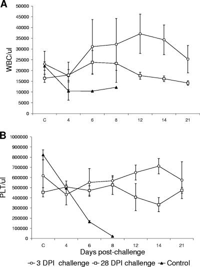
The limited in vivo replication kinetics of N1v is similar to that observed with CSICv (29), a CSFV vaccine strain. However, restricted viral in vivo replication could also impair protection against wild-type-virus infection. Thus, the ability of N1v to induce protection against virulent BICv was assessed in early- and late-vaccination exposure experiments. Groups of pigs (n = 4) were intranasally inoculated with N1v and challenged at 3 or 28 dpi. Mock-vaccinated control pigs receiving BICv only (n = 3) developed anorexia, depression, and fever by 4 days postchallenge (dpc) and a marked reduction of circulating leukocytes and platelets by 4 dpc (Fig. 6) and died or were euthanized in extremis by 9 dpc. Notably, N1v induced complete protection by 3 dpi. All pigs survived infection and remained clinically normal, without significant changes in their hematological values (Fig. 6). Pigs challenged at 28 days after N1v infection were protected, remaining clinically normal, with no alterations of hematological profiles after challenge (Fig. 6). Viremia and virus shedding of vaccinated-exposed animals were examined at 4, 6, 8, 12, 14, and 21 dpc (Table 4). As expected, in mock-vaccinated control animals, viremia was observed by 4 dpc, with virus titers remaining high by 8 dpc (107.8 TCID50/ml) in the surviving pigs. Furthermore, virus was detected in nasal swabs and tonsil scrapings of these animals after 4 dpc. Conversely, viremia was detected only at 4 dpc in animals challenged at 3 dpi (Table 4), but no virus was detected in nasal or tonsil samples in this group during the observation period. Virus was not detected in clinical samples obtained from pigs challenged at 28 dpi. Even though N1v showed limited in vivo growth, a solid protection was induced shortly after vaccination.
FIG. 6.
(A) Peripheral white blood cell (WBC) and (B) platelet (PLT) counts in pigs mock vaccinated or vaccinated with N1v and challenged at 3 or 28 dpi with BICv. Values for control, mock-vaccinated, and challenged animals are represented by filled triangles. Counts are expressed as numbers/μl and represent the means and standard deviations from four individuals.
TABLE 4.
Detection of virus in nasal swabs, tonsil scrapings, and blood samples obtained after challenge of N1v-vaccinated animals with virulent BICv
| Group | Sample | No. of animals positive for virus isolation/ total no. of challenged animals at daya:
|
||||||
|---|---|---|---|---|---|---|---|---|
| C | 4 | 6 | 8 | 12 | 14 | 21 | ||
| Group challenged | Nasal | 0/4 | 0/4 | 0/4 | 0/4 | 0/4 | 0/4 | 0/4 |
| 3 dpi | Tonsil | 0/4 | 0/4 | 0/4 | 0/4 | 0/4 | 0/4 | 0/4 |
| Blood | 0/4 | 4/4 (3.96) | 0/4 | 0/4 | 0/4 | 0/4 | 0/4 | |
| Group challenged | Nasal | 0/4 | 0/4 | 0/4 | 0/4 | 0/4 | 0/4 | 0/4 |
| 28 dpi | Tonsil | 0/4 | 0/4 | 0/4 | 0/4 | 0/4 | 0/4 | 0/4 |
| Blood | 0/4 | 0/4 | 0/4 | 0/4 | 0/4 | 0/4 | 0/4 | |
| Control group | Nasal | 0/3 | 0/3 | 1/1 (6.63) | 1/1 (4.3) | |||
| Tonsil | 0/3 | 1/3 (1.97) | 1/1 (4.8) | 1/1 (1.97) | ||||
| Blood | 0/3 | 1/3 (2.47) | 1/1 (7.8) | 1/1 (7.8) | ||||
Day C, day of challenge. Other days are days postchallenge. Numbers in parentheses indicate average virus titers expressed as log10 TCID50/ml.
DISCUSSION
Virus glycoproteins are crucial in key steps of the virus cycle, such as attachment to host cell receptors, entry, assembly of newly produced viral progeny, and exit. In vivo viral glycoproteins have been shown to influence infectivity (2), virulence (13), and host immune response (24, 25). Added oligosaccharides confer proper function to viral glycoproteins since alteration of those glycosylation sites has shown dramatic consequences for viruses affecting protein folding (10, 17, 36) and protein active conformation (20). In this study, we analyzed the role of glycosylation of the CSFV E2 glycoprotein in virus virulence in swine. All putative glycosylation sites in E2 were modified by site-directed mutagenesis using a full-length cDNA infectious clone of virulent strain Brescia as the target sequence (29). Here, we showed that some of these sites have a major role in virulence and protection and that some of the sites seem to be critical for the production of viable virus. Interestingly, not all of the sites seem to be essential for in vitro or in vivo infectivity.
CSFV strain Brescia E2 glycoprotein contains five or six N-linked sites and one O-linked putative glycosylation site (http://www.cbs.dtu.dk/services/) (23). Sequence analysis of CSFV E2 glycoproteins showed that five of the N-linked glycosylation sites are highly conserved (N116, N121, N185, N229, and N260); three of them, at CSFV E2 positions N116 (N1), N185 (N3), and N229 (N4), are also highly conserved among BVDV types I and II and border disease virus (data not shown), implying an important role for these sites in all pestiviruses. However, very little is known about the role of glycosylation in the function of pestivirus glycoproteins. A previous study that examined the closely related pestivirus BVDV E2 glycoprotein expressed in a baculovirus/insect cell system (26) showed that the pattern of glycosylation affects the ability of the isolated glycoprotein to prevent infection of calf testis cells with BVDV, an otherwise inhibitory effect observed with wild-type E2 protein (14). The same study also showed that modification of N1 and N3 sites in BVDV E2 impaired expression and secretion of the protein in insect cells, suggesting that glycosylation at those sites is essential for correct folding and subsequent secretion of E2. Although these experiments were performed with a baculovirus expression system and in our study we used recombinant viruses, the gathered evidence supports the idea that glycosylation at N1 and N3 sites is critical for E2 activity.
Electrophoretic mobility analysis of the E2 glycoprotein in lysates obtained from infected SK6 cells shows that amino acid residues N116 (N1v), N121 (N2v), N185 (N3v), N229 (N4v), and N260 (N5v) are used for carbohydrate addition (Fig. 4). In vitro growth characteristics and virus progeny yields of these mutants assessed in primary swine macrophage cultures, a CSFV natural target cell, were comparable to that of parental BICv, except mutant N3v (with the N185A mutation), which demonstrated delayed growth kinetics (Fig. 2A). Suggestive of a role for CSFV E2 glycosylation patterns in virus attachment, entry, and/or exit from infected cells was the small-plaque phenotype exhibited by single mutants N1v (with the N116A mutation) and N3v (with the N185A mutation) and multiple mutant ΔO1N1-N6/N3v, in which a substantial plaque size reduction relative to that of parental BICv was observed to occur in infected SK6 cells (Fig. 3). Similarly, loss of one specific N-linked glycosylation site (G4) from the HN protein of NDV yielded plaques in cultured cells of considerably smaller size (25), while three other single mutants (G1, G2, and G3) produced plaques comparable to the size of the parental virus. The G4 glycosylation site in the NDV HN protein has been shown to be necessary for correct folding and transport of the protein to the cell surface of infected cells (19, 25). Interestingly, G4 virus was considerably attenuated in chickens (25). In the case of CSFV, we have observed previously that other BICv-derived viruses containing recombinant E2 protein (29) showed reduced plaque size as well. Like N1v and ΔO1N1-N6/N3v here, those recombinant viruses were also attenuated in swine. In this study we also observed that mutant N3v showed delayed growth kinetics in primary swine macrophage cultures. Further, mutant viruses showing plaque sizes smaller than those of parental BICv on SK6 cells (Fig. 2A and 3) retained the capability of causing severe disease in swine (Table 3). These data suggest that changes in the E2 protein due to the mutation N185A in N3v, unlike the N116A mutation in N1v, are not sufficient to alter virus range within the host.
Cleavage and glycosylation patterns of the hemagglutinin gene of H5 avian influenza viruses have been shown to affect pathogenicity in chickens (4, 11). More recently it has been shown that glycosylation patterns of the neuraminidase gene of highly pathogenic H5N1 avian flu viruses are important for increased virulence in chickens (13). The mechanisms by which these patterns affect avian flu virulence are unknown. Similarly, a single mutation (mutant N1v) or multiple mutations (mutant ΔO1N1-N6/N3v) within E2 rendered attenuated viruses with restricted in vivo replication ability (Tables 3 and 4). Unlike the acute fatal disease induced by BICv, infections caused by these mutants were subclinical in swine and characterized by decreased viral replication in target organs and reduced virus shedding. Interestingly, mutants O1v, N2v, N3v, N4v, N5v, and N6v retained the same capability of causing severe disease in swine as parental BICv, showing that in vivo E2 functions are retained and not influenced by the lack of glycans at positions N116, N121, N185, N229, and N260. As with avian flu, the genetic basis and the molecular mechanisms underlying CSFV virulence remain unknown.
As shown in this study, single mutations of E2 putative glycosylation sites have no effect on in vitro or in vivo infectivity of CSFV, with the exception of residue N116 in the N1v mutant. However, when multiple site mutations were introduced in E2, we observed that certain residue changes (O1N1N2N3, N1N2N3, N3N4N5, and O1N1-N6) rendered nonviable viruses (data not shown). Conversely, when residue N185 (N3) was left unmodified in those clones, virus viability was not affected.
In summary, our studies determined the utilization of five potential N-glycosylation sites in the CSFV strain Brescia E2 protein. Individual N-linked glycosylation sites are not essential for viral particle formation or virus infectivity in cultured swine macrophages or the natural host, with one individual site, N116, involved in attenuation of the virulent parental virus. This study also showed that in the context of three or more putative glycosylation site modifications, residue N185 is critical for virus viability. The effective protective immunity elicited by N1v suggests that glycosylation of E2 could be modified for the development of live attenuated vaccines. An improved understanding of the genetic basis of virus virulence and host range will permit future rational design of efficacious biological tools for controlling CSF.
Acknowledgments
We thank the Plum Island Animal Disease Center Animal Care Unit staff for excellent technical assistance. We thank Melanie Prarat for editing the manuscript.
Footnotes
Published ahead of print on 15 November 2006.
REFERENCES
- 1.Abe, Y., E. Takashita, K. Sugawara, Y. Matsuzaki, Y. Muraki, and S. Hongo. 2004. Effect of the addition of oligosaccharides on the biological activities and antigenicity of influenza A/H3N2 virus hemagglutinin. J. Virol. 78:9605-9611. [DOI] [PMC free article] [PubMed] [Google Scholar]
- 2.Ansari, I. H., B. Kwon, F. A. Osorio, and A. K. Pattnaik. 2006. Influence of N-linked glycosylation of porcine reproductive and respiratory syndrome virus GP5 on virus infectivity, antigenicity, and ability to induce neutralizing antibodies. J. Virol. 80:3994-4004. [DOI] [PMC free article] [PubMed] [Google Scholar]
- 3.Becher, P., R. Avalos Ramirez, M. Orlich, S. Cedillo Rosales, M. Konig, M. Schweizer, H. Stalder, H. Schirrmeier, and H. J. Thiel. 2003. Genetic and antigenic characterization of novel pestivirus genotypes: implications for classification. Virology 311:96-104. [DOI] [PubMed] [Google Scholar]
- 4.Deshpande, K. L., V. A. Fried, M. Ando, and R. G. Webster. 1987. Glycosylation affects cleavage of an H5N2 influenza virus hemagglutinin and regulates virulence. Proc. Natl. Acad. Sci. USA 84:36-40. [DOI] [PMC free article] [PubMed] [Google Scholar]
- 5.Doms, R. W., R. A. Lamb, J. K. Rose, and A. Helenius. 1993. Folding and assembly of viral membrane proteins. Virology 193:545-562. [DOI] [PubMed] [Google Scholar]
- 6.Edwards, S., V. Moennig, and G. Wensvoort. 1991. The development of an international reference panel of monoclonal antibodies for the differentiation of hog cholera virus from other pestiviruses. Vet. Microbiol. 29:101-108. [DOI] [PubMed] [Google Scholar]
- 7.Gavel, Y., and G. von Heijne. 1990. Sequence differences between glycosylated and non-glycosylated Asn-X-Thr/Ser acceptor sites: implications for protein engineering. Protein Eng. 3:433-442. [DOI] [PMC free article] [PubMed] [Google Scholar]
- 8.Gupta, R., and S. Brunak. 2002. Prediction of glycosylation across the human proteome and the correlation to protein function. Pac. Symp. Biocomput. 7:310-322. [PubMed] [Google Scholar]
- 9.Hanna, S. L., T. C. Pierson, M. D. Sanchez, A. A. Ahmed, M. M. Murtadha, and R. W. Doms. 2005. N-linked glycosylation of West Nile virus envelope proteins influences particle assembly and infectivity. J. Virol. 79:13262-13274. [DOI] [PMC free article] [PubMed] [Google Scholar]
- 10.Hebert, D. N., J. X. Zhang, W. Chen, B. Foellmer, and A. Helenius. 1997. The number and location of glycans on influenza hemagglutinin determine folding and association with calnexin and calreticulin. J. Cell Biol. 139:613-623. [DOI] [PMC free article] [PubMed] [Google Scholar]
- 11.Horimoto, T., and Y. Kawaoka. 1994. Reverse genetics provides direct evidence for a correlation of hemagglutinin cleavability and virulence of an avian influenza A virus. J. Virol. 68:3120-3128. [DOI] [PMC free article] [PubMed] [Google Scholar]
- 12.Huang, X., and A. Madan. 1999. CAP3: a DNA sequence assembly program. Genome Res. 9:868-877. [DOI] [PMC free article] [PubMed] [Google Scholar]
- 13.Hulse, D. J., R. G. Webster, R. J. Russell, and D. R. Perez. 2004. Molecular determinants within the surface proteins involved in the pathogenicity of H5N1 influenza viruses in chickens. J. Virol. 78:9954-9964. [DOI] [PMC free article] [PubMed] [Google Scholar]
- 14.Hulst, M. M., and R. J. Moormann. 1997. Inhibition of pestivirus infection in cell culture by envelope proteins E(rns) and E2 of classical swine fever virus: E(rns) and E2 interact with different receptors. J. Gen. Virol. 78:2779-2787. [DOI] [PubMed] [Google Scholar]
- 15.Konig, M., T. Lengsfeld, T. Pauly, R. Stark, and H. J. Thiel. 1995. Classical swine fever virus: independent induction of protective immunity by two structural glycoproteins. J. Virol. 69:6479-6486. [DOI] [PMC free article] [PubMed] [Google Scholar]
- 16.Kornfeld, R., and S. Kornfeld. 1985. Assembly of asparagine-linked oligosaccharides. Annu. Rev. Biochem. 54:631-664. [DOI] [PubMed] [Google Scholar]
- 17.Land, A., and I. Braakman. 2001. Folding of the human immunodeficiency virus type 1 envelope glycoprotein in the endoplasmic reticulum. Biochimie 83:783-790. [DOI] [PubMed] [Google Scholar]
- 18.Liang, D., I. F. Sainz, I. H. Ansari, L. H. Gil, V. Vassilev, and R. O. Donis. 2003. The envelope glycoprotein E2 is a determinant of cell culture tropism in ruminant pestiviruses. J. Gen. Virol. 84:1269-1274. [DOI] [PubMed] [Google Scholar]
- 19.McGinnes, L. W., and T. G. Morrison. 1995. The role of individual oligosaccharide chains in the activities of the HN glycoprotein of Newcastle disease virus. Virology 212:398-410. [DOI] [PubMed] [Google Scholar]
- 20.Meunier, J. C., A. Fournillier, A. Choukhi, A. Cahour, L. Cocquerel, J. Dubuisson, and C. Wychowski. 1999. Analysis of the glycosylation sites of hepatitis C virus (HCV) glycoprotein E1 and the influence of E1 glycans on the formation of the HCV glycoprotein complex. J. Gen. Virol. 80:887-896. [DOI] [PubMed] [Google Scholar]
- 21.Meyers, G., A. Saalmüller, and M. Buttner. 1999. Mutations abrogating the RNase activity in glycoprotein Erns of the pestivirus classical swine fever virus lead to virus attenuation. J. Virol. 73:10224-10235. [DOI] [PMC free article] [PubMed] [Google Scholar]
- 22.Mittelholzer, C., C. Moser, J. D. Tratschin, and M. A. Hofmann. 2000. Analysis of classical swine fever virus replication kinetics allows differentiation of highly virulent from avirulent strains. Vet. Microbiol. 74:293-308. [DOI] [PubMed] [Google Scholar]
- 23.Moormann, R. J., P. A. Warmerdam, B. van der Meer, and M. M. Hulst. 1990. Nucleotide sequence of hog cholera virus RNA: properties of the polyprotein encoded by the open reading frame spanning the viral genomic RNA. Vet. Microbiol. 23:185-191. [DOI] [PubMed] [Google Scholar]
- 24.Mori, K., Y. Yasutomi, S. Ohgimoto, T. Nakasone, S. Takamura, T. Shioda, and Y. Nagai. 2001. Quintuple deglycosylation mutant of simian immunodeficiency virus SIVmac239 in rhesus macaques: robust primary replication, tightly contained chronic infection, and elicitation of potent immunity against the parental wild-type strain. J. Virol. 75:4023-4028. [DOI] [PMC free article] [PubMed] [Google Scholar]
- 25.Panda, A., S. Elankumaran, S. Krishnamurthy, Z. Huang, and S. K. Samal. 2004. Loss of N-linked glycosylation from the hemagglutinin-neuraminidase protein alters virulence of Newcastle disease virus. J. Virol. 78:4965-4975. [DOI] [PMC free article] [PubMed] [Google Scholar]
- 26.Pande, A., B. V. Carr, S. Y. Wong, K. Dalton, I. M. Jones, J. W. McCauley, and B. Charleston. 2005. The glycosylation pattern of baculovirus expressed envelope protein E2 affects its ability to prevent infection with bovine viral diarrhoea virus. Virus Res. 114:54-62. [DOI] [PubMed] [Google Scholar]
- 27.Reed, L. J., and H. A. Muench. 1938. A simple method of estimating fifty per cent endpoints. Am. J. Hyg. 27:493-497. [Google Scholar]
- 28.Rice, C. M. 1996. Flaviviridae: the viruses and their replication, p. 931-959. In D. M. Knipe, B. N. Fields, and P. Howley (ed.), Fundamental virology, 3rd ed. Lippincott Raven, Philadelphia, PA.
- 29.Risatti, G. R., M. V. Borca, G. F. Kutish, Z. Lu, L. G. Holinka, R. A. French, E. R. Tulman, and D. L. Rock. 2005. The E2 glycoprotein of classical swine fever virus is a virulence determinant in swine. J. Virol. 79:3787-3796. [DOI] [PMC free article] [PubMed] [Google Scholar]
- 30.Risatti, G. R., J. D. Callahan, W. M. Nelson, and M. V. Borca. 2003. Rapid detection of classical swine fever virus by a portable real-time reverse transcriptase PCR assay. J. Clin. Microbiol. 41:500-505. [DOI] [PMC free article] [PubMed] [Google Scholar]
- 31.Risatti, G. R., L. G. Holinka, Z. Lu, G. F. Kutish, E. R. Tulman, R. A. French, J. H. Sur, D. L. Rock, and M. V. Borca. 2005. Mutation of E1 glycoprotein of classical swine fever virus affects viral virulence in swine. Virology 343:116-127. [DOI] [PubMed] [Google Scholar]
- 32.Sanger, F., S. Nicklen, and A. R. Coulson. 1977. DNA sequencing with chain-terminating inhibitors. Proc. Natl. Acad. Sci. USA 74:5463-5467. [DOI] [PMC free article] [PubMed] [Google Scholar]
- 33.Shakin-Eshleman, S. H., A. T. Remaley, J. R. Eshleman, W. H. Wunner, and S. L. Spitalnik. 1992. N-linked glycosylation of rabies virus glycoprotein. Individual sequons differ in their glycosylation efficiencies and influence on cell surface expression. J. Biol. Chem. 267:10690-10698. [PubMed] [Google Scholar]
- 34.Shi, X., K. Brauburger, and R. M. Elliott. 2005. Role of N-linked glycans on Bunyamwera virus glycoproteins in intracellular trafficking, protein folding, and virus infectivity. J. Virol. 79:13725-13734. [DOI] [PMC free article] [PubMed] [Google Scholar]
- 35.Shi, X., and R. M. Elliott. 2004. Analysis of N-linked glycosylation of Hantaan virus glycoproteins and the role of oligosaccharide side chains in protein folding and intracellular trafficking. J. Virol. 78:5414-5422. [DOI] [PMC free article] [PubMed] [Google Scholar]
- 36.Slater-Handshy, T., D. A. Droll, X. Fan, A. M. Di Bisceglie, and T. J. Chambers. 2004. HCV E2 glycoprotein: mutagenesis of N-linked glycosylation sites and its effects on E2 expression and processing. Virology 319:36-48. [DOI] [PubMed] [Google Scholar]
- 37.Terpstra, C., R. Woortmeyer, and S. J. Barteling. 1990. Development and properties of a cell culture produced vaccine for hog cholera based on the Chinese strain. Dtsch. Tieraerztl. Wochenschr. 97:77-79. [PubMed] [Google Scholar]
- 38.Thiel, H. J., R. Stark, E. Weiland, T. Rumenapf, and G. Meyers. 1991. Hog cholera virus: molecular composition of virions from a pestivirus. J. Virol. 65:4705-4712. [DOI] [PMC free article] [PubMed] [Google Scholar]
- 39.van Gennip, H. G., A. Bouma, P. A. van Rijn, M. N. Widjojoatmodjo, and R. J. Moormann. 2002. Experimental non-transmissible marker vaccines for classical swine fever (CSF) by trans-complementation of E(rns) or E2 of CSFV. Vaccine 20:1544-1556. [DOI] [PubMed] [Google Scholar]
- 40.van Gennip, H. G., P. A. van Rijn, M. N. Widjojoatmodjo, A. J. de Smit, and R. J. Moormann. 2000. Chimeric classical swine fever viruses containing envelope protein E(RNS) or E2 of bovine viral diarrhoea virus protect pigs against challenge with CSFV and induce a distinguishable antibody response. Vaccine 19:447-459. [DOI] [PubMed] [Google Scholar]
- 41.van Gennip, H. G. P., A. C. Vlot, M. M. Hulst, A. J. de Smit, and R. J. M. Moormann. 2004. Determinants of virulence of classical swine fever virus strain Brescia. J. Virol. 78:8812-8823. [DOI] [PMC free article] [PubMed] [Google Scholar]
- 42.van Rijn, P. A., G. K. Miedema, G. Wensvoort, H. G. van Gennip, and R. J. Moormann. 1994. Antigenic structure of envelope glycoprotein E1 of hog cholera virus. J. Virol. 68:3934-3942. [DOI] [PMC free article] [PubMed] [Google Scholar]
- 43.Wang, Z., Y. Nie, P. Wang, M. Ding, and H. Deng. 2004. Characterization of classical swine fever virus entry by using pseudotyped viruses: E1 and E2 are sufficient to mediate viral entry. Virology 330:332-341. [DOI] [PubMed] [Google Scholar]
- 44.Weiland, E., R. Stark, B. Haas, T. Rumenapf, G. Meyers, and H. J. Thiel. 1990. Pestivirus glycoprotein which induces neutralizing antibodies forms part of a disulfide-linked heterodimer. J. Virol. 64:3563-3569. [DOI] [PMC free article] [PubMed] [Google Scholar]
- 45.Weiland, F., E. Weiland, G. Unger, A. Saalmuller, and H. J. Thiel. 1999. Localization of pestiviral envelope proteins E(rns) and E2 at the cell surface and on isolated particles. J. Gen. Virol. 80:1157-1165. [DOI] [PubMed] [Google Scholar]
- 46.Zsak, L., Z. Lu, G. F. Kutish, J. G. Neilan, and D. L. Rock. 1996. An African swine fever virus virulence-associated gene NL-S with similarity to the herpes simplex virus ICP34.5 gene. J. Virol. 70:8865-8871. [DOI] [PMC free article] [PubMed] [Google Scholar]


