The principal causative agent of cat scratch disease (CSD) is Bartonella henselae, and the natural host of the bacteria is the domestic cat, with the bacteria located in erythrocytes (6). Another species, B. clarridgeiae, can be isolated from the blood of healthy cats, and based on serological evidence, this species has been associated with CSD in humans (3). A newly described species, B. koehlerae, was recovered for the first time in the United States during a prevalence study of Bartonella sp. bacteremia in domestic cats (2). It is not known whether this new Bartonella species can infect humans.
Case report.
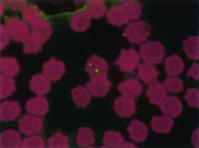
A 30-year-old female was admitted to the hospital for fever, mastitis, and axillary lymphadenopathy. Lymph node biopsy and laboratory data were nonspecific except for the Bartonella serology, which was positive on screening but below our cutoff (<1:100) when quantified, suggesting the possibility of an atypical bartonellosis. The patient was bitten by her kitten on her breasts before the onset of the mastitis. Samples were taken from the cat, and the blood was cultured on blood agar at 37°C in a 5% CO2 atmosphere and grew Bartonella-like colonies after 14 days of culture. After PCR amplification, DNA sequencing of the 16S-23S intergenic spacer region (its) and the citrate synthase gene (gltA) (1, 7) were 100% identical to those of B. koehlerae ATCC 700693 (GenBank accession numbers AF 312490 and AF 176091, respectively). Thin blood smears made from the fresh blood, fixed with methanol, and stained using a locally prepared rabbit polyclonal antibody were viewed under a laser confocal microscope as previously described (6) and showed the presence of intraerythrocytic Bartonella (Fig. 1).
FIG. 1.
Digital section of feline red blood cell infected with Bartonella koehlerae as viewed by laser confocal microscopy. Sections were taken in 0.5-μm increments from top to bottom. The presence of B. koehlerae was revealed with a locally prepared rabbit polyclonal antibody.
The presence of B. koehlerae in a European cat is reported for the first time, since only two isolates have been described in the United States (2). Nevertheless, we were unable to confirm the presence of the bacteria in the patient either in the lymph node biopsy or sample. Moreover, serology of the patient performed with antigens from the cat's isolate was negative. Since the cells targeted by Bartonella species are erythrocytes in cats (B. henselae) (6) and in humans (B. quintana) (4), we developed an immunofluorescence assay to also detect B. koehlerae in the blood of the cat of the present study and found that 4% of erythrocytes were sometimes infected with two bacteria in the same cell (Fig. 1). These results are in accordance with previous reports obtained for bacteremic cats infected with B. henselae, with 3 to 8% of infected cells (6). The main vector of B. henselae infections in cats is most likely the cat flea, whereas the vector of B. koehlerae remains unknown. However, we have recently detected DNA sequences of B. koehlerae in cat fleas from France, suggesting that this bacterium might be transmitted by cat fleas (5). Theprevalence of this new Bartonella species in cats remains unknown and is probably underestimated, since the bacterium is extremely fastidious.
REFERENCES
- 1.Birtles, R. J., and D. Raoult. 1996. Comparison of partial citrate synthase gene (gltA) sequences for phylogenetic analysis of Bartonella species. Int. J. Syst. Bacteriol. 46:891-897. [DOI] [PubMed] [Google Scholar]
- 2.Droz, S., B. Chi, E. Horn, A. G. Steigerwalt, A. M. Whitney, and D. J. Brenner. 1999. Bartonella koehlerae sp. nov., isolated from cats. J. Clin. Microbiol. 37:1117-1122. [DOI] [PMC free article] [PubMed] [Google Scholar]
- 3.Kordick, D. L., E. J. Hilyard, T. L. Hadfield, K. H. Wilson, A. G. Steigerwalt, D. J. Brenner, and E. B. Breitschwerdt. 1997. Bartonella clarridgeiae, a newly recognized zoonotic pathogen causing inoculation papules, fever, and lymphadenopathy (cat scratch disease). J. Clin. Microbiol. 35:1813-1818. [DOI] [PMC free article] [PubMed] [Google Scholar]
- 4.Rolain, J. M., C. Foucault, R. Guieu, B. La Scola, P. Brouqui, and D. Raoult. 2002. Bartonella quintana in human erythrocytes. Lancet 360:226-228. [DOI] [PubMed] [Google Scholar]
- 5.Rolain, J. M., M. Franc, B. Davoust, and D. Raoult. Molecular detection of pathogenic Bartonella and Rickettsia in cat fleas from France. Emerg. Infect. Dis., in press. [DOI] [PMC free article] [PubMed]
- 6.Rolain, J. M., B. La Scola, Z. Liang, B. Davoust, and D. Raoult. 2001. Immunofluorescent detection of intraerythrocytic Bartonella henselae in naturally infected cats. J. Clin. Microbiol. 39:2978-2980. [DOI] [PMC free article] [PubMed] [Google Scholar]
- 7.Roux, V., and D. Raoult. 1995. Inter- and intraspecies identification of Bartonella (Rochalimea) species. J. Clin. Microbiol. 33:1573-1579. [DOI] [PMC free article] [PubMed] [Google Scholar]


