Systemic sclerosis (SSc) is known to be characterised by a diffuse microvascular pathological process leading to cutaneous and visceral changes and to related clinical manifestations.
Both necropsy studies1,2 and in vivo investigations3,4,5 have shown that in a number of patients with SSc there is evidence of a coronary microvascular disease, while coronary artery disease does not exceed that seen in a control group. In particular, myocardial perfusion defects on thallium‐201 scintigraphy usually occur in the absence of angiographic evidence of coronary stenosis.3
Recently, we used a new and non‐invasive method of contrast enhanced, transthoracic, second harmonic echo Doppler in patients with SSc to evaluate the coronary flow reserve (CFR), a functional variable measuring the ability of the coronary microvasculature to adapt its lumen to a vasodilating stimulus.6 We detected a significant reduction of the CFR in 14/27 patients with SSc. In that study we did not examine the extramural tract of the coronary arteries; therefore, we could not exclude formally the possibility of a coronary stenosis which might have impaired the CFR.
To verify this possibility we assessed the status of the epicardial coronary arteries by a direct imaging test—the coronary contrast angiography with myocardial multidetector computed tomography (MDCT)7,8,9—in seven of the patients with SSc previously found to have a markedly reduced CFR (⩽2.5).
As previously reported, all these patients were asymptomatic for cardiac ischaemic manifestations.6 Table 1 gives the demographic and clinical data of these seven patients.
Table 1 Demographic and clinical features of the seven patients with SSc.
| Demographic and clinical features | Data |
|---|---|
| Age (years), mean (SD), range | 56.3 (25.4), 38–74 |
| F/M ratio | 7/0 |
| Disease duration (years), mean (SD), range | 12.9 (7.0), 5–21 |
| Clinical forms, No (%) | |
| lcSSc | 2 (29) |
| dcSSc | 5 (71) |
| Clinical manifestations, No (%) | |
| Raynaud's phoenomenon | 7 (100) |
| Lung disease | 6 (86) |
| Oesophagus involvement | 6 (86) |
| Telangiectasia | 5 (71) |
| Trophic ulcers | 5 (71) |
| Calcinosis | 1 (14) |
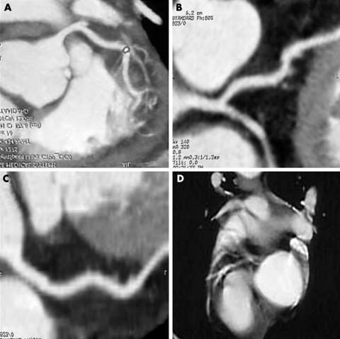
The myocardial MDCT examination was carried out by a multidetector computerised tomograph with eight lines of detectors and rotation time of 500 ms on 360° and with a slice depth of 1.3 mm (Light Speed Ultra, General Electric Medical Systems, Milwaulkee, Illinois, USA); retrospective gating was used. Non‐ionic contrast medium (120 ml) was injected at the speed of 4 ml/s, and the images were processed as appropriate on a SUN‐80 ULTRA workstation. The entire examination took 20–30 minutes. Image evaluation was carried out by an expert radiologist who knew the diagnosis of the patients. Figures 1A‐D show the images obtained in this study.
Figure 1 (A) Patient 1: Normal left coronary artery; “electronic caliper” in the left anterior descendent coronary artery. (B) Patient 2: Left coronary artery sections: absence of parietal lesions or stenosis. (C) Patient 3: Normal wall and diameter in the left coronary artery. (D) Patient 4: Isolated parietal spotty calcifications in the left anterior descendent coronary artery.
No defect in the coronary size and lumen was detected in any of the patient in this series. Parietal spots of calcium deposition were detected (fig 1D) only in one female patient (patient 4, aged 60 years) with high cholesterol serum levels (7.40 mmol/l; normal 2.60–5.20). The abnormal CFR value determined in this patient (CFR value 2.26) was within the range (2.37–1.78) of that of the other six patients tested. This patient was not affected by cutaneous calcinosis.
The results of this study enable us to exclude the presence of coronary stenosis in our patients with SSc with severe CFR impairment. This investigation therefore demonstrates that the CFR impairment in these patients is not due to a primary stenosis of an epicardial coronary artery but to a primary dysfunction of the coronary microvasculature, as previously reported.9
The non‐invasive MDCT test was preferred to conventional coronary angiography as it avoided cardiac catheterisation. Furthermore, the myocardial MDCT technique enabled the evaluation carried out by contrast echocardiography to be extended to all three coronary arteries. Moreover, this radiological diagnostic procedure is less expensive than conventional coronary angiography with catheterisation.10
We believe that the two non‐invasive techniques employed may be complementary and combined in a sequential manner: CFR should be investigated first, then the MDCT scanning can be applied to patients with CFR pathological values.
In conclusion, this study demonstrates that the reduced CFR seen in this series of asymptomatic patients with SSc is not secondary to stenotic lesions of the major epicardial coronary arteries.
Acknowledgments
This study was supported in part by grants No 2478/2002 and No 2411/2003 for a research project of AM, IInd Chair of Rheumatology, University of Cagliari, founded by the Health Administration of the Regione Autonoma della Sardegna, Italy.
References
- 1.D'Angelo W A, Fries J F, Masi A T, Shulman L E. Pathologic observations in systemic sclerosis (scleroderma): a study of fifty‐eight autopsy cases and fifty‐eight matched controls. Am J Med 196946428–440. [DOI] [PubMed] [Google Scholar]
- 2.Follansbee W P, Miller T R, Curtiss E I, Orie J E, Bernstein R L, Kiernan J M.et al A controlled clinicopathologic study of myocardial fibrosis in systemic sclerosis (scleroderma). J Rheumatol 199017656–662. [PubMed] [Google Scholar]
- 3.Kahan A, Devaux J Y, Amor B, Menkes C J, Weber S, Nitemberg A.et al Nifedipine and thallium‐201 myocardial perfusion in progressive systemic sclerosis. N Engl J Med 19863141307–1402. [DOI] [PubMed] [Google Scholar]
- 4.Alexander E L, Firestein G S, Weiss J L, Heuser R R, Leitl G, Wagner H N., Jret al Reversible cold‐induced abnormalities myocardial perfusion and function in systemic sclerosis. Ann Intern Med 1986105661–668. [DOI] [PubMed] [Google Scholar]
- 5.Anvari A, Graninger W, Schneider B, Sochor H, Weber H, Schmidinger H. Cardiac involvement in systemic sclerosis. Arthritis Rheum 1992351356–1361. [DOI] [PubMed] [Google Scholar]
- 6.Montisci R, Vacca A, Garau P, Colonna P, Ruscazio M, Passiu G.et al Detection of early impairment of coronary flow reserve in patients with systemic sclerosis. Ann Rheum Dis 200362890–893. [DOI] [PMC free article] [PubMed] [Google Scholar]
- 7.Achenbach S, Giesler T, Ropers D, Ulzheimer S, Derlien H, Schulteet al Detection of coronary artery stenoses by contrast‐enhanced, retrospectively electrocardiographically‐gated, multislice spiral computed tomography. Circulation 20011032535–2538. [DOI] [PubMed] [Google Scholar]
- 8.Nieman K, Cademartiri F, Lemos P A, Raaijmakers R, Pattynama P M, de Feyter P J. Reliable non invasive coronary angiography with multislice spiral computed tomography. Circulation 20021062051–2054. [DOI] [PubMed] [Google Scholar]
- 9.Kahan A, Nitenberg A, Foult J M, Amor B, Menkes C ‐ J, Devaux J Y.et al Decreased coronary reserve in primary scleroderma myocardial disease. Arthritis Rheum 198528637–646. [DOI] [PubMed] [Google Scholar]
- 10.Budoff M J, Achenbach S, Duerinckx A. Clinical utility of computed tomography and magnetic resonance techniques for noninvasive coronary angiography. J Am Coll Cardiol 2003421867–1878. [DOI] [PubMed] [Google Scholar]


