Abstract
Objective
To evaluate the outcomes of arthrocentesis with the new highly controllable, one handed reciprocating procedure syringe compared with a conventional syringe.
Methods
100 arthrocentesis procedures were randomised between the reciprocating syringe and the conventional syringe. Outcome measures included patient pain, procedure duration, operator satisfaction, synovial fluid volume, cell counts, and complications.
Results
50 arthrocentesis procedures with the conventional syringe resulted in a mean (SD) procedure time of 3.39 (1.88) minutes, a mean VAPS (patient pain) score of 5.35 (3.15), and a mean VASS (operator satisfaction) score of 4.88 (1.92); 30 of the 50 subjects experienced moderate to severe pain (VAPS score 5 or greater) during arthrocentesis. In contrast, the reciprocating syringe resulted in a reduced procedure time of 1.94 (1.14) minutes (p<0.001), a reduced VAPS (patient pain) score of 2.54 (1.60) (p<0.001), and an increased VASS (operator satisfaction) score of 8.91 (0.79) (p<0.001). Only five of the 50 of subjects experienced moderate to severe pain with the reciprocating syringe. Synovial cell counts were similar between the two syringes (p>0.05), but there was a trend toward greater volume (greater synovial fluid yield) and fewer red blood cells with the reciprocating syringe.
Conclusions
Arthrocentesis with a conventional syringe results in moderate to severe pain in 60% of subjects. The reciprocating syringe prevents significant pain, reduces procedure time, and improves physician performance of arthrocentesis. The reciprocating syringe is superior to the conventional syringe in arthrocentesis.
Keywords: syringe, joint procedure, arthrocentesis, reciprocating, injection
Arthrocentesis is the single most important invasive procedure in musculoskeletal medicine.1,2 Arthrocentesis is essential for the diagnosis of septic arthritis and inflammatory joint disease, and is the basic underlying procedure for intra‐articular treatment, including therapeutic arthrocentesis, needle lavage, and intra‐articular injection of therapeutic substances.3,4,5,6,7,8,9,10,11,12,13
Recently the Food and Drug Administration (FDA) has formally approved the highly controllable, one handed reciprocating procedure syringe.14 The reciprocating syringe incorporates a reciprocating plunger mechanism that permits the index and middle fingers to remain in one position during aspiration and injection, while the thumb moves horizontally to the alternative plunger in order to change the direction of aspiration or injection. Because of these favourable characteristics, we hypothesised that the reciprocating syringe would improve the physician's performance of arthrocentesis.
Methods
Subjects
This project was approved by the institutional review board (IRB). Twenty six physicians who regularly undertake syringe procedures carried out 100 arthrocentesis procedures on 46 individual patients who required a diagnostic or therapeutic arthrocentesis for their usual and customary medical care. The mean (SD) age of the physicians was 38.8 (15.7) years, indicating that the physicians were generally in early to mid‐career, but the group as a whole had considerable syringe experience, with a mean of 13.6 (13.9) years of syringe experience and 1002 (1390) syringe procedures each. The physicians undertook a mean of 8.4 (7.1) syringe procedures per week, indicating that the test group was an active, practised group of physicians. There were more male (65%) than female physicians (35%), representative of the local physician population. In each case, patients individually consented both to the arthrocentesis, as required for all procedures, and to the IRB approved research protocol. The mean (SD) age of the subjects was 47.3 (15.0) years. The great majority of subjects (76%) had rheumatoid arthritis and the remainder other diagnoses. The 100 syringe procedures included arthrocentesis of the knee (39%), small joints of the fingers (26%) (proximal interphalangeal, metacarpophalangeal, and carpometacarpal joint), the shoulder (17%), and other joints (18%). In each case, each procedure was randomised to either the conventional or the reciprocating syringe. If the subject had more than one joint requiring arthrocentesis, then each joint was randomised between the reciprocating and the conventional syringes. Ninety one per cent (41/46 subjects) had had a previous arthrocentesis with a conventional syringe before entry into the study, 31% (14/46) had more than one joint aspirated and were randomised between the two syringes during the same visit, and 19% (9/46) had more than one arthrocentesis on two different occasions randomised between the two syringes. The final proportions of arthrocentesis procedures in individual joints within each treatment group were statistically equivalent.
Syringes
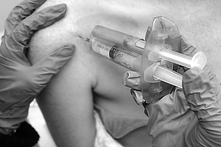
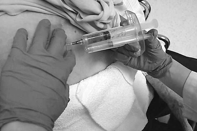
The conventional syringe was a 10 ml Luer‐Lok™ BD syringe (Ref No 309604, Becton Dickinson Co, Franklin Lakes, New Jersey, USA). The reciprocating procedure syringe used in these experiments was the recently FDA approved 10 ml reciprocating syringe (The RECIPROCATOR Procedur‐10, AVANCA Medical Devices Inc, Albuquerque, New Mexico, USA; www.AVANCAMedical.com). Illustrations of the reciprocating syringe in use are provided with the patients' permission (figs 1 and 2).

Figure 1 One handed use of the reciprocating syringe for musculoskeletal procedures. This photograph shows the reciprocating syringe being used in a one handed fashion for aspiration and injection of the glenohumeral joint. The larger plunger is depressed with the thumb for injection and the smaller plunger is depressed with the thumb for aspiration. The free hand is used to steady the patient, feel the surface anatomy, or operate other devices.

Figure 2 One handed use of the reciprocating syringe for aspiration of a large shoulder effusion. This photograph shows the reciprocating syringe being used in a one handed fashion for aspiration and drainage of a shoulder effusion. The larger plunger is depressed with the thumb for injection and the smaller plunger is depressed with the thumb for aspiration. As shown here the smaller plunger is depressed for continuous aspiration. The free hand is used to feel, steady, and apply pressure to the effusion or operate an ultrasound transducer.
Arthrocentesis
Arthrocentesis was carried out in a standardised manner and in a customary fashion.15,16,17,18,19,20,21
Outcome data of clinical procedures
A non‐operating observer timed each clinical procedure (in minutes), and questioned the patient in real time about pain; after the procedure the observer questioned the physician about satisfaction with the syringe used in the procedure. Patient pain was determined with the standardised and validated visual analogue pain scale (VAPS) where 0 cm = no pain and 10 cm = unbearable pain.22,23 The VAPS was obtained twice during the procedure—after the anaesthesia portion and directly after the arthrocentesis portion, and a mean VAPS score was obtained by averaging the two scores. Moderate to severe pain was defined as VAPS ⩾5. Operator satisfaction with the syringe after the procedure was determined with the visual analogue satisfaction scale (VASS), where 0 cm = completely dissatisfied with the performance of the procedure syringe and 10 cm = completely satisfied.24,25 Final clinical outcomes were determined first, directly at the conclusion of the procedure, and second, at two weeks after the procedure. Synovial fluid outcome measures included culture results, cell count, cell differential counts, crystal examination, and volume determination.
Statistical analysis
Data were entered into Microsoft Excel (version 5), and analysed in SAS (SAS/STAT software, release 6.11, Cary, North Carolina, USA). Differences between parametric two‐group data were determined by t test, differences in categorical data by Fisher's exact test, and differences between multiple parametric datasets by Fisher's least significant difference method. Corrections were made for multiple comparisons. Correlations between parametric data were determined by logistic regression, and between non‐parametric data by Spearman correlation and Kendall rank method.
Results
At the conclusion of the study, the physicians had substantially more experience with the conventional syringe (1002 (1390) total conventional syringe procedures (mean (SD)) than with the reciprocating syringe (3.6 (4.6) total reciprocating syringe procedures, p<0.001)
The overall outcomes of the clinical syringe procedures are shown in table 1. One hundred arthrocentesis procedures were randomised to either the reciprocating syringe or the conventional syringe, such that 50 procedures of each type were completed. In arthrocentesis procedures as a whole, the reciprocating syringe resulted in:
Table 1 Outcome of 100 arthrocentesis procedures randomised to either the conventional syringe or the reciprocating syringe.
| Conventional | Reciprocating | Significance | |
|---|---|---|---|
| Number of procedures | 50 | 50 | NS |
| Procedure time (minutes) | 3.39 (1.88) | 1.94 (1.14) | p<0.001 |
| Patient pain (VAPS) | 5.35 (3.15) | 2.54 (1.60) | p<0.001 |
| Physician satisfaction (VASS) | 4.88 (1.92) | 8.91 (0.79) | p<0.001 |
| Successful immediate outcome | 100% | 100% | NS |
| (good to excellent) | |||
| Successful outcome at 2 weeks | 100% | 100% | NS |
| (good to excellent) |
Values are mean (SD).
VAPS, visual analogue scale, pain; VASS, visual analogue scale, operator satisfaction.
reduced procedure time compared with the conventional syringe (reciprocating syringe, 1.94 (1.14) min; conventional syringe, 3.39 (1.88) min; p<0.001);
reduced patient pain (reciprocating syringe VAPS score, 2.54 (1.60); conventional syringe VAPS score, 5.35 (3.15); p<0.001);
improved physician satisfaction (reciprocating syringe VASS score, 8.91 (0.79); conventional syringe VASS score, 4.88 (1.92); p<0.001).
Thus, relative to a conventional syringe, the reciprocating syringe produced a 43% reduction in procedure duration (p<0.001), a 53% reduction in patient pain (p<0.001), and an 83% increase in operator satisfaction with syringe performance (p<0.001). Sixty per cent of the subjects (30/50) experienced moderate to severe pain during arthrocentesis with the conventional syringe, while only 10% (5/50) experienced moderate to severe pain with the reciprocating syringe.
Immediately after these procedures and at two weeks, there were no complications in any patient, and outcomes were good to excellent in all patients with either reciprocating or conventional syringes (table 1). There were no significant differences in cell counts, including white blood cells, red blood cells, neutrophils, lymphocytes, and monocytes (table 2). However, there was a trend to greater synovial fluid yield (volume) and fewer red blood cells with the reciprocating syringe. One subject with the conventional syringe had a positive synovial fluid culture for Neisseria gonorrhoeae.
Table 2 Synovial fluid analysis from the knee with the reciprocating and conventional syringes.
| White blood | Neutrophils (%) | Monocytes (%) | Lymphocytes (%) | Red blood | Volume (ml) | No of knee fluid samples | |
|---|---|---|---|---|---|---|---|
| cells/mm3 | cells/mm3 | ||||||
| Conventional syringe | 11 439 (9786) | 60.6 (35.5) | 27.8 (25.4) | 11.7 (10.7) | 49 222 (79 513) | 8.89 (2.47) | 9 |
| Reciprocating syringe | 16 950 (14 089) | 59.7 (25.8) | 29.7 (27.8) | 8.29 (5.54) | 14 832 (14 906) | 13.26 (6.47) | 9 |
| Significance | p = 0.20 | p = 0.91 | p = 0.65 | p = 0.49 | p = 0.24 | p = 0.06 |
Values are mean (SD).
Discussion
This study is the first large randomised clinical trial with the reciprocating syringe in invasive syringe procedures, and shows measurably better outcomes in the case of arthrocentesis. The reciprocating syringe resulted in a 43% reduction in procedure duration (p<0.001), a 53% reduction in pain (p<0.001), and an 83% increase in operator satisfaction with syringe performance (p<0.001) (table 1). Significant pain was reduced from 60% to 10%, indicating an 84% effectiveness in preventing moderate to severe pain during arthrocentesis. Synovial fluid characteristics were similar between the two syringes (table 2), but there was a trend towards greater synovial fluid yield and fewer red blood cells with the reciprocating syringe. The improvement in physician performance in terms of procedure duration and reduced patient pain with the reciprocating syringe could not be attributed to practice effects, as the physicians had on average 278 times more practice with the conventional syringe.
An important finding to this study is the unexpectedly high degree of pain that patients experience during arthrocentesis, with mean pain scores (VAPS scores) ⩾5, indicating moderate to severe pain in many patients (table 1). In this study the local anaesthetic used was lignocaine (lidocaine), which has been shown to be superior to ethyl chloride21; nevertheless, the patients experienced considerable pain. With the conventional syringe and individual patients, 30 of 50 subjects (60%) reported individual pain scores (VAPS) of 5 or more. This is far more pain that most musculoskeletal experts commonly believe that patients experience with arthrocentesis. However, pain with arthrocentesis has not been rigorously measured before this study, and the rigorous characterisation of pain is one of the most important features of our study. Poor control of the needle may be a significant cause of pain in arthrocentesis,26,27,28,29 and this degree of pain is certainly a major reason why paediatric patients abhor arthrocentesis.30,31,32 Because of the significant reduction in pain, the reciprocating syringe may be of particular value in paediatric syringe procedures, in individuals with known needle phobia or vasovagal responses to pain, and in those allergic to local anaesthetics.33,34,35
The conventional syringe is still commonly used for even the most difficult syringe procedures in most fields of medicine. Despite the recognised instability and danger of conventional syringes, the major reason for persistence of conventional syringes in procedures is the low cost of conventional syringes and the lack of an effective alternative. However, as noted in this study, the conventional syringe is associated with significantly greater patient pain, longer procedure times, and reduced physician satisfaction—all indicating a fundamental design inadequacy for syringe procedures.
The reciprocating syringe is formed around the core of a conventional syringe barrel and plunger, but has a parallel accessory plunger and an accessory barrel or track to control the motion of the accessory plunger. The two plungers are mechanically linked in an opposing fashion, resulting in a set of reciprocating plungers. Thus when one plunger is depressed with the thumb the syringe injects, and when the accessory plunger is depressed with the same thumb, the syringe aspirates. This permits the index and middle fingers to remain in one position during both aspiration and injection, while the thumb only needs to move in a horizontal plane to the alternative plunger in order to change the direction of aspiration or injection. This permits the powerful and exquisitely well controlled flexor musculature of the hand and forearm to be used for both injection and aspiration. These characteristics of stable finger positioning and the exclusive use of the flexor musculature create a powerful and finely controlled one handed device. A one handed procedure syringe would have obvious applications in ultrasound guided arthrocentesis where a free hand is needed for the ultrasound transducer, and in applying vacuum for synovial and deep tissue biopsy.36,37,38,39,40,41,42,43,44,45,46
In terms of procedure time, patient pain, and operator satisfaction during arthrocentesis, the reciprocating syringe is clearly superior to the conventional syringe (table 1). Synovial fluid analysis (table 2) suggests that a larger study may also show significantly greater synovial fluid yield (volume) and higher quality (less blood contamination) with the reciprocating syringe. Further study of reciprocating interventional devices in specific procedures will be required to determine specific indications and future applications of this new technology to the broad field of syringe procedures in musculoskeletal medicine.
Acknowledgements
We would like to thank AVANCA Medical Devices Inc for donating the syringes for this study. We also thank Dr Wilmer L Sibbitt Jr of AVANCA Medical Devices Inc, for instructing the participants in the use of the reciprocating syringe and providing the authors with advice as needed. This paper was submitted in abstract form to American College of Rheumatology Annual Meeting, San Diego, California, 2005.
Abbreviations
VAPS - visual analogue scale, pain
VASS - visual analogue scale, operator satisfaction
References
- 1.Aceves‐Avila F J, Delgadillo‐Ruano M A, Ramos‐Remus C, Gomez‐Vargas A, Gutierrez‐Urena S. The first descriptions of therapeutic arthrocentesis: a historical note. Rheumatology (Oxford) 200342180–183. [DOI] [PubMed] [Google Scholar]
- 2.Johnson M W. Acute knee effusions: a systematic approach to diagnosis. Am Fam Physician 2000612391–2400. [PubMed] [Google Scholar]
- 3.Guggi V, Calame L. Contribution of digit joint aspiration to the diagnosis of rheumatic diseases. Joint Bone Spine 20026958–61. [DOI] [PubMed] [Google Scholar]
- 4.Lane J G, Falahee M H, Wojtys E M, Hankin F M, Kaufer H. Pyarthrosis of the knee. Treatment considerations. Clin Orthop Relat Res 199014198–204. [PubMed] [Google Scholar]
- 5.Manadan A M, Block J A. Daily needle aspiration versus surgical lavage for the treatment of bacterial septic arthritis in adults. Am J Ther 200411412–415. [DOI] [PubMed] [Google Scholar]
- 6.Dooley P, Martin R. Corticosteroid injections and arthrocentesis. Can Fam Physician 200248285–292. [PMC free article] [PubMed] [Google Scholar]
- 7.Samuelson C O, Cannon G W, Ward J R. Arthrocentesis. J Fam Pract 198520179–184. [PubMed] [Google Scholar]
- 8.Brown P W. Arthrocentesis for diagnosis and therapy. Surg Clin North Am 1969491269–1278. [PubMed] [Google Scholar]
- 9.Lee A H, Chin A E, Ramanujam T, Thadhani R I, Callegari P E, Freundlich B. Gonococcal septic arthritis of the hip. J Rheumatol 1991181932–1933. [PubMed] [Google Scholar]
- 10.Kesteris U, Wingstrand H, Forsberg L, Egund N. The effect of arthrocentesis in transient synovitis of the hip in the child: a longitudinal sonographic study. J Pediatr Orthop 19961624–29. [DOI] [PubMed] [Google Scholar]
- 11.Weidner S, Kellner W, Kellner H. Interventional radiology and the musculoskeletal system. Best Pract Res Clin Rheumatol 200418945–956. [DOI] [PubMed] [Google Scholar]
- 12.Bureau N J, Ali S S, Chhem R K, Cardinal E. Ultrasound of musculoskeletal infections. Semin Musculoskelet Radiol 19982299–306. [DOI] [PubMed] [Google Scholar]
- 13.Grassi W, Farina A, Filippucci E, Cervini C. Sonographically guided procedures in rheumatology. Semin Arthritis Rheum 200130347–353. [DOI] [PubMed] [Google Scholar]
- 14.Food and Drug Administration Substantial equivalence determination: 510 (K), summary. FDA document K042487. pdf, January 21 2005
- 15.Schaffer T C. Joint and soft‐tissue arthrocentesis. Prim Care 199320757–770. [PubMed] [Google Scholar]
- 16.Pfenninger J L. Injections of joints and soft tissue: part II. Guidelines for specific joints. Am Fam Physician 1991441690–1701. [PubMed] [Google Scholar]
- 17.Zuber T J. Knee joint aspiration and injection. Am Fam Physician. 2002 15;66: 1497–500, 1503–4, 1507 [PubMed]
- 18.Tallia A F, Cardone D A. Diagnostic and therapeutic injection of the shoulder region. Am Fam Physician. 2003 15 671271–1278. [PubMed] [Google Scholar]
- 19.Tallia A F, Cardone D A. Diagnostic and therapeutic injection of the wrist and hand region. Am Fam Physician 200367745–750. [PubMed] [Google Scholar]
- 20.Cardone D A, Tallia A F. Diagnostic and therapeutic injection of the elbow region. Am Fam Physician. 2002 1 662097–2100. [PubMed] [Google Scholar]
- 21.Armstrong P, Young C, McKeown D. Ethyl chloride and venepuncture pain: a comparison with intradermal lidocaine. Can J Anaesth 199037656–658. [DOI] [PubMed] [Google Scholar]
- 22.Katz J, Melzack R. Measurement of pain. Surg Clin North Am 199979231–252. [DOI] [PubMed] [Google Scholar]
- 23.Bhachu H S, Kay B, Healy T E, Beatty P. Grading of pain and anxiety. Comparison between a linear analogue and a computerised audiovisual analogue scale. Anaesthesia 198338875–878. [DOI] [PubMed] [Google Scholar]
- 24.Sutherland H J, Lockwood G A, Minkin S, Tritchler D L, Till J E, Llewellyn‐Thomas H A. Measuring satisfaction with health care: a comparison of single with paired rating strategies. Soc Sci Med 19892853–58. [DOI] [PubMed] [Google Scholar]
- 25.Miller M D, Ferris D G. Abstract Measurement of subjective phenomena in primary care research: the visual analogue scale. Fam Pract Res J 19931315–24. [PubMed] [Google Scholar]
- 26.Partington P F, Broome G H. Diagnostic injection around the shoulder: hit and miss? A cadaveric study of injection accuracy. J Shoulder Elbow Surg 19987147–150. [DOI] [PubMed] [Google Scholar]
- 27.Roberts W N, Hayes C W, Breitbach S A, Owen D S. Dry taps and what to do about them: a pictorial essay on failed arthrocentesis of the knee. Am J Med 1996100461–464. [DOI] [PubMed] [Google Scholar]
- 28.Solomon D H, Bates D W, Panush R S, Katz J N. Costs, outcomes, and patient satisfaction by provider type for patients with rheumatic and musculoskeletal conditions: a critical review of the literature and proposed methodologic standards. Ann Intern Med 199712752–60. [DOI] [PubMed] [Google Scholar]
- 29.Hicks C M, Gonzalez R, Morton M T, Gibbons R V, Wigton R S, Anderson R J. Procedural experience and comfort level in internal medicine trainees. J Gen Intern Med 200015716–722. [DOI] [PMC free article] [PubMed] [Google Scholar]
- 30.Uziel Y, Berkovitch M, Gazarian M, Koren G, Silverman E D, Schneider R.et al Evaluation of eutectic lidocaine/prilocaine cream (EMLA) for steroid joint injection in children with juvenile rheumatoid arthritis: a double blind, randomized, placebo controlled trial. J Rheumatol 200330594–596. [PubMed] [Google Scholar]
- 31.Fassler D. The fear of needles in children. Am J Orthopsychiatry 198555371–377. [DOI] [PubMed] [Google Scholar]
- 32.Majstorovic M, Veerkamp J S. Relationship between needle phobia and dental anxiety. J Dent Child (Chicago) 200471201–205. [PubMed] [Google Scholar]
- 33.Eberhard B A, Sison M C, Gottlieb B S, Ilowite N T. Comparison of the intra‐articular effectiveness of triamcinolone hexacetonide and triamcinolone acetonide in treatment of juvenile rheumatoid arthritis. J Rheumatol 2004312507–2512. [PubMed] [Google Scholar]
- 34.Wilson N I, Di Paola M. Acute septic arthritis in infancy and childhood. 10 years' experience. J Bone Joint Surg Br 198668584–587. [DOI] [PubMed] [Google Scholar]
- 35.Padeh S, Passwell J H. Intra‐articular corticosteroid injection in the management of children with chronic arthritis. Arthritis Rheum 1998411210–1214. [DOI] [PubMed] [Google Scholar]
- 36.Balint P V, Kane D, Hunter J, McInnes I B, Field M, Sturrock R D. Ultrasound guided versus conventional joint and soft tissue fluid aspiration in rheumatology practice: a pilot study. J Rheumatol 2002292209–2213. [PubMed] [Google Scholar]
- 37.Lim‐Dunham J E, Ben‐Ami T E, Yousefzadeh D K. Septic arthritis of the elbow in children: the role of sonography. Pediatr Radiol 199525556–559. [DOI] [PubMed] [Google Scholar]
- 38.Zwar R B, Read J W, Noakes J B. Sonographically guided glenohumeral joint injection. Am J Roentgenol 200418348–50. [DOI] [PubMed] [Google Scholar]
- 39.Tuite M J. Facet joint and sacroiliac joint injection. Semin Roentgenol 20043937–51. [DOI] [PubMed] [Google Scholar]
- 40.Raza K, Lee C Y, Pilling D, Heaton S, Situnayake R D, Carruthers D M.et al Ultrasound guidance allows accurate needle placement and aspiration from small joints in patients with early inflammatory arthritis. Rheumatology (Oxford) 200342976–979. [DOI] [PubMed] [Google Scholar]
- 41.el‐Khoury G Y, Renfrew D L, Walker C W. Interventional musculoskeletal radiology. Curr Probl Diagn Radiol 199423161–203. [PubMed] [Google Scholar]
- 42.Moon M S, Kim I, Kim J M, Lee H S, Ahn Y P. Synovial biopsy by Franklin‐Silverman needle. Clin Orthop Relat Res 1980(Jul‐Aug)224–228. [PubMed]
- 43.Rege J, Shet T, Naik L. Fine needle aspiration of tophi for crystal identification in problematic cases of gout. A report of two cases. Acta Cytol 200044433–436. [DOI] [PubMed] [Google Scholar]
- 44.Hopper K D, Abendroth C S, Sturtz K W, Matthews Y L, Shirk S J. Fine‐needle aspiration biopsy for cytopathologic analysis: utility of syringe handles, automated guns, and the nonsuction method. Radiology 1992185819–824. [DOI] [PubMed] [Google Scholar]
- 45.Arayssi T K, Schumacher H R. Evaluation of a modified needle. J Rheumatol 199825876–878. [PubMed] [Google Scholar]
- 46.Goldenberg D L, Cohen A S. Synovial membrane histopathology in the differential diagnosis of rheumatoid arthritis, gout, pseudogout, systemic lupus erythematosus, infectious arthritis and degenerative joint disease. Medicine (Baltimore) 197857239–252. [DOI] [PubMed] [Google Scholar]


