The occurrence of secondary hepatogenous photosensitization associated with pastures of Brachiaria decumbens date from the mid 70s (1). Since then, many outbreaks have been described in cattle, water buffaloes, sheep, and horses (1).
The only reports of photosensitization in camelids have been those of Rosychuk (2), who described hepatogenous photosensitization in llamas, and Coulton et al (3), who reported the occurrence of secondary hepatogenous photosensitization in alpacas in Australia due to exposure to the mycotoxin sporidesmin.
In the last 2 y, the frequency of ruminants being affected by photosensitization has increased in the ruminant practice at the Faculdade de Medicina Veterinária e Zootecnia, Universidade de São Paulo, and there has been a significant increase in the diagnosis of the disease in sheep — in 2003, 5.7% (14/244) of the sheep sent to the service were diagnosed as having secondary hepatogenous photosensitization.
Among the animals with photosensitization, an adult female llama (Lama glama) was sent to the service in August 2003. The animal had a history of apathy, anorexia, and weight loss, and had been kept on a Brachiaria decumbens pasture.
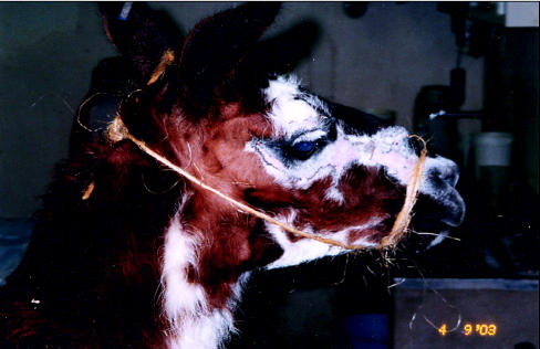
Apart from a decrease in ruminal movements, the other vital signs (heart rate, respiratory frequency, and body temperature) were within the normal range. On clinical examination, the ocular mucus membranes were slightly yellow and the skin in nonpigmented areas was swollen. After 10 d, the animal showed mummification of the skin over the head (Figure 1). The leathery surface and scaling were characteristic of solar dermatitis. Rosychuk (2), in describing photosensitization in llamas, emphasized the presence of erythema and edema in various degrees and of scaling restricted to nonpigmented areas of the skin, predominantly in the head. According to Coulton et al (3), there may be abdominal distension, fever, and a reduced quantity of feces.
Figure 1.
Skin lesions with mummified appearance due to photosensitization.
Biochemical analyses revealed increased values of aspartate aminotransferase (319 U/L, normal range, 100–250 U/L [4]), gamma glutamiltransferase (313 U/L, normal range, 5–29 U/L [4]), direct bilirubin (6.33 μmol/L normal range — no reference value available) and total bilirubin (7.69 μmol/L, normal range, 0.0–1.71 μmol/L [4]), similar to those reported by Coulton et al (3). After 30 d, associated with recovery of the animal, serum levels of these biochemical parameters had returned to normal.
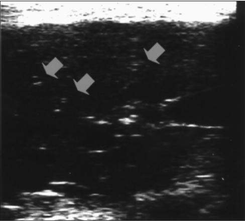
Ultrasonographic visualization of the liver with a 5 mHz transducer was possible between the 10th and 11th right intercostal spaces. Small hyperechoic points, characteristic of organ congestion, were observed diffused in the hepatic parenchyma and associated with the thickening of bile canaliculi (Figure 2). In the morphometric evaluation of the liver, the following characteristics were measured: size of the liver — from 10.0 to 11.0 cm; thickness of the liver — from 6.5 to 7.5 cm; angle of the ventral edge — from 46 to 49°; diameter of the portal vein — from 0.9 and 1.1 cm; and diameter of the vena cava — from 1.2 and 1.9 cm.
Figure 2.
Ultrasonographic image of the liver. The arrows show thickening of bile canaliculi.
The thickening of bile canaliculi was considered to be a relevant finding because, in animals with hepatogenous photosensitization, anatomopathological analysis shows a firm consistency of the liver and fibrosis of the bile ducts. Histologically, these alterations have been described as pericholangitis with proliferation of epithelial cells in bile ducts (1). No previous reports of ultrasonographic findings in animals with photosensitization were found in the literature, so, possibly, this is the first description of these findings (Figure 2).
During treatment, the animal was kept away from direct sunlight, fed hay, and submitted to the following protocol: fluid therapy, glucose, methionine, B complex vitamins, and calcium.
Based on the skin lesions, the changes in hepatic function, the ultrasonographic findings, and the improvement in clinical signs after the animal was kept away from the Brachiaria decumbens pasture and from direct sunlight, secondary hepatogenous photosensitization was diagnosed.
References
- 1.Tokarnia CH, Döbereiner J, Peixoto PV. Plantas fotossensiblilizantes. In: Plantas Tóxicas do Brasil. Rio de Janeiro: Editora Helianthus, 2000:151–177.
- 2.Rosychuk RAW. Llama dermatology. Vet Clin North Am Food Anim Pract. 1994;10:228–239. doi: 10.1016/s0749-0720(15)30557-0. [DOI] [PubMed] [Google Scholar]
- 3.Coulton MA, Dart AJ, McLintock SA, Hodgson DR. Sporodesmin toxicosisin in an alpaca. Aust Vet J. 1997;75:136–137. doi: 10.1111/j.1751-0813.1997.tb14174.x. [DOI] [PubMed] [Google Scholar]
- 4.Garry F, Weiser G, Belknap E. Clinical pathology of the llama. Vet Clin North Am Food Anim Pract. 1994;10:201–209. doi: 10.1016/s0749-0720(15)30555-7. [DOI] [PubMed] [Google Scholar]


