Abstract
Postpartum splinting can completely correct congenital ear deformities and obviate the need for later surgery
Congenital ear deformities are common and usually corrected surgically in childhood. Ear deformities are often first noticed by parents or non-specialist personnel such as midwives, general practitioners, and health visitors. Splinting of ear deformities in the early neonatal period has been shown to be a safe and effective non-surgical treatment.1,2,3,4,5,6,7,8 The splint is made from a wire core segment in a 6-French silastic tube and held in place with adhesive skin closure strips. It is applied with no anaesthesia for three to four weeks.1 We present three cases that show how different congenital ear deformities can be treated non-surgically, thereby obviating the need for surgery.
Case reports
Case 1: constricted ear
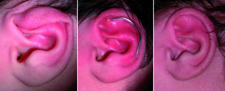
A male child was born at full term with bilateral constricted ears. No family history of ear deformity existed. In this deformity, the rim of the ear looks as if it has been tightened, rather like a purse string that has been pulled closed.1,9 We initiated splinting three days after birth and the programme was continued for one month. By 10 days the upper pole had expanded and a good result was seen at six months' follow-up (fig 1).
Fig 1 Case 1 (constricted ear) at 3 days postpartum (left), with splint in situ (middle, and at 10 days (right)
Case 2: Stahl's ear
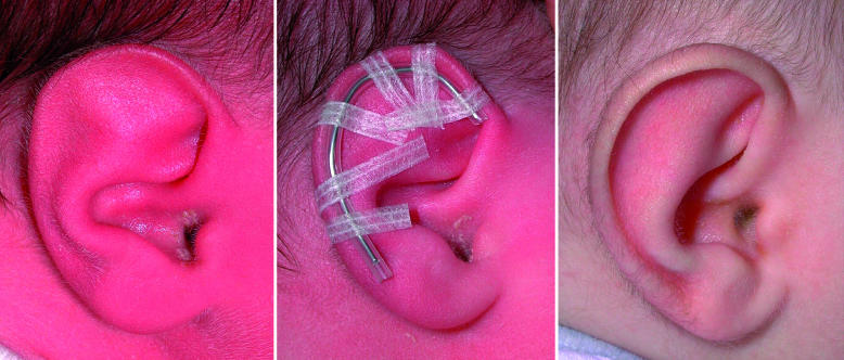
A male child was born at full term with a unilateral Stahl's ear deformity. Stahl's ear is a helical rim deformity characterised by a third crus, flat helix, and malformed scaphoid fossa (fig 2).10 We initiated splinting three days after birth and the programme was continued for three weeks. By 10 days the correction was already apparent with disappearance of the third crus and a normal helical rim. The good initial result was maintained at six months (fig 2).
Fig 2 Case 2 (Stahl's ear) at 3 days postpartum (left), with splint in situ (middle), and at 6 months (right)
Case 3: prominent ears
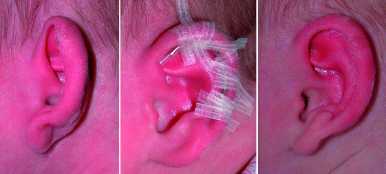
A female child was born at full term with bilateral prominent ears. This deformity is defined by excessive height of the conchal wall or a wide conchoscaphal angle (>90 degrees). We initiated splinting three days after birth and continued with the programme for four weeks. Initially the ear was protuberant with an increased conchoscaphal angle, but after splinting the angle was reduced and the ear sat in a more natural position (fig 3).
Fig 3 Case 3 (prominent ears) at 3 days postpartum (left), with splint in situ (middle), and at 30 days (right)
Discussion
Congenital ear deformities are defined as either malformations (microtia, cryptotia) or deformations. Ear deformation implies a normal chondrocutaneous component with an abnormal architecture.10 Deformed ears are categorised as constricted (fig 1), Stahl's (fig 2), or prominent (fig 3). The causes of these deformities are variable. Abnormal development and functioning of the intrinsic and extrinsic muscles of the ear may generate deforming forces. External forces applied to the ears, such as malpositioning of the head during the prenatal and neonatal periods, may also contribute.10
Although ear deformities are anecdotally common, their true incidence is unknown. Around 5% of the white population are thought to have prominent ears, but this may be an underestimate as most reports do not include less severe anomalies.
Although some of these deformities resolve spontaneously, a large proportion do not. In today's society, which puts great emphasis on appearance, the pressure on parents to seek surgical treatment if their child has an ear deformity can be great.
Several surgical techniques are available to treat these conditions. Although the results are often good, they can be unpredictable, especially for more complex deformities.
Splinting of ears in the early neonatal period has been advocated as an effective non-surgical treatment1,2,3,4,5,6,7,8 that often produces better results than surgery. The best results are achieved and the shortest period of splintage is needed when treatment is started immediately after birth. Moulding of the ears is possible then because maternal oestrogens render the ear of the neonate soft and malleable.4,5 After the first few days of life the ear becomes stiffer and less amenable to moulding, which makes splinting less effective.
Many kinds of splints and moulding materials have been described (table). Methods other than the one we used include self adhering foam designed to prevent skin damage from splints,3 temporary stopping (dental material) in combination with surgical tapes,4 dental bite and impression waxes,5 lead-free soldering wire inserted within an 8-French suction catheter, and thermoplastic material.11 Splint kits are now also available from various online sources.
Splinting materials and methods
| Material | Method |
|---|---|
| Wire core segment in 6-French silastic tubing1 | The splint is shaped and positioned in the groove between the helix and the antihelix and held in place with 3-5 skin closure strips |
| self adhering foam designed to prevent skin damage from splints3 | Applied at the bottom of the fold of the auricle and in the conchal fossa itself |
| Temporary stopping (dental material, a kind of gutta percha latex)4 | Used to press and correct abnormal folding from anterolateral or posteromedial surface; kept in place with skin closure strips |
| Dental bite and impression waxes5 | Heated under hot tap water and moulded to achieve the desired normal contour and held in place with skin closure strips |
| Thermoplastic material11 | Elastic and hard at room temperature but becomes soft in seconds at a temperature of >60°C; warmed and softened material is applied with light pressure from the anterior and posterior side of the ear—it hardens in minutes |
| Ear Buddies (Fresca Commerce; http://earbuddies.fresca.co.uk/pws/Content.ice?page=Home&pgForward=content) | Commercially available kit |
Splinting is a simple, effective, and cheap way of treating even the most complex congenital ear deformity. It is non-invasive and avoids the risks associated with surgery and anaesthesia. It prevents later psychological distress by treating the deformity before it is perceived as a problem by the child.
The potential for splinting congenital ear deformities in early neonatal life needs to be better publicised. Tan and Gault12 reported that parents are the first to notice the deformity at birth in 61% of babies with prominent ears. They should be offered the possibility of splinting to correct these deformities. Postpartum clinical screening and non-surgical treatment are effective for congenital dislocation of the hip joint and congenital club feet. We recommend that similar measures should be taken for congenital ear deformities to obviate the need for surgical correction later in childhood. It is vital that neonatal paediatricians, obstetricians, general practitioners, and midwives are educated about early detection and how to initiate treatment themselves.
The delay incurred by referring to a plastic surgeon may result in a missed opportunity to treat these deformities. If successful, an effective splinting programme could consign the surgical correction of all but the most severe ear deformities to the past.
Contributors: AJL, SH, and FS thought of the idea for the paper. FS cared for the three patients. AJL and SH reviewed the literature and wrote the article. SH is guarantor.
Funding: None.
Competing interests: None declared.
References
- 1.Schonauer F, La Rusca I, Fera G, Molea G. A splint for correction of congenital ear deformities. Eur J Plast Surg 2004;47:575. [Google Scholar]
- 2.Matsuo K, Hayashi R, Kiyono M, Hirose T, Netsu Y. Nonsurgical correction of congenital auricular deformities. Clin Plast Surg 1990;17:383-95. [PubMed] [Google Scholar]
- 3.Kurozumi N, Ono S, Ishida H. Non-surgical correction of a congenital lop ear deformity by splinting with Reston foam. Br J Plast Surg 1982;35:181-2. [DOI] [PubMed] [Google Scholar]
- 4.Matsuo K, Hirose T, Tomono T, Iwasawa M, Katohda S, Takahashi N, et al. Nonsurgical correction of congenital auricular deformities in the early neonate: a preliminary report. Plast Reconstr Surg 1984;73:38-51. [DOI] [PubMed] [Google Scholar]
- 5.Brown FE, Colen LB, Addante RR, Graham JM Jr. Correction of congenital auricular deformities by splinting in the neonatal period. Pediatrics 1986;78:406-11. [PubMed] [Google Scholar]
- 6.Merlob P, Eshel Y, Mor N. Splinting therapy for congenital auricular deformities with the use of soft material. J Perinatol 1995;15:293-6. [PubMed] [Google Scholar]
- 7.Ullmann Y, Blazer S, Ramon Y, Blumenfeld I, Peled IJ. Early nonsurgical correction of congenital auricular deformities. Plast Reconstr Surg 2002;109:907-13. [DOI] [PubMed] [Google Scholar]
- 8.Tan ST, Shibu M, Gault DT. A splint for correction of congenital ear deformities. Br J Plast Surg 1994;47:575. [DOI] [PubMed] [Google Scholar]
- 9.Tanzer RC. The constricted (cup and lop) ear. Plast Reconstr Surg 1975;55:406. [PubMed] [Google Scholar]
- 10.Porter CJ, Tan ST. Congenital auricular anomalies: topographic anatomy, embryology, classification, and treatment strategies. Plast Reconstr Surg 2005;115:1701-12. [DOI] [PubMed] [Google Scholar]
- 11.Yotsuyanagi T, Yokoi K, Urushidate S, Sawada Y. Nonsurgical correction of congenital auricular deformities in children older than early neonates. Plast Reconstr Surg 1998;101:907-14. [DOI] [PubMed] [Google Scholar]
- 12.Tan ST, Gault DT. When do ears become prominent? Br J Plast Surg 1994;47:573-4. [DOI] [PubMed] [Google Scholar]


