Abstract
Purpose
The purpose of this investigation was to determine if the mechanical environment of the mandible is changed by osteotomy and fixation, as assessed by the measurement of bone strain on the condylar neck and mandibular corpus.
Materials and Methods
Immediately following unilateral mandibular osteotomy and distractor placement, strain gauges were attached directly to the corpus and condylar neck in a sample of domestic pigs. Bone strains were recorded during mastication and muscle stimulation. Comparisons of principal strain magnitudes and orientations were made between sides and between the osteotomy sample and a control database.
Results
The animals preferred to chew on the non-osteotomy side. Corpus strains were higher for osteotomy-side chewing but were comparable to the control database, regardless of chewing side. For the condyle, compared to the control database and the non-osteotomy side, the osteotomy side was underloaded in compression. Furthermore, the orientation of compressive strain was highly variable and more horizontally oriented than that of control and non-osteotomy condyles. Stimulation of the masseter and medial pterygoid loaded the mandible to normal levels.
Conclusions
Masticatory behavior was altered, probably as a combined result of disruption to the occlusion, changes in muscle recruitment, and probable loss of sensory feedback. However, neither these changes nor damage to the muscles explain the decrease and reorientation of compressive strain on the condylar neck. Alternatively, the modified strain pattern could have arisen from positional instability of the proximal bone fragment.
Keywords: condyle, mandible, bone strain, distraction osteogenesis, pig
INTRODUCTION
Distraction osteogenesis is an increasingly popular method for the treatment of hemifacial microsomia and other disorders1-2. The distraction appliance serves to fix the osteotomy while also gradually opening the gap to promote the apposition of new bone. While tensile force is applied to both the proximal and distal segments, tissue contact between the mandibular condyle and articular eminence prevents posterior displacement of the proximal segment. As a consequence, it is expected that the process of distraction puts compressive loads on the condyle. In the short term, degenerative alterations in the temporomandibular joint have been observed in several animal models, including dogs3, rabbits4, sheep5 and pigs6 following mandibular distraction. Changes in the mandibular condyle may also result from mechanical changes associated with the presence of an osteotomy and appliance, and/or the partial resection of masticatory muscles.
We have previously demonstrated that an acutely placed distraction appliance allows micromovements of the osteotomy gap on a scale of several hundred micrometers7. Mastication was sufficient to cause these movements. In patients also, the mandible continues to experience occlusal and joint reaction loads throughout the process of jaw lengthening and healing. In this study we explore mandibular loading in a sample of animals with acute osteotomy through the use of strain gauges applied to the mandibular condyle and corpus. An actual distraction was not accomplished. Rather, our objective is to determine if the mechanical environment of the mandible is changed by osteotomy and fixation, and if so, how the magnitudes and orientations of bone strains are altered.
MATERIALS AND METHODS
The samples were a subset of animals described previously7 and included twenty-four domestic pigs, ranging in age between six and eight weeks and comprised of both sexes. All animal procedures were approved by the University of Washington Institutional Animal Care and Use Committee.
Over a period of a week the pigs were handled and fed in the laboratory environment, and electromyographic (EMG) activity of the masseter and temporalis muscles was recorded during one of the feeding sessions. This preoperative EMG served as a normal baseline for comparison with muscle activity patterns following osteotomy.
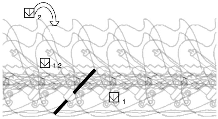
On the day of the terminal surgical procedure, the animals were anesthetized with isoflurane and nitrous oxide. An incision was made along the inferior border of the mandible. The anterior portion of the masseter was exposed and reflected from the mandible. An oblique corticotomy (60° to the occlusal plane) was made at the intersection of the mandibular body and ramus, and a Synthes® (Monument, CO) or a KLS-Martin® (Jacksonville, FL) distraction appliance was attached with self tapping screws (Figure 1). The fixation of the distractor prior to completion of the osteotomy prevented shifting of the mandibular fragments, preserving occlusion as much as possible. Following completion of the osteotomy, instrumentation was placed to measure bone strain on the mandible.
Figure 1.
Osteotomy location, distractor placement, and strain gauge locations. Numbers refer to Group 1, which had rosette strain gauges on the right condyle and mandibular corpus, and Group 2, which had rosette strain gauges on the right and left condyles.
In Group 1 (n=13), rosette strain gauges (SK-06-030WR-120, Measurements Group, Raleigh, NC) were installed on the right (osteotomy side) condylar neck and mandibular body, whereas Group 2 pigs (n=11) received gauges on both the right and left (non-osteotomy side) condylar necks, but not on the mandibular body (Figure 1). Standard procedures of site preparation and application were followed8. Analgesics (Ketorolac, buprenorphine) were administered and incisions were infiltrated with a topical anesthetic (lidocaine).
The gas anesthesia was removed and the animals were allowed to awaken briefly and to eat pig chow. The animals typically ate enthusiastically, during which time strain gauge and EMG data were recorded, along with data from other instrumentation7. Strain signals were conditioned and amplified (2100 System, Measurements Group, Raleigh, NC) and output to the computer using Biopac Systems, Inc. (Santa Barbara, CA) hardware and AcqKnowledge software. One or two series of 15-20 consecutive chews were selected per animal for analysis. The chew cycles were selected with the major criterion being clear strain signals. The principal strain magnitudes (εMAX and εMIN, tension and compression, respectively) and orientations were calculated in Microsoft Excel using standard formulae (Tech Note 515, Measurements Group Inc.). The peak principal strains for each power stroke were identified as those coinciding with γPEAK, the maximum shear strain (γPEAK = εMAX − εMIN).
The pigs were reanesthetized after about 10 minutes of feeding and muscle stimulations were carried out. Stimulating electrodes were placed in the bilateral masseter, medial pterygoid, digastric and lateral pterygoid muscles. Tetani were produced by 600ms trains of 5ms pulses delivered at 60 pulses per second (model S48 and SIU, Grass, Quincy MA), and voltages were adjusted to achieve supramaximal contractions. Only the masseter and medial pterygoid muscles produced significant bone strains. Following completion of these procedures, pigs were euthanized.
Stimulation strain data were collected and analyzed similarly to mastication strain data. Stimulations of the masseter and medial pterygoid muscles provide information on the extent to which these muscles are capable of loading the condyle following the surgical placement of the distraction appliance. The most relevant comparisons are thus between right (osteotomy) side condyle strains produced by the right masseter and medial pterygoid and left (non-osteotomy) side condyle strains produced by left side muscles.
Descriptive statistics (mean and standard deviation) were calculated for the principal strain magnitudes. Comparisons between strains produced by right and left side chews, and between osteotomy and non-osteotomy side condyles were made using paired t-tests on individuals with data from both sides. Comparisons were made between each side (osteotomy, non-osteotomy) and a control database using two-sample t-tests. The control database for the condyle strain consisted of a previously published group of four 12 to 16 week old Hanford minipigs9, supplemented by one 8 week and two 12 to 16 week old minipigs (data unpublished). The only available comparative control sample for mandibular corpus strain consisted of much larger eight month old Hanford minipigs10. For the stimulation data, paired t-tests were used to compare osteotomy side condyle strains produced by the same side masseter and medial pterygoid to non-osteotomy side condyle strains produced by non-osteotomy side muscles. Correlations between masticatory and stimulation shear strain in the same animals were calculated for each condyle using Pearson's correlation coefficient.
RESULTS
Mastication
1. Chewing side preference
The chewing side for each strain peak was determined for most animals using the onset and duration of the EMG signals. Postoperatively approximately half (n=11) of the animals demonstrated both right (osteotomy side) and left (non-osteotomy side) chewing patterns with a tendency for more left side chews. About a quarter (n=6) of the animals showed a strong bias for left side chews, with few unambiguous examples of clear right side chews. The remaining subset of animals (n=5) had poor EMG and thus chewing side could not be determined. Chewing patterns were also further examined in a subset of five animals with good pre and post-operative EMG (data not shown). The frequency of right versus left side chews was nearly even preoperatively (48% right, 44% left, and 8% indeterminate) but became skewed in favor of the non-osteotomy side (15% right, 79% left, and 5% indeterminate). For many of the animals the timing of the osteotomy side masseter was unusual, making it difficult to diagnose chewing side. Normally, the masseter on the chewing side (and presumably the side of the main food bolus) is activated slightly later and turns off later than the contralateral. The ipsilateral masseter not only contributes to adducting the jaws and crushing the bolus, but also by virtue of its timing serves to swing the mandible toward the midline (in concert with the contralateral temporalis). The common absence of this pattern suggests a more “chopping” type of osteotomy side chewing.
2. Corpus Strain
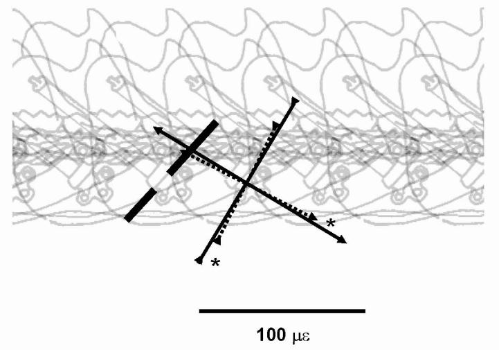
Results for mandibular corpus strain are presented in Table 1. The strain magnitudes on the right (osteotomy) side mandibular body were significantly larger during same side chews than during opposite (left) side chews (p < 0.02, paired t-test, Table 1, Figure 2). Strain orientation, which did not vary according to chewing side, was rostral-dorsal for compression and caudal-dorsal for tension, relative to the occlusal plane.
Table 1.
Peak principal corpus strains (osteotomy side) during mastication on different sides.
| Non-osteotomy side chews |
Osteotomy side chews |
||||||
|---|---|---|---|---|---|---|---|
| I.D. # | # of cycles L/R/U | εMAX (με) | εMIN (με) | angle (degrees) | εMAX (με) | εMIN (με) | angle (degrees) |
| 3 | 31/20/0 | 77 (32) | −66 (38) | 72 (16) | 159 (47) | −116 (12) | 80 (12) |
| 6 | 13/13/0 | 84 (7) | −116 (17) | 21 (3) | 131 (50) | −169 (56) | 24 (4) |
| 7 | 11/9/0 | 71 (46) | −52 (29) | 58 (6) | 172 (62) | −81 (37) | 64 (7) |
| 8 | 19/9/0 | 78 (25) | −114 (26) | 26 (6) | 105 (44) | −109 (40) | 30 (8) |
| 9 | 17/3/0 | 180 (62) | −62 (34) | 43 (20) | 304 (62) | −191 (36) | 54 (8) |
| 12 | 12/10/0 | 74 (43) | −78 (30) | −21 (10) | 99 (32) | −87 (26) | −11 (3) |
| 13 | 13/12/0 | 56 (13) | −56 (10) | −5 (7) | 45 (13) | −79 (21) | −27 (22) |
| 14 | 13/11/0 | 54 (23) | −75 (30) | 14 (2) | 44 (28) | −82 (38) | 18 (3) |
| 15 | 17/12/0 | 182 (40) | −79 (22) | 27 (5) | 208 (62) | −105 (36) | 31 (6) |
| 16 | 11/13/0 | 75 (25) | −101 (24) | 35 (12) | 94 (60) | −124 (58) | 42 (15) |
| Mean | 93* | −80* | 27 | 136* | −114* | 31 | |
| S.D. | (47) | (23) | (28) | (79) | (38) | (33) | |
Values are means, (standard deviations).
L/R/U are number of left, right and unknown side chew cycles analyzed.
εMAX, peak tensile principal strain; εMIN, peak compressive principal strain; angle is degrees of εMIN above the occlusal plane.
p < 0.02, paired t-test between chewing side strains.
Figure 2.
Magnitude and orientation of peak principal strains on the mandibular corpus during mastication. Solid lines represent right (osteotomy) side chews and dashed lines represent left side chews. Outward directed arrows depict principal tensile strain, and inward directed arrows depict principal compressive strain. The asterisks indicate significant differences (p<0.05) in strain magnitude of right and left chews based on a paired t-test (data in Table 1). No differences were found in strain orientation.
3. Condylar neck strain
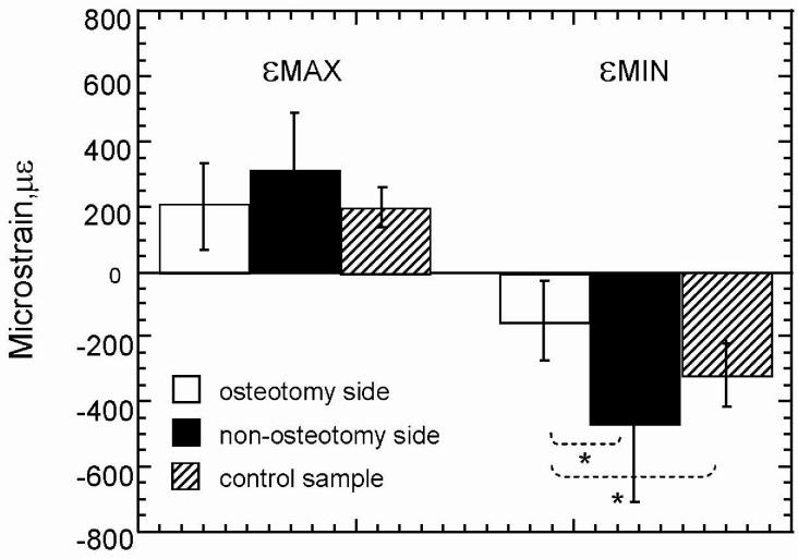
For the condylar neck, chewing side made no difference in strain pattern in Group I (Table 2). These data are therefore combined and pooled with data from animals that chewed only on the left side or had unknown chewing side patterns, to create a larger sample (). Comparisons between osteotomy side, non-osteotomy side, and control sample condylar neck strains are presented in Figure 3. Osteotomy and non-osteotomy side strain comparisons were conducted in two ways. First, the total samples were compared using two-sample Student's t-tests. Second, a subset of animals (n=8) that had both right and left condylar gauges were compared using a paired t-test (see shaded area in Table 2). As the findings were similar, only the results of the more conservative paired sample are shown (Figure 3). The osteotomy side experienced less (absolute) principal compressive strain than the non-osteotomy side. While tensile principal strain was also less on the osteotomy side, this difference did not reach significance.
Table 2.
Peak principal condyle strains during mastication on different sides.
| Right condyle strain (osteotomy side) |
Left condyle strain |
||||||||||||
|---|---|---|---|---|---|---|---|---|---|---|---|---|---|
| Non-osteotomy side chews |
Osteotomy side chews |
Chewing sides combined |
Chewing sides combined |
||||||||||
| I.D. # | L/R/U | εMAX (με) | εMIN (με) | angle (degrees) | εMAX (με) | εMIN (με) | angle (degrees) | εMAX (με) | εMIN (με) | angle (degrees) | εMAX (με) | εMIN (με) | angle (degrees) |
| 3 | 31/20/0 | 207 (62) | −178 (37) | 137(6) | 391 (136) | −216 (46) | 148 (7) | 280 (133) | −193 (44) | 141 (8) | |||
| 5 | 0/0/12 | 119 (7) | −78 (4) | 225 (1) | |||||||||
| 6 | 13/13/0 | 90 (21) | −47 (8) | 189 (23) | 79 (43) | −48 (20) | 173 (34) | 85 (33) | −48 (15) | 181 (25) | |||
| 7 | 11/9/0 | 286 (138) | −82 (34) | 170 (7) | 426 (145) | −115 (40) | 169 (7) | 349 (155) | −97 (39) | 170 (7) | |||
| 9 | 17/3/0 | 401 (81) | −128 (28) | 201 (4) | 352 (33) | −104 (3) | 188 (12) | 394 (77) | −124 (27) | 199 (7) | |||
| 10 | 0/0/20 | 218 (72) | −152 (45) | 229 (7) | |||||||||
| 11 | 0/0/32 | 96 (29) | −71 (21) | 119 (25) | |||||||||
| 12 | 12/10/0 | 578 (158) | −225 (60) | 161 (2) | 485 (98) | −187 (37) | 164 (1) | 535 (140) | −208 (53) | 162 (2) | |||
| 13 | 13/12/0 | 113 (35) | −31 (10) | 143 (7) | 88 (35) | −33 (22) | 154(9) | 101 (37) | −32 (16) | 148 (10) | |||
| 15 | 17/12/0 | 197 (40) | −223 (56) | 234 (3) | 317 (50) | −133 (7) | 151 (2) | 247 (74) | −186 (62) | 200 (41) | |||
| 16 | 11/13/0 | 188 (65) | −119 (48) | 119(15) | 149 (73) | −113 (60) | 116 (16) | 167 (73) | −116 (55) | 118 (14) | |||
| 19 | 9/4/11 | 108 (36) | −57 (21) | 158 (4) | 104 (27) | −47 (14) | 157 (5) | 98 (28) | −47 (17) | 157 (4) | 345 (51) | −600 (99) | 87 (5) |
| 20 | 0/0/14 | 182 (52) | −104 (21) | 175 (9) | 232 (44) | −619 (114) | 112 (2) | ||||||
| 21 | 25/0/9 | 140 (92) | −59 (36) | 167 (23) | 156 (115) | −66 (39) | 169 (23) | ||||||
| 22 | 20/0/18 | 275 (141) | −89 (33) | 175 (8) | 325 (152) | −103 (34) | 174 (9) | 163 (70) | −133 (63) | 119(28) | |||
| 23 | 0/0/37 | 446 (191) | −389 (160) | 219 (4) | 67 (34) | −113 (58) | 115 (9) | ||||||
| 25 | 13/3/0 | 157 (84) | −128 (35) | 135 (9) | 107 (56) | −105 (33) | 124 (15) | 148 (81) | −124 (35) | 133 (11) | 563 (136) | −701 (169) | 123 (2) |
| 26 | 17/5/0 | 170 (54) | −71 (23) | 184 (24) | 308 (125) | −105 (38) | 171 (14) | 203 (95) | −79 (30) | 181 (22) | 525 (101) | −670 (134) | 127 (3) |
| 27 | 22/0/0 | 95 (27) | −254 (59) | 236 (2) | 95 (27) | −254 (59) | 236 (2) | 209 (76) | −375 (114) | 124 (4) | |||
| 28 | 11/4/0 | 115 (27) | −65 (22) | 201 (5) | 40 (31) | −37 (17) | 196 (31) | 95 (44) | −58 (24) | 200 (15) | 269 (82) | −527 (125) | 116 (8) |
| Mean | 208 | −117 | 174 | 237 | −104 | 159 | 217 | −126* | 177*** | 297 | −467* | 115*** | |
| S.D. | (133) | (72) | (34) | (158) | (58) | (23) | (132) | (86) | (35) | (172) | (235) | (13) | |
| Mean | 218 | −113 | 169 | 237 | −104 | 159 | 199 | −145* | 184*** | 297 | −467* | 115*** | |
| S.D. | (143) | (66) | (34) | (158) | (58) | (23) | (126) | (118) | (33) | (172) | (235) | (13) | |
Values are means (standard deviations).
Shaded area is subset of individuals with data from right and left chews or from right and left gauges (paired t-test performed).
εMAX, peak tensile principal strain; εMIN, peak compressive principal strain; angle is degrees of εMIN above the occlusal plane.
p < 0.05
p <0.001
Figure 3.
Condylar neck peak principal strain magnitudes during mastication. Positive values are principal tensile strain and negative values are principal compressive strain. Osteotomy side = no-fill, non-osteotomy side = black, control database = pattern. The asterisks indicate significant differences (p<0.05) in strain magnitude between osteotomy and non-osteotomy sides (paired t-test) and between these samples and the control (two sample t-test).
Comparisons with the control database further elucidate these differences. Principal compressive strain was significantly less in the osteotomy side condylar necks compared to the control data (Figure 3). Principal tensile strain was not significantly different between the osteotomy side and control data. For the non-osteotomy side condylar neck the principal compressive and tensile strain values were elevated, but not significantly so, in comparison to the control sample strains (Figure 3). Thus, the significant difference in compressive strain between sides is accounted for primarily by a decrease in compressive strain on the osteotomy side. However, the slight increase in compressive strain on the non-osteotomy side contributed to the side difference. No significant side differences were found in tensile strain due to the fact that both sides showed increased tension (non-significant) compared to the non-surgical control database.
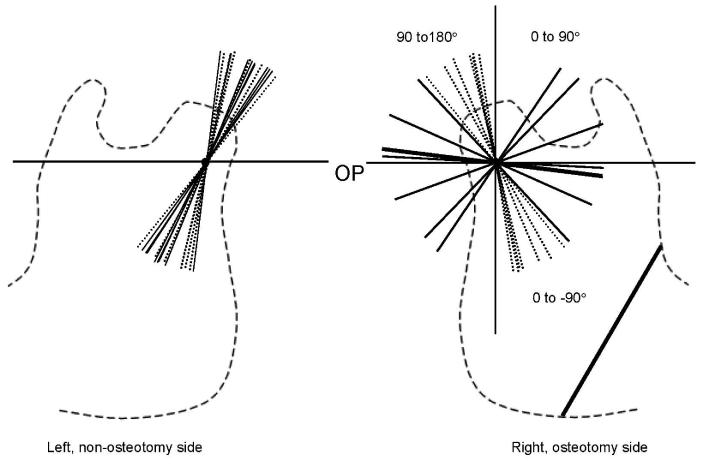
The distribution of condylar compressive strain orientations for the right (osteotomy) and left (non-osteotomy) sides of each individual is shown in Figure 4. The strain orientation on the osteotomy side condyle varied greatly among individuals, whereas it was quite consistent on the non-osteotomy side condyle, which was essentially identical to the control database. The wide distribution of angles on the osteotomy side makes it problematic to calculate a mean angle because it is necessary to decide if angles between 0° and 90° should be “flipped” 180° to larger angles below the occlusal plane (180° to 270°)(see Figure 4). The tightest cluster of orientations consisted of those that fell within 90-270° (mean was approximately 180°, parallel to the occlusal plane). The distribution of compressive strain orientations on the osteotomy side condyle do not overlap with the controls. Biologically, the important findings are that, first, the non-osteotomy (left) side strain orientation is “normal.” The compressive strain is directed dorso-caudally (at 116°), the same as in the control database. Second, the strain orientation on the osteotomy (right) side condyle is highly variable among the same individuals. Third, even considering the variability, compressive strain orientation on the osteotomy side differs from the non-osteotomy side/control database in being more horizontal.
Figure 4.
Compressive strain orientation on the condylar neck during mastication. Dotted lines are the strain orientations in the control database and solid lines are the strain orientations in the osteotomy (right) and non-osteotomy (left) samples. Angles are the orientation of principal compressive strain above the occlusal plane.
Muscle Stimulation
Muscle stimulation data are compared in Table 3. The major interest of this comparison was to determine whether reflection of a portion of the right muscles during surgery had a detrimental effect on their ability to load the condyle. If so, then stimulation of right muscles should have resulted in lower strains on the right condyle compared to those produced on the left condyle by the fully intact left muscles. The paired t-tests revealed no significant differences in magnitude or orientation between osteotomy and non-osteotomy side condyle strains produced by same side muscle stimulations. Seven animals had smaller osteotomy side shear strains (from osteotomy side masseter contractions) compared to the untreated side/untreated side muscle contractions. However, four animals had the opposite condition, effectively canceling any group differences. Similarly, for the medial pterygoid, there was an equal effect, with fully half the animals having larger strains on the osteotomy side. Moreover, there was no correlation between the magnitude of the stimulation strains and the magnitude of the masticatory strains for the same condyle in the same animals (right condyle muscle stimulation strain versus right condyle mastication strain p=0.253, and left condyle masseter stimulation strain versus left condyle mastication strain p=0.250). Thus, the decreased magnitude of compressive strain on the osteotomy side during mastication cannot be ascribed to muscle injury.
Table 3.
Condyle strains during same side masseter and medial pterygoid contraction.
| Masseter |
Medial Pterygoid |
|||||||
|---|---|---|---|---|---|---|---|---|
| R Muscle, R condyle |
L Muscle, L condyle |
R Muscle, R condyle |
L Muscle, L condyle |
|||||
| Indiv. # | γMAX (με) | angle (degrees) | γMAX (με) | angle (degrees) | γMAX (με) | angle (degrees) | γMAX (με) | angle (degrees) |
| 18 | 284 | 79 | 149 | 0 | no data | no data | no data | no data |
| 19 | 76 | 74 | 437 | 59 | no data | no data | no data | no data |
| 20 | 117 | 58 | 455 | 95 | 363 | 155 | 244 | 185 |
| 21 | 191 | 59 | 1911 | 64 | 361 | 161 | 760 | 147 |
| 22 | 71 | 49 | 239 | 78 | 711 | 155 | 202 | 157 |
| 23 | 76 | 7 | 163 | 28 | 586 | 127 | 78 | 199 |
| 24 | 246 | 59 | 468 | 47 | 62 | 143 | 47 | 132 |
| 25 | 300 | 70 | 684 | 61 | 671 | 159 | 912 | 139 |
| 26 | 186 | 79 | 137 | 97 | 249 | 144 | 562 | 148 |
| 27 | 380 | 60 | 127 | 110 | 210 | 140 | 406 | 150 |
| 28 | 614 | 75 | 349 | 79 | no data | no data | no data | no data |
| Mean | 231 | 61 | 465 | 65 | 402 | 148 | 401 | 157 |
| S.D. | 163.4 | 20.4 | 511.6 | 32.1 | 233.2 | 11.6 | 318.4 | 23.1 |
Values are means. γPEAK, shear strain; angle is degrees of εMIN above the occlusal plane.
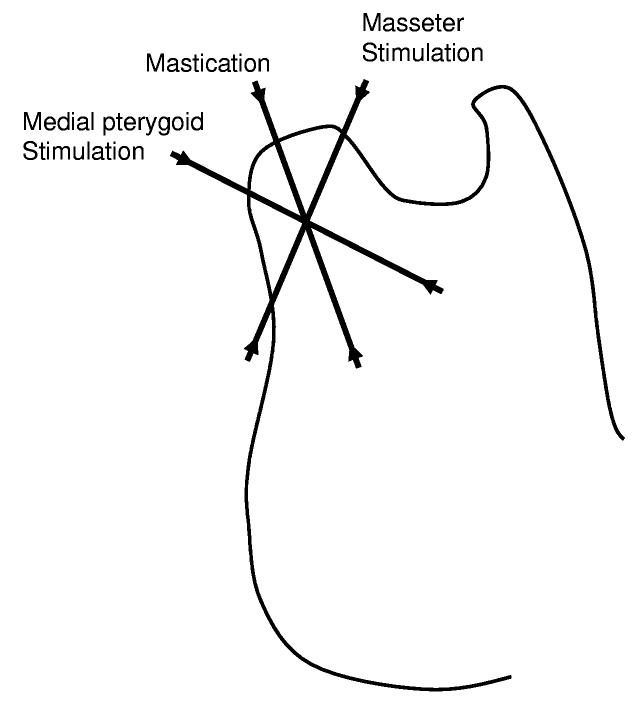
While there were no side differences in strain orientation, it is interesting to note that the compressive orientation on the condyle in response to masseter stimulation was 63°, which is more rostral than the “normal” orientation produced by mastication (116°)(Figure 5). On the other hand, the orientation of compressive strain on the lateral condylar neck resulting from stimulations of the medial pterygoid, which has not previously been reported, is more caudal (154°)(Figure 5).
Figure 5.
Summary of compressive strain orientation on the condylar neck during stimulation of the medial pterygoid, masseter, and during mastication. Averages of data presented in Tables 2 and 3.
DISCUSSION
The strain gauges placed in this study recorded overall patterns of bone deformation rather than the site-specific effects of the surgery and appliance. Unusual strains associated with loads at fixation points (screws) are likely to be highly localized and clearly are not detected by the strain gauges that were located well over 1 cm away. The strains observed indicate that the distractor appliance did not provide a stress-shielding effect. Although strain magnitudes were lower on the osteotomy side condylar neck than control values during mastication, the mandibular corpus showed no such decrease. Furthermore, osteotomy side condylar strains were the same as those of the non-osteotomy side for muscle stimulations.
Although there were changes in masticatory strain following the procedures, these relate to functional changes. Chewing itself was altered to some degree, because a chewing preference for the non-osteotomy side developed post-surgery. Nevertheless, most animals continued to chew, at least some of the time, on the side of the osteotomy. Surprisingly, the side of chewing had only a minor impact on mandibular strain, affecting solely the magnitude (and not the orientation) of strain on the corpus. Condylar strain was unaffected by chewing side.
The present study and the control database10 both showed rostral-dorsal compression and caudal-dorsal tension on the mandibular corpus. Bone strain on the mandibular corpus is thought to reflect torsion resulting from the opposite rotations applied to the molars by jaw movement and to the ramus by muscle contraction11-12. Assuming this model is correct, the absence of a change in the present study shows that the action of the muscles is not essential or that the muscles could still exert their effect through the rigid distractor. The fact that the osteotomy side corpus showed larger strains during working side chewing than balancing cycles is surprising. Although working-side mandibular strains are larger than balancing side strains in many species13-15, the previous study on pigs did not show such a difference10, probably because pigs have bilateral occlusion. While we did not advance the distractor in this acute study and the occlusion was intact, the loss of tissue from the saw cut and the instability of the osteotomy7 must have slightly altered the alignment of the tooth row. In particular, the typical bilateral occlusion may have been disrupted such that the teeth on the right side were well intercuspated only when the pig chewed on that side, accounting for the larger working side strains.
The most notable differences in strain pattern occurred on the condylar neck. In particular, the osteotomy side had lower strain magnitudes than the non-osteotomy side, significantly so for compressive strain. This could be explained by either an increased magnitude on the non-osteotomy side, a decreased magnitude on the osteotomy side, or a combination of both. When compared with a control database, osteotomy side compressive strains were significantly lower (absolute magnitude), while non-osteotomy side compressive strains were not significantly higher. Thus, reduced loading of the osteotomy side condyle probably accounts for most of the side discrepancy. It is interesting that the osteotomy side condylar loading pattern was also altered in terms of the ratio of tension to compression and in strain orientation. Unlike the non-osteotomy side and the control data, tensile strain was greater in absolute magnitude than compression. The orientation of the principal strains was also highly variable on the osteotomy side. This variability was most pronounced among individuals but was also evident within some individuals. Overall, compressive strain was more horizontally oriented than in the non-osteotomy side or control database.
Two possible explanations for the decreased strain magnitude and altered orientation on the condyle of the osteotomy side are 1) decreased loading due to partial reflection of the masseter and medial pterygoid muscles, and 2) decreased loading due to changes in masticatory behavior. Despite the partial removal of the right side masseter and medial pterygoid, stimulation of these muscles did not consistently produce lower strains than that of the contralateral condyle/muscle. Moreover, there was no correlation between the magnitude of strain produced by mastication and the magnitude of strain produced by muscle stimulation. Thus, the first possibility is rejected. With regard to the second possibility, injury to the right side mandible, change in occlusion with appliance placement, and loss of sensory feedback due to damage to the inferior alveolar nerve are all possible reasons why the animals might chew differently or avoid the osteotomy side. Because the pigs were avoiding the osteotomy side, it seems reasonable to expect that this change in masticatory behavior was responsible for decreased and altered strain. However, this argument has limited explanatory power given the large working side strains on the mandibular corpus. Furthermore, strains on the osteotomy side condyle were identical for left and right chewing cycles. Thus, the reduced loading on the osteotomy side condyle does not result from the preference for left side chewing. Although difficult to analyze quantitatively, examination of EMG signals revealed that the timing of the osteotomy side masseter was unusual, suggesting a more “chopping” style of osteotomy side chewing. However, this change in chewing behavior also does not account for the condylar strain patterns, because there was no difference in condyle strain between right and left chewing sides but rather a difference between right and left condyles. Because the temporalis and deep masseter were not recorded, it is not possible to make a full assessment of the muscular contributions to mandibular loading. Nevertheless, neither muscle damage nor changes in chewing behavior can explain the fundamental change in osteotomy side condylar loading.
The most likely explanation for reduced and altered osteotomy-side condylar loading is the instability of the fixation itself, as previously documented7. Poor control of the small mandibular fragment on the right side could account for the variability of strain orientation. Furthermore, mobility during the power stroke would act to decrease strain magnitude (which increases for more isometric contractions). Finally, the predominant pattern of sagittal bending of the mandible7 would tend to rotate the osteotomy side condyle backward, and thus lead to compressive strains that were less vertically and more horizontally oriented, as were observed.
This study was done on an acute basis and cannot answer questions regarding the long term consequences of mandibular distraction on condylar loading. Chronic studies on mandibular distraction in which strain is measured at different endpoints are needed in order to determine if the osteotomy side condyle continues to be underloaded in compression and to have altered strain orientations.
ACKNOWLEDGMENTS
We thank Dr. Zi-Jun Liu and Mr. Frank Starr for their assistance with experiments, and Israel Fuentes for analysis of EMG data. This project was funded by PHS awards R01 DE 14336 and P60 DE 13061.
LITERATURE CITED
- 1.McCarthy JG. Distraction of the Craniofacial skeleton. Springer-Verlag; New York: 1999. [Google Scholar]
- 2.McCarthy JG, Katzen JT, Hopper, et al. The First Decade of Mandibular Distraction: Lessons We Have Learned. Plast Reconstr Surg. 2002;110:1704. doi: 10.1097/01.PRS.0000036260.60746.1B. [DOI] [PubMed] [Google Scholar]
- 3.McCormick SU, McCarthy JG, Grayson BH, et al. Effect of mandibular distraction on the temporomandibular joint, part I: Canine study. J Craniofac Surg. 1995;6:358. doi: 10.1097/00001665-199509000-00005. [DOI] [PubMed] [Google Scholar]
- 4.Kruse-Lösler B, Meyer U, Floren C, et al. Influence of distraction rates on the temporomandibular joint position and cartilage morphology in a rabbit model of mandibular lengthening. J Oral Maxillofac Surg. 2001;59:1452. doi: 10.1053/joms.2001.28281. [DOI] [PubMed] [Google Scholar]
- 5.Karaharju-Suvanto T, Peltonen J, Laitinen O, et al. The effect of gradual distraction of the mandible on the sheep temporomandibular joint. Int J Oral Maxillofac Surg. 1996;25:152. doi: 10.1016/s0901-5027(96)80063-4. [DOI] [PubMed] [Google Scholar]
- 6.Thürmuller P, Troulis MJ, Rosenberg A, et al. Changes in the condyle and disc in response to distraction osteogenesis of the minipig mandible. J Oral Maxillofac Surg. 2002;60:1327. doi: 10.1053/joms.2002.35733. [DOI] [PubMed] [Google Scholar]
- 7.Sun Z, Rafferty KL, Egbert MA, et al. Mandibular mechanics following ostetomy and distraction placement: Part I. Mobility of the osteotomy site. J Oral Maxillofac Surg. doi: 10.1016/j.joms.2005.12.008. Submitted. [DOI] [PMC free article] [PubMed] [Google Scholar]
- 8.Rafferty KL, Herring SW, Artese F. Three-dimensional loading and growth of the zygomatic arch. J Exp Biol. 2000;203:2093. doi: 10.1242/jeb.203.14.2093. [DOI] [PMC free article] [PubMed] [Google Scholar]
- 9.Marks L, Teng S, Årtun J, et al. Reaction strains on the condylar neck during mastication and maximum muscle stimulation in different condylar positions: An experimental study in the miniature pig. J Dent Res. 1997;76:1412. doi: 10.1177/00220345970760071201. [DOI] [PubMed] [Google Scholar]
- 10.Liu ZJ, Herring SW. Masticatory strains on osseous and ligamentous components of the jaw joint in miniature pigs. J Orofac Pain. 2000;14:265. [PubMed] [Google Scholar]
- 11.Hylander WL. Mandibular function in galago crassicaudatus and macaca fascicularis: An in vivo approach to stress analysis of the mandible. J Morph. 1979;159:253. doi: 10.1002/jmor.1051590208. [DOI] [PubMed] [Google Scholar]
- 12.Herring SW, Rafferty KL, Liu ZJ, et al. Jaw muscles and the skull in mammals: The biomechanics of mastication. Comp Biochem Physiol A Mol Integr Physiol. 2001;131:207. doi: 10.1016/s1095-6433(01)00472-x. [DOI] [PubMed] [Google Scholar]
- 13.Weijs WA, de Jongh HJ. Strain in mandibular alveolar bone during mastication in the rabbit. Arch Oral Biol. 1977;22:667. doi: 10.1016/0003-9969(77)90096-6. [DOI] [PubMed] [Google Scholar]
- 14.Crompton AW. Functional morphology in vertebrate paleontology. Cambridge University Press; New York, NY: 1995. p. 55. [Google Scholar]
- 15.Hylander WL, Ravosa MJ, Ross CF, et al. Mandibular corpus strain in primates: Further evidence for a functional link between symphyseal fusion and jaw-adductor muscle force. Amer J Phys Anthropol. 1998;107:257. doi: 10.1002/(SICI)1096-8644(199811)107:3<257::AID-AJPA3>3.0.CO;2-6. [DOI] [PubMed] [Google Scholar]


