Abstract
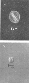
The proportion of oocysts of Cryptosporidium parvum showing a fold on oocyst walls when incubated with either fluorescent monoclonal antibody or a surface-reactive fluorescent dye was increased by incubating suspensions of oocysts with dimethyl sulfoxide, sucrose, or Hanks' balanced salt solution. Further incubation of sucrose-incubated oocysts with water showed this to be a reversible phenomenon. Oocysts demonstrating this fold after incubation in dimethyl sulfoxide were of the same viability as control oocysts and followed the same excystation dynamics. Despite this fold having been previously described as a suture, we were unable to find any evidence that this pattern of fluorescence highlighted the same suture that has been described in ultrastructural studies. Furthermore, oocysts were observed in which this fold was not always continuous with the gape in the oocyst wall through which the sporozoites had emerged. We propose that this fluorescently highlighted region or fold should no longer be described as a suture and question its validity as a diagnostic feature. When environmental and other samples are being examined for the presence of C. parvum oocysts, objects of appropriate size, shape, and fluorescence which do not demonstrate a surface fold should not necessarily be excluded.
Full text
PDF


Images in this article
Selected References
These references are in PubMed. This may not be the complete list of references from this article.
- Campbell A. T., Robertson L. J., Smith H. V. Viability of Cryptosporidium parvum oocysts: correlation of in vitro excystation with inclusion or exclusion of fluorogenic vital dyes. Appl Environ Microbiol. 1992 Nov;58(11):3488–3493. doi: 10.1128/aem.58.11.3488-3493.1992. [DOI] [PMC free article] [PubMed] [Google Scholar]
- Casemore D. P. ACP Broadsheet 128: June 1991. Laboratory methods for diagnosing cryptosporidiosis. J Clin Pathol. 1991 Jun;44(6):445–451. doi: 10.1136/jcp.44.6.445. [DOI] [PMC free article] [PubMed] [Google Scholar]
- Reduker D. W., Speer C. A., Blixt J. A. Ultrastructure of Cryptosporidium parvum oocysts and excysting sporozoites as revealed by high resolution scanning electron microscopy. J Protozool. 1985 Nov;32(4):708–711. doi: 10.1111/j.1550-7408.1985.tb03106.x. [DOI] [PubMed] [Google Scholar]
- Robertson L. J., Campbell A. T., Smith H. V. In vitro excystation of Cryptosporidium parvum. Parasitology. 1993 Jan;106(Pt 1):13–19. doi: 10.1017/s003118200007476x. [DOI] [PubMed] [Google Scholar]
- Rose J. B., Landeen L. K., Riley K. R., Gerba C. P. Evaluation of immunofluorescence techniques for detection of Cryptosporidium oocysts and Giardia cysts from environmental samples. Appl Environ Microbiol. 1989 Dec;55(12):3189–3196. doi: 10.1128/aem.55.12.3189-3196.1989. [DOI] [PMC free article] [PubMed] [Google Scholar]
- Smith H. V., McDiarmid A., Smith A. L., Hinson A. R., Gilmour R. A. An analysis of staining methods for the detection of Cryptosporidium spp. oocysts in water-related samples. Parasitology. 1989 Dec;99(Pt 3):323–327. doi: 10.1017/s0031182000059023. [DOI] [PubMed] [Google Scholar]


