Abstract
CD9 is a tetraspan protein that associates with several β1 integrins, including α6β1. Because α6β1 is present on murine eggs and interacts with the sperm-surface glycoprotein ADAM 2 (fertilin β), we first asked whether CD9 is present on murine eggs and whether it functions in sperm–egg binding and fusion. CD9 is present on the plasma membrane of oocytes in the ovary as well as on eggs isolated from the oviduct. The anti-CD9 mAb, JF9, potently inhibits sperm–egg binding and fusion in vitro in a dose-dependent manner. JF9 also disrupts binding of fluorescent beads coated with native fertilin or a recombinant fertilin β disintegrin domain. (Both ligands bind to the egg via α6β1.) Immunohistochemistry showed that CD9 is undetectable in the uterine epithelium, appears basolaterally and as prominent apical patches on the epithelium in the region between the uterus and the oviduct, and then persists apically in the oviduct. The integrin α6A subunit is found in similar apical patches in the region between the uterus and oviduct, but is confined to the basal aspect of the epithelium in the uterus and oviduct. Hence, α6A and CD9 both are expressed on the apical epithelial surface at the uterine–oviduct junction. These findings correlate with the observation that fertilin β “knockout” sperm traverse the uterus but do not progress into the oviduct, contributing to the infertility of fertilin β−/− male mice. Our results suggest that high-avidity binding between fertilin β (ADAM 2) and α6β1 requires cooperation between α6β1 and CD9. Such cooperation may assist sperm passage into the oviduct as well as sperm–egg interactions.
Keywords: tetraspan, fertilin β, cell adhesion
CD9 belongs to the tetraspan superfamily (TM4SF) of integral plasma membrane proteins. These proteins possess four transmembrane domains, two extracellular loops, and three short cytoplasmic segments. Tetraspan proteins have been proposed to act as “molecular facilitators” by bringing together and stabilizing molecular complexes. Tetraspan proteins have been shown to associate physically with members of the integrin family (1–3), with MHC class II glycoproteins (4–6), as well as with each other (2, 7–10). Although tetraspan proteins have been implicated in growth control, intracellular signaling, migration, and adhesion, their precise mechanisms of action remain unclear (2, 11).
The tetraspan protein CD9 interacts with the integrins α3β1, α6β1, α4β1, and αIIbβ3 (2). CD9 has been reported to function in integrin-mediated signaling, migration, and adhesion to extracellular matrix substrates (12–14). Expression of CD9 also has been shown to enhance membrane fusion and infection by two enveloped viruses, the paramyxovirus, canine distemper virus (15), and the retrovirus, feline immunodeficiency virus (16). Another integrin-associated protein, CD98, has been shown to modulate fusion by the HIV (17), as well as fusion of monocytes to form osteoclast-like cells (18).
Among the integrins that associate with CD9, the integrin α6β1 has been implicated as the partner, on the murine egg, for fertilin β [a disintegrin and metalloprotease (ADAM) 2], a sperm-surface glycoprotein implicated in sperm–egg binding and fusion (for reviews see refs. 19–21). Binding of fertilin β to α6β1 involves its disintegrin domain (refs. 22–26; D.B., Y.T., M.S.C., E. A. C. Almeida, L. Osbourne, and J.M.W., unpublished data). Recent studies indicate that fertilin β−/− sperm show greatly reduced ability to bind to the egg plasma membrane and, unexpectedly, to enter the oviduct from the uterus (27).
Given the association of CD9 with the α6β1 integrin as well as with certain membrane fusion events, we asked whether CD9 is present on the mouse egg plasma membrane and whether it is involved in sperm–egg binding and fusion. Given the phenotype of the fertilin β “knockout” sperm, we also investigated the localization of CD9 and the α6 integrin subunit along the female reproductive tract.
Materials and Methods
Egg Isolation.
Mature mouse eggs were collected from superovulated 8- to 12-week-old ICR female mice (Harlan Breeders, Indianapolis; Hilltop Labs, Philadelphia) as described previously (22, 26). Zonae pellucidae were removed by treating the eggs for 1 min in acidic Tyrode’s solution, pH 2.5 (28), or, where indicated, for 3 min with 10 μg/ml α-chymotrypsin followed by mechanical shearing. Similar results for Figs. 2 and 3 were obtained with either method of zona removal (data not shown). Eggs were washed through three 100-μl drops of fresh M199 medium and incubated in M199 medium overlaid with light mineral oil for 2–3 h at 37°C in a 5% CO2 incubator before use.
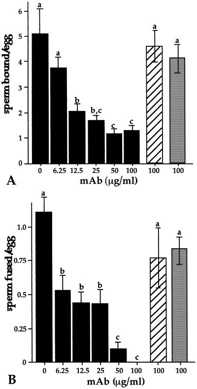
Figure 2.
Effect of an anti-CD9 mAb on mouse sperm–egg binding and fusion. Zonae pellucidae from mature mouse eggs were removed by brief incubation in chymotrypsin followed by mechanical shearing. Zona-free eggs were incubated with the indicated concentrations of the anti-CD9 mAb (solid bars) or with 100 μg/ml of the non-function-blocking anti-α6 mAb, J1B5 (hatched bars), or anti-MLV mAb (shaded bars) in M199 medium. Eggs were processed and analyzed for sperm–egg binding (A) and fusion (B). Data represent the mean ± SE from three pooled experiments. Values with different letters are statistically different (P < 0.05). Sperm binding was inhibited similarly when eggs were pretreated with JF9 and then washed before sperm binding (data not shown).
Figure 3.
Effects of JF9 on binding of beads coated with fertilin β, anti-α6 mAb, and laminin E8 to zona-free eggs. (A) Yellow-green fluorescent latex beads were coated with native fertilin captured from a sperm lysate (Fβ) or with a recombinant fertilin β disintegrin domain (rFβ). Zona-free eggs were either untreated (Ct) or preincubated with 200 μg/ml of a function-blocking anti-α6 mAb (GoH3), a non-function-blocking anti-α6 mAb (J1B5), or the anti-CD9 mAb, JF9, for 15 min. (B) Yellow-green fluorescent latex beads were coated with GoH3 or J1B5. Zona-free eggs were either untreated (Ct) or preincubated with 200 μg/ml JF9 for 15 min. (C) Yellow-green fluorescent latex beads were coated with laminin E8. Eggs were placed in Puck’s saline A with 1 mM CaCl2 or 1 mM MnCl2 as indicated and then left untreated (Ct) or incubated with 200 μg/ml JF9 for 15 min. Eggs then were incubated with beads for 1 h, washed, and analyzed by confocal microscopy. Fluorescent images are shown.
Antibodies.
Antibodies were obtained from the following sources: rat anti-α6 mAb GoH3 (Immunotech) and rat anti-α6 mAb J1B5 (C. Damsky, University of California, San Francisco). The rat mAb JF9 (IgG2b) against murine CD9 was prepared as for the preparation of the mAb KMC8.8 (29). Briefly, Wistar rats were immunized three times with J774A.1 macrophage cells. Spleens were removed and fused with SP2/0 myeloma cells (American Type Culture Collection), and mAbs were prepared by standard procedures. Polyclonal rabbit antiserum against the cytoplasmic tail of fertilin β was prepared and purified as described previously (22). A rat mAb against the gag protein of the Moloney murine leukemia virus (MLV) was obtained from the American Type Culture Collection. The anti-HA mAb, 12CA5, was used as described previously (30). A polyclonal goat antiserum against the cytoplasmic tail of the mouse integrin α6A and the corresponding immunizing peptide were obtained from Santa Cruz Biotechnologies.
Immunohistochemistry and Tissue Immunofluorescence.
Normal adult (C57BL/6 × A/J) F1 female mice (25–30 g) were euthanized, and the entire reproductive tract was fixed in 4% paraformaldehyde, cryopreserved in 30% sucrose, and embedded in OCT compound (Miles). Alternatively, tissue was directly frozen in liquid N2. For staining of CD9 by mAbs KMC 8.8 or JF9, paraformaldehyde-fixed frozen sections were incubated with mAb for 1 h, washed twice with PBS, and incubated with biotinylated rabbit antibody to rat IgG. The slides then were incubated with biotin/streptavidin/peroxidase, developed with a colorimetric substrate, and counterstained with methylene blue (31). For detection of α6, frozen tissue sections were postfixed in acetone, blocked with 10% rabbit serum in PBS for 30 min at 25°C, and incubated for 45 min at 25°C with 10 μg/ml antibody against the α6A cytoplasmic domain or mAb GoH3 in PBS containing 3% BSA (PBS/BSA). Where indicated, the antibody was immunoabsorbed for 2 h at 25°C with 50 μg/ml of the immunizing peptide. Tissues were blocked for 15 min at 25°C with avidin and 15 min with biotin (Avidin/Biotin Blocking Kit; Vector Laboratories). Biotinylated secondary antibody (15 μg/ml; Southern Biotechnology Associates) was added for 30 min at 25°C in PBS/BSA, followed by incubation with 5 μg/ml Texas red-conjugated streptavidin (Southern Biotechnology Associates) in PBS/BSA for 30 min at 25°C. Tissues were rinsed twice with PBS between each incubation step. Slides were coated with 0.7% Elvanol (DuPont) in 0.01 K2HPO4 (pH 8.5) with 0.14 M NaCl and 33% glycerol (31).
Egg Immunofluorescence.
Live, mature zona-free eggs were incubated with 100 μg/ml rat mAbs for 1 h at 4°C in M199 medium. Eggs were washed, incubated with a 1:100 dilution of Cy3-conjugated anti-rat IgG (Zymed) for 30 min at 4°C (in M199 medium), washed, and mounted in M199 onto glass slides. Eggs were visualized at ×200 magnification by using a Zeiss Axioplan 2 microscope. Images were captured by using openlab (Improvision, Coventry, England) software.
Immunoprecipitation.
Two hundred and fifty zona-free mouse eggs were treated twice with 2 mg/ml EZ-Link sulfo-NHS-LC-biotin (Pierce) for 20 min at 4°C in 300 μl BSA-free M199 medium with 10 μg/ml polyvinyl alcohol (PVA), washed with M199/PVA, and lysed with 1% Nonidet P-40 in 50 mM Tris⋅HCl, pH 7.4/1 mM CaCl2/1 mM MgCl2/150 mM NaCl for 30 min at 4°C. Lysates were centrifuged (16,000 × g for 15 sec at 4°C), and the supernatants were precleared for 30 min at 4°C with 20 μl anti-rat agarose beads (ICN). Cleared supernatants were passed through a 0.2-μm syringe filter and incubated for 30 min at 4°C with 10 μl anti-rat agarose beads precoupled with 1 μg anti-MLV gag IgG. Beads were pelleted and the supernatants were immunoprecipitated with 10 μl anti-rat agarose beads precoupled with 1 μg GoH3 or JF9 mAbs for 1 h at 4°C. Beads were washed four times with Nonidet P-40 lysis buffer, resuspended in 15 μl of 2× SDS gel sample buffer (22), incubated at 95°C for 5 min, and subjected to nonreducing 10% PAGE and avidin blotting as described (22).
In Vitro Fertilization.
Sperm–egg binding and fusion were assayed as described previously (26, 32). Briefly, zona-free eggs, prepared with chymotrypsin and mechanical shearing, were preloaded with 1 μg/ml Hoechst dye no. 33342 (Sigma) for 10 min, washed, and pretreated with mAbs for 15–30 min in M199 medium at 37°C in a 5% CO2 incubator. Sperm were collected from the cauda epididymus of retired male breeders and capacitated for 3 h (22), at which time approximately 75% were spontaneously acrosome-reacted (data not shown). Sperm were added at a final concentration of ≈1 × 10⁵ sperm/ml and coincubated with eggs for 40 min at 37°C in a 5% CO2 incubator. Eggs were washed and mounted on glass slides. Twenty to 40 eggs were analyzed per condition, and the average sperm bound per egg was determined by phase-contrast microscopy. To determine sperm fused per egg, the number of Hoechst-stained, decondensed sperm nuclei within the eggs was averaged.
Bead–Egg Binding.
Yellow-green fluorescent sulfate-derivatized latex beads (0.2 μm; Molecular Probes) were coated with fertilin β isolated from caput epididymal sperm or a recombinant protein containing the fertilin β disintegrin domain (D.B., et al., unpublished data) as described previously (22). Beads (1.0 μm) were coated with 0.4 mg/ml GoH3 or J1B5 mAb or with 1 mg/ml laminin E8 (22). Twenty to 40 zona-free eggs were pretreated for 15 min with mAbs in 20-μl drops of Puck’s saline A (Sigma) supplemented with 1 mM Ca2+ (or with 1 mM Mn2+, where indicated) and 0.4% BSA. Bead binding was performed as described previously (22).
Results
Expression of CD9 on Oocytes.
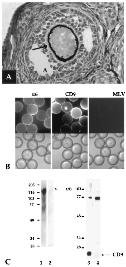
We first used immunohistochemistry to ask whether CD9 is present on the plasma membrane of mouse oocytes. In the ovary, CD9 expression was concentrated at the plasma membrane of oocytes in developing follicles, with the most intense staining found on oocytes in mature follicles (Fig. 1A, arrow). Fibrillar staining in the surrounding zona pellucida likely was due to membrane extensions from the oocyte plasma membrane because surrounding follicular cells were negative for CD9 staining. CD9 also was observed on some cells in the theca layer at the periphery of the mature follicle (Fig. 1A, T) and in immature oocytes, but not in surrounding ovarian tissue nor in late atretic follicles (data not shown). The presence of CD9 in immature oocytes ranging from 30 to 100 μm in diameter was confirmed by indirect immunofluorescence of oocytes isolated from mouse ovaries (data not shown) and correlates with α6β1 expression in oocytes at these stages (ref. 33; data not shown). We next asked whether CD9 is present on the plasma membrane of eggs taken from the oviduct. As seen in Fig. 1B, both α6 and CD9 were detected by immunofluorescence analysis of zona-free mouse eggs. Surface expression of α6 and CD9 was confirmed further by immunoprecipitation from lysates of biotinylated eggs (Fig. 1C, lanes 1 and 3, respectively).
Figure 1.
Expression of α6 and CD9 on the surface of oocytes and mature, fertilization-competent eggs (A). Ovarian sections from mature mice were processed and stained for CD9. A secondary follicle is shown. Arrow indicates staining with the mAb KMC 8.8 on the oocyte plasma membrane and fine fibrillar staining extending into the zona pellucida. T, thecal layer; F, follicular cells; A, antrum. Staining was also seen on oocytes in Graafian follicles (data not shown). (B) Mature zona-free mouse eggs were incubated with GoH3 (Left), JF9 (Center), or a control rat mAb, anti-MLV (Right), and processed for immunofluorescence; fluorescence (Upper) and phase-contrast (Lower). (C) Zona-free biotinylated mouse eggs were lysed and processed for immunoprecipitation with anti-α6 mAb, GoH3 (lane 1), or anti-CD9 mAb, JF9 (lane 3). Samples were first immunoprecipitated with an anti-virus mAb, anti-MLV (lanes 2 and 4). Samples were analyzed by nonreducing 10% SDS/PAGE. We found no evidence (by Western analysis or immunofluorescence) for CD9 expression on mature sperm (data not shown).
Effect of Anti-CD9 mAb on Sperm–Egg Interactions.
We next examined whether CD9 plays a role in fertilization in vitro. As seen in Fig. 2 (solid bars), the anti-CD9 mAb, JF9, inhibited sperm–egg binding (Fig. 2A) and fusion (Fig. 2B) in vitro in a dose-dependent manner. Binding and fusion in the presence of the non-function-blocking anti-α6 mAb, J1B5 (Fig. 2, hatched bars), or a control anti-virus mAb (Fig. 2, shaded bars) were not significantly perturbed. The JF9 mAb did not activate the egg as evidenced by the lack of cortical granule exocytosis or polar body extrusion. Another anti-CD9 mAb against a different epitope, KMC8.8 [Oritani et al. (29)], also inhibited sperm binding and fusion to zona-free eggs (data not shown).
Effect of Anti-CD9 mAbs on Fertilin–Egg Interactions.
We next asked whether the anti-CD9 mAb, JF9, influences binding of fertilin-coated beads to eggs. As seen in Fig. 3A (Upper Right), JF9 inhibited binding of beads coated with native fertilin β captured from a sperm lysate. The anti-CD9 mAb, JF9, also inhibited binding of beads coated with a recombinant protein containing the disintegrin domain of fertilin β (Fig. 3A Lower Right). Binding of both intact fertilin β as well as a recombinant disintegrin domain-containing construct of fertilin β was inhibited by the function-blocking anti-α6 mAb, GoH3, but not by the non-function-blocking anti-α6 mAb, J1B5 (ref. 22; D.B., et al., unpublished data).
JF9 did not inhibit binding to eggs of beads coated with either of the anti-α6 mAbs, GoH3 or J1B5 (Fig. 3B). JF9 also did not prevent binding of beads coated with the laminin E8 digestion fragment (in the presence of Mn2+). We showed previously that laminin E8 binds to eggs in buffers containing Mn2+ but not Ca2+ (Fig. 3C) (22). Hence, JF9 does not alter surface expression of the α6 integrin, nor does it mask either the GoH3 or J1B5 epitopes. In addition JF9 does not activate binding of laminin E8-coated beads in the presence of Ca2+.
D9 and α6 Expression in the Female Reproductive Tract.
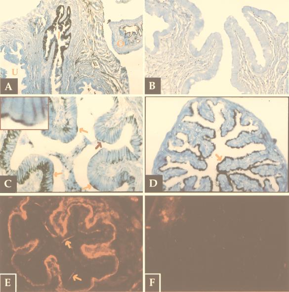
We next examined expression of CD9 in the mouse female reproductive tract. Immunohistochemical analysis showed that CD9 is absent from the epithelial cells of the uterus (Fig. 4A, U, and 4B). Beginning at the junction between the uterus and the oviduct, the utero–tubal junction, CD9 is present on the epithelial cells (Fig. 4A). In the isthmus of the fallopian tube, CD9 is present basolaterally and also at the apical region of patches of epithelial cells (Fig. 4C Inset and arrows). In the upper portion of the fallopian tube, including the ampulla, the entire apical region, including the cilia, is strongly positive for CD9, whereas the basolateral regions lack CD9 (Fig. 4A, O, and 4D, arrow).
Figure 4.
Immunohistochemistry of regions of the female reproductive tract with anti-CD9 and anti-α6 antibodies. Sections of the reproductive tract from mature female mice were stained with JF9. (A) A low-magnification cross-section of the utero–tubal junction (J) flanked by uterine epithelium (U) and oviduct (O) and higher-magnification sections of the uterus (B), the isthmus (C), and the oviduct (D) are shown. (C Inset) A section of C indicated by a red arrow is magnified four times. Sections of the isthmus were stained with an anti-α6A cytoplasmic tail antibody in the absence (E) or presence (F) of the immunizing peptide. Arrows in C–F indicate locations of positive apical staining.
We next examined expression of the integrin α6 subunit in the female reproductive tract. Staining with an α6A cytoplasmic tail antibody showed that, like CD9, α6A is present on the apical surface of patches of epithelial cells at the isthmus of the fallopian tube (Fig. 4E, arrows); it is also detected on the basal surface. In the oviduct and uterus, staining for α6A was confined to the basal region of the epithelial cells and, possibly, to stromal glands (data not shown). Similar staining was seen with the anti-α6 mAb GoH3 (data not shown). Our attempt to colocalize CD9 and the α6 integrin subunit was not possible because of differential sensitivity of the two antigens to the fixatives required for immunostaining by the respective mAbs.
Discussion
Given previous associations of the tetraspan protein CD9 with the α6β1 integrin (1, 3, 5, 34, 35) and with certain membrane fusion events (15, 16), we hypothesized that CD9 plays a role in the interaction of fertilin β (ADAM 2), a sperm protein involved in sperm–egg binding and fusion (27, 36), with α6β1 on the egg. We found that CD9 is expressed on the plasma membrane of oocytes and fertilization-competent eggs. A mAb to CD9 potently inhibits, in parallel, sperm–egg binding and fusion in a dose-dependent manner. The same anti-CD9 mAb inhibits binding of beads coated with intact fertilin or a recombinant fertilin β disintegrin domain to eggs. In addition, immunohistochemical analysis indicated that both CD9 and the α6A integrin subunit are present at the apical region of patches of epithelial cells in the region between the uterus and the oviduct. This is the only site in the female reproductive tract at which we have seen apical expression of both CD9 and α6A on a luminal surface, a surface accessible to sperm during transport to the egg.
Models for CD9-Assisted Binding of Sperm ADAM 2 (Fertilin β) to Egg α6β1.
Binding of fluorescent beads coated with intact fertilin to α6β1 on the egg is inhibited by treatments with either latrunculin, a cytoskeletal disrupting agent, or with phorbol 12-myristate 13-acetate, a phorbol ester that activates the protein kinase C pathway (22). Both of these treatments can disrupt associations between integrins and the actin cytoskeleton (37, 38). Hence, our previous data suggested that the ADAM protein fertilin β interacts with α6β1 that is tethered to the actin cytoskeleton of the egg (22).
Here we have shown that binding of fertilin-coated beads (and sperm) to eggs is also inhibited by a mAb against the tetraspan protein, CD9. Two possible models to explain our findings are depicted in Fig. 5. In the first (Model I), fertilin binds to α6β1 that is physically associated with CD9 and tethered to the actin cytoskeleton. (Fertilin β may contact only the integrin or a composite surface including parts of the integrin and parts of CD9.) In this model, the anti-CD9 mAb JF9 may either sterically block fertilin binding or it may somehow cause dissociation between α6β1 and the actin cytoskeleton, thereby inhibiting fertilin binding. In the second model (Model II), fertilin binds to α6β1 that is tethered to the actin cytoskeleton but is not physically associated with CD9. Binding of the anti-CD9 mAb may send a signal into the egg that weakens the association of α6β1 with the actin cytoskeleton (thereby inhibiting fertilin binding). Of note, tetraspan family members have been observed to link integrins to PI 4-kinase, an upstream effector of actin polymerization (7). Through an influence on the cytoskeleton CD9 may either sequester a population of α6β1 or establish a critical concentration or array of α6β1 on the egg surface for sperm/fertilin binding. An association between integrins, signaling molecules, and tetraspan proteins also may influence immediate postfertilization events, such as elevation in intracellular Ca2+ and tyrosine phosphorylation (39).
Figure 5.
Models for the role of CD9 in binding of fertilin to α6β1. In Model I, fertilin β (ADAM 2) binds to α6β1 that is physically associated with CD9 and tethered to the actin cytoskeleton. In Model II, fertilin β binds to α6β1 that is tethered to the actin cytoskeleton; CD9 indirectly influences the association of α6β1 with the actin cytoskeleton and, therefore, its ability to bind fertilin β.
Our data support the hypothesis that multiple egg surface molecules, including (but not limited to) the integrin α6β1 and the tetraspan protein CD9, are involved in sperm–egg binding and fusion. In this context it is of note that the MHC class II molecule HLA-DR was reported previously to support binding and entry of human sperm to somatic cells (40, 41) and that CD4 has been found on the head of human and mouse sperm (42, 43). Several tetraspans, including CD9, have been observed to form complexes with MHC class II molecules, as well as with the integrin α6β1 (5). Other integrin-associated membrane proteins as well as novel proteins (32) may also contribute to sperm–egg binding.
Possible Role for CD9 in Sperm Passage into the Oviduct.
In addition to its putative role in sperm–egg interactions, CD9 may play a role in earlier fertilization-related events in vivo. There is a well known but poorly understood requirement for sperm to transiently adhere to the epithelium at the junction between the uterus and the oviduct and at the isthmus of the oviduct to continue their passage through the oviduct to reach the egg (44–47). The apical appearance of both α6A and CD9 in patches of epithelial cells in the isthmus correlates with this necessary sperm–epithelial cell interaction. Apical localization of α6A and CD9 at this location (Fig. 4 C and E) further correlates with the significantly reduced numbers of sperm from fertilin β “knock-out” mice observed in the oviduct (27). This phenotype suggests that fertilin β is involved in sperm–apical epithelial interactions at the utero–tubal junction and at the isthmus of the oviduct, which are crucial for further sperm transport.
The only place where we have thus far observed both CD9 and α6A at the apical region of epithelial cells in the female reproductive tract is at the uterine–oviduct junction. Perhaps CD9 assists α6β1 at the surface of this junction in a high-avidity binding interaction with fertilin β that is required for sperm to adhere to the epithelium and progress through the oviduct. A cooperative interaction between α6 and CD9 then may be employed again when sperm reach the egg plasma membrane. In this context it is interesting that the region of the egg where sperm bind, which contains both α6 and CD9, is extensively covered with microvilli and exhibits dense cortical actin (48); this fertilization surface therefore may have apical, epithelial-like properties. The uterine–oviduct junction and the microvillar surface of the egg may be uniquely suited for CD9-assisted tethering of α6β1 to the underlying actin cytoskeleton, which may, in turn, promote binding of sperm via fertilin β (Fig. 5).
Conclusions
In conclusion, the first major finding of this study is that the tetraspan protein CD9 facilitates α6β1-mediated binding of fertilin β (ADAM 2) to eggs. Perhaps CD9, or other integrin-associated proteins, facilitates binding of other ADAMs to their respective integrin coreceptors (49). The second major finding of this study is that the pattern of apical epithelial expression of CD9 and α6A at the utero–tubal junction and isthmus of the oviduct correlates well with the known requirement of sperm to transiently adhere to the utero–tubal junction and isthmus, as well as with the failure of fertilin β−/− sperm to transit these regions. Our data also indicate that CD9 plays a role in sperm–egg binding and fusion, activities that are also decreased in fertilin β−/− sperm. A prediction of our findings is that female mice lacking CD9 will show diminished fertilization because of difficulties in either sperm passage into the oviduct or in sperm–egg binding or fusion.
Acknowledgments
We thank Dr. Caroline Damsky, University of California at San Francisco, for mAb J1B5, Dr. Peter Yurchenco, University of Medicine and Dentistry of New Jersey, for laminin E8, Jason Borillo for technical assistance, and the Cell Science Core at the University of Virginia for tissue sectioning. This work was supported by Grant GM48739 to J.M.W. and Grant U54 HD 29099 to J.C.H. and K.S.K.T. from the National Institutes of Health. M.S.C. was supported by training grants from the Medical Scientist Training Program and the Cell and Molecular Biology Program (University of Virginia). Y.T. was supported by a fellowship from the Lalor Foundation. The Cell Science Core was supported by Grant U54 HD 28934.
Abbreviations
- ADAM
a disintegrin and metalloprotease
- MLV
Moloney murine leukemia virus
Footnotes
This paper was submitted directly (Track II) to the PNAS office.
References
- 1.Berditchevski F, Zutter M M, Hemler M E. Mol Biol Cell. 1996;7:193–207. doi: 10.1091/mbc.7.2.193. [DOI] [PMC free article] [PubMed] [Google Scholar]
- 2.Hemler M E. Curr Op Cell Biol. 1998;10:578–585. doi: 10.1016/s0955-0674(98)80032-x. [DOI] [PubMed] [Google Scholar]
- 3.Hadjiargyrou M, Kaprielian Z, Kato N, Patterson P H. J Neurochem. 1996;67:2505–2513. doi: 10.1046/j.1471-4159.1996.67062505.x. [DOI] [PubMed] [Google Scholar]
- 4.Angelisova P, Hilgert I, Horejs V. Immunogenetics. 1994;39:249–256. doi: 10.1007/BF00188787. [DOI] [PubMed] [Google Scholar]
- 5.Rubinstein E, Le Naour F, Lagaudriere-Gesbert C, Billard M, Conjeaud H, Boucheix C. Eur J Immunol. 1996;26:2657–2665. doi: 10.1002/eji.1830261117. [DOI] [PubMed] [Google Scholar]
- 6.Schick M R, Levy S. J Immunol. 1993;151:4090–4097. [PubMed] [Google Scholar]
- 7.Berditchevski F, Tolias K F, Wong K, Carpenter C L, Hemler M E. J Biol Chem. 1997;272:2595–2598. doi: 10.1074/jbc.272.5.2595. [DOI] [PubMed] [Google Scholar]
- 8.Porter J C, Hogg N. Trends Cell Biol. 1998;8:390–396. doi: 10.1016/s0962-8924(98)01344-0. [DOI] [PubMed] [Google Scholar]
- 9.Faull R, Ginsberg M H. Stem Cells. 1995;13:38–46. doi: 10.1002/stem.5530130106. [DOI] [PubMed] [Google Scholar]
- 10.Brown E, Hogg N. Immunol Lett. 1996;54:189–193. doi: 10.1016/s0165-2478(96)02671-5. [DOI] [PubMed] [Google Scholar]
- 11.Maecker H T, Todd S C, Levy S. FASEB J. 1997;11:428–442. [PubMed] [Google Scholar]
- 12.Shaw A R E, Domanska A, Mak A, Gilchrist A, Dobler K, Visser L, Poppema S, Fliegel L, Letarte M, Willett B J. J Biol Chem. 1995;270:24092–24099. doi: 10.1074/jbc.270.41.24092. [DOI] [PubMed] [Google Scholar]
- 13.Masellis-Smith A, Shaw A R E. J Immunol. 1994;152:2768–2778. [PubMed] [Google Scholar]
- 14.Jones P H, Bishop L A, Watt F M. Cell Adhes Comm. 1996;4:297–305. doi: 10.3109/15419069609010773. [DOI] [PubMed] [Google Scholar]
- 15.Loffler S, Lottspeich F, Lanza F, Azorsa D O, ter Meulen J, Schneider-Schaulies J. J Virol. 1997;71:42–49. doi: 10.1128/jvi.71.1.42-49.1997. [DOI] [PMC free article] [PubMed] [Google Scholar]
- 16.Willet B J, Hosie M J, Jarrett O, Neil J C. Immunology. 1994;81:228–233. [PMC free article] [PubMed] [Google Scholar]
- 17.Ohta H, Tsurodome M, Matsumura H, Koga Y, Morikawa S, Kawano M, Kusugawa S, Komada H, Nishio M, Ito Y. EMBO J. 1994;13:2044–2055. doi: 10.1002/j.1460-2075.1994.tb06479.x. [DOI] [PMC free article] [PubMed] [Google Scholar]
- 18.Higuchi S, Tabata N, Tajima M, Ito M, Tsurudome M, Sudo A, Uchida A, Ito Y. J Bone Mineral Res. 1998;13:44–49. doi: 10.1359/jbmr.1998.13.1.44. [DOI] [PubMed] [Google Scholar]
- 19.Bigler D, Chen M, Waters S, White J M. Trends Cell Biol. 1997;7:220–225. doi: 10.1016/S0962-8924(97)01058-1. [DOI] [PubMed] [Google Scholar]
- 20.Wasserman P M. Cell. 1999;96:175–183. doi: 10.1016/s0092-8674(00)80558-9. [DOI] [PubMed] [Google Scholar]
- 21.Myles D G, Primakoff P. Biol Reprod. 1997;56:320–327. doi: 10.1095/biolreprod56.2.320. [DOI] [PubMed] [Google Scholar]
- 22.Chen M S, Almeida E A C, Huovila A-P J, Takahashi Y, Shaw L M, Mercurio A M, White J M. J Cell Biol. 1999;144:549–561. doi: 10.1083/jcb.144.3.549. [DOI] [PMC free article] [PubMed] [Google Scholar]
- 23.Evans J P, Kopf G S, Schultz R M. Dev Biol. 1997;187:79–93. doi: 10.1006/dbio.1997.8611. [DOI] [PubMed] [Google Scholar]
- 24.Yuan R, Primakoff P, Myles D G. J Cell Biol. 1997;137:105–112. doi: 10.1083/jcb.137.1.105. [DOI] [PMC free article] [PubMed] [Google Scholar]
- 25.Chen H, Sampson N S. Chem Biol. 1999;6:1–10. doi: 10.1016/S1074-5521(99)80015-5. [DOI] [PubMed] [Google Scholar]
- 26.Almeida E A C, Huovila A-P J, Sutherland A E, Stephens L E, Calarco P G, Shaw L M, Mercurio A M, Sonnenberg A, Primakoff P, Myles D G, et al. Cell. 1995;81:1095–1104. doi: 10.1016/s0092-8674(05)80014-5. [DOI] [PubMed] [Google Scholar]
- 27.Cho C, Bunch D O, Faure J-E, Goulding E H, Eddy E, Primakoff P, Myles D G. Science. 1998;281:1857–1859. doi: 10.1126/science.281.5384.1857. [DOI] [PubMed] [Google Scholar]
- 28.Hogan B, Beddington R, Constantini F, Lacy E. Manipulating the Mouse Embryo: A Laboratory Manual. Plainview, NY: Cold Spring Harbor Lab. Press; 1994. p. 415. [Google Scholar]
- 29.Oritani K, Wu X, Medina K, Hudson J, Miyake K, Gimble J M, Burstein S A, Kincade P W. Blood. 1996;87:2252–2261. [PubMed] [Google Scholar]
- 30.White J M, Wilson I A. J Cell Biol. 1987;105:2887–2896. doi: 10.1083/jcb.105.6.2887. [DOI] [PMC free article] [PubMed] [Google Scholar]
- 31.Luo A M, Garza K M, Hunt D, Tung K S K. J Clin Invest. 1993;92:2117–2123. doi: 10.1172/JCI116812. [DOI] [PMC free article] [PubMed] [Google Scholar]
- 32.Coonrod S, Naaby-Hansen S, Shetty J, Shibahara H, Chen M, White J, Herr J. Dev Biol. 1999;207:334–349. doi: 10.1006/dbio.1998.9161. [DOI] [PubMed] [Google Scholar]
- 33.Zuccotti M, Rossi P G, Fiorillo E, Garagna S, Forabosco A, Redi C A. Dev Biol. 1998;200:27–34. doi: 10.1006/dbio.1998.8923. [DOI] [PubMed] [Google Scholar]
- 34.Schmidt C, Kunemud V, Wintergerst E S, Schmitz B, Schachner M. J Neurosci Res. 1996;43:12–31. doi: 10.1002/jnr.490430103. [DOI] [PubMed] [Google Scholar]
- 35.Takao Y, Fujiwara H, Yamada S, Hirano T, Maeda M, Fujii S, Ueda M. Mol Human Reprod. 1999;5:303–310. doi: 10.1093/molehr/5.4.303. [DOI] [PubMed] [Google Scholar]
- 36.Blobel C P, Wolfsberg T G, Turck C W, Myles D G, Primakoff P, White J M. Nature (London) 1992;356:248–252. doi: 10.1038/356248a0. [DOI] [PubMed] [Google Scholar]
- 37.Kucik D F, Dustin M L, Miller J M, Brown E J. J Clin Invest. 1996;97:2139–2144. doi: 10.1172/JCI118651. [DOI] [PMC free article] [PubMed] [Google Scholar]
- 38.Fox J E, Shattil S J, Kinlough-Rathbone R L, Richardson M, Packham M A, Sanan D A. J Biol Chem. 1996;271:7004–7011. doi: 10.1074/jbc.271.12.7004. [DOI] [PubMed] [Google Scholar]
- 39.Jaffe L A. In: Molecular Biology of Membrane Transport Disorders. Schultz S G, Andreoli T, Brown A, Fambrough D, Hoffman J, Welsh M, editors. New York: Plenum; 1996. pp. 367–378. [Google Scholar]
- 40.Scofield V L, Clisham R, Bandyopadhyay L, Gladstone P, Zamboni L, Raghupathy R. J Immunol. 1992;148:1718–1724. [PubMed] [Google Scholar]
- 41.Ashida E R, Scofield V L. Proc Natl Acad Sci USA. 1987;84:3395–3399. doi: 10.1073/pnas.84.10.3395. [DOI] [PMC free article] [PubMed] [Google Scholar]
- 42.Dimitrova-Dikanarova D K, Marinova T T, Fichorova R N. Andrologia. 1998;30:147–151. doi: 10.1111/j.1439-0272.1998.tb01391.x. [DOI] [PubMed] [Google Scholar]
- 43.Lavitrano M, Maione B, Forte E, Francolini M, Sperandio S, Testi R, Spadafora C. Exp Cell Res. 1997;233:56–62. doi: 10.1006/excr.1997.3534. [DOI] [PubMed] [Google Scholar]
- 44.Smith T T. Biol Reprod. 1990;42:450–457. doi: 10.1095/biolreprod42.3.450. [DOI] [PubMed] [Google Scholar]
- 45.Suarez S. Biol Reprod. 1987;36:203–210. doi: 10.1095/biolreprod36.1.203. [DOI] [PubMed] [Google Scholar]
- 46.Fazeli A, Duncan A E, Watson P F, Holt W V. Biol Reprod. 1999;60:879–886. doi: 10.1095/biolreprod60.4.879. [DOI] [PubMed] [Google Scholar]
- 47.Yanagimachi R. In: The Physiology of Reproduction. Knobil E, Neill J D, editors. New York: Raven; 1994. pp. 189–317. [Google Scholar]
- 48.Wilson N F, Snell W J. Trends Cell Biol. 1998;8:93–96. doi: 10.1016/s0962-8924(98)01234-3. [DOI] [PubMed] [Google Scholar]
- 49.Nakamura K, Iwamoto R, Mekada E. J Cell Biol. 1995;129:1691–1705. doi: 10.1083/jcb.129.6.1691. [DOI] [PMC free article] [PubMed] [Google Scholar]


