Abstract
The discovery of the molecular basis of sickle cell disease was an important landmark in molecular medicine. The modern tools of molecular and cellular biology have refined our understanding of its pathophysiology and facilitated the development of new therapies. In this review, we discuss some of the important advances in this field and the impediments that limit the impact of these advances.
Historical perspective
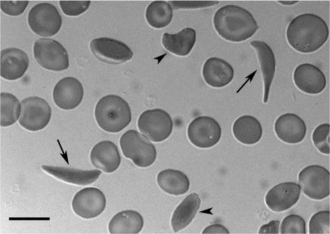
Sickle cell disease (SCD) was first described in 1910, in a dental student who presented with pulmonary symptoms (1). Herrick coined the term “sickle-shaped” to describe the peculiar appearance of the rbc of this patient (Figure 1). However, given the patient’s symptoms, he was not sure at the time whether the blood condition was a disease sui generis or a manifestation of another disease (2). In the next 15 years, several similar cases were described, supporting the idea that this was a new disease entity and providing enough evidence for a preliminary clinical and pathological description (3). Shortly thereafter, Hahn and Gillespie suggested that anoxia caused rbc sickling by demonstrating that shape changes could be induced by saturating a cell suspension with carbon dioxide (4). Scriver and Waugh, in experiments that would undoubtedly not receive institutional review board approval today, proved this concept in vivo by inducing venous stasis in a finger using a rubber band. They showed that stasis-induced hypoxia dramatically increased the proportion of sickle-shaped cells from approximately 15% to more than 95% (5). These seminal studies were noted by Linus Pauling, who was the first to hypothesize in 1945 that the disease might originate from an abnormality in the hemoglobin molecule (6). This hypothesis was validated in 1949 by the demonstration of the differential migration of sickle versus normal hemoglobin as assessed by gel electrophoresis (7). That same year, the autosomal recessive inheritance of the disease was elucidated (8). Around the same time, Watson et al. predicted the importance of fetal hemoglobin (Hb F) by suggesting that its presence could explain the longer period necessary for sickling of newborn rbc compared with those from mothers who had “sicklemia” (9). Ingram and colleagues demonstrated shortly thereafter that the mutant sickle hemoglobin (Hb S) differed from normal hemoglobin A by a single amino acid (10). This was followed by studies that analyzed the structure and physical properties of Hb S, which formed intracellular polymers upon deoxygenation (11). These studies placed SCD at the leading edge of investigations to elucidate the molecular basis of human diseases.
Figure 1. Sickle erythrocytes.
Peripheral blood smear from a patient with SCD obtained during a routine clinic visit. The smear shows classical sickle-shaped (arrows) and various other misshaped erythrocytes (arrowheads). The image was obtained from an air-dried smear using differential interference contrast (DIC) microscopy with an Olympus BX61WI work station equipped with a LUMPlanFI ×60 numerical aperture 0.90 ∞ objective (Olympus) and a CoolSnap HQ camera (6.6 μm2 pixel, 1,392 × 1,040 pixel format) (Roper Scientific). Scale bar: 10 μm.
Natural history of SCD
During the time of these pioneering laboratory investigations, further clinical observations brought to light the wide-ranging manifestations of SCD. In retrospect, it is clear that no amount of anecdotal reports would have substituted for the tremendous contributions of the study of the natural history of this disease known as the Cooperative Study of Sickle Cell Disease (CSSCD). The CSSCD was commissioned in 1978 by the National Heart, Lung, and Blood Institute to characterize prospectively the clinical course of SCD in a cohort of more than 4,000 patients from 23 centers across the United States (12). The CSSCD cohort defined the incidence and characteristics of virtually every known complication of SCD. One of the earliest contributions of the CSSCD was to identify the persistent high mortality rate of severe pneumococcal infections in children with SCD, despite the widespread use of pneumococcal vaccination. This led to the development of the landmark Prophylactic Penicillin Study (PROPS), which highlighted the importance of neonatal screening for SCD followed by prophylactic penicillin therapy in children between the ages of 4 months and 5 years (13). Other CSSCD studies that evaluated the mortality of the disease found that 50% of patients died before the fifth decade, and most of those who died did not have overt chronic organ failure but succumbed during an episode of acute pain, acute chest syndrome, or stroke (14). Prospective follow-up made it possible to determine the incidence (approximately 13 per 100 patient-years), risk factors, presentation, and prognosis of the acute chest syndrome (15, 16). Parallel studies established that approximately 11% of patients with SCD will go on to develop a clinically apparent stroke by the age of 20 years, and 24% by the age of 45 years (17). This high risk of a life-threatening complication generated the momentum for the Stroke Prevention Trial in Sickle Cell Anemia (STOP), which demonstrated the benefit of prophylactic transfusions in preventing a first stroke in patients with an elevated flow rate by transcranial Doppler ultrasonography (18). Moreover, the observation that more than 50% of patients with SCD have at least one crisis per year and the association between multiple pain episodes and early death in young adults (14, 19) provided the impetus for the Multicenter Study of Hydroxyurea (MSH), which is discussed below. The CSSCD also shed some important light on the incidence and risk of other complicating conditions, including alloimmunization (20), pregnancy (21), and surgery (22). Improvements in survival of patients with SCD have been associated with a marked increase in the incidence of chronic organ dysfunction, especially pulmonary hypertension (23). The lessons learned from the study of the natural history of SCD underscored the fact that this disease, which is caused by a single missense mutation in a gene whose expression is restricted to the hematopoietic system, can have wide-ranging manifestations and complications that affect every aspect of the life of afflicted patients.
Genetics of SCD
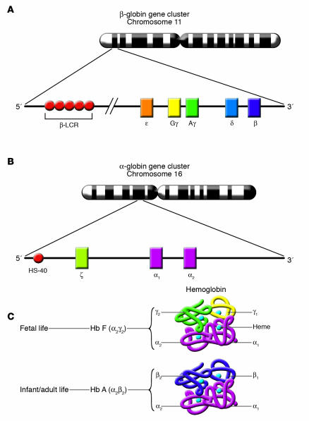
As mentioned above, pioneering studies by Pauling et al. established that SCD results from a defect in the hemoglobin molecule (7). During the same year, the mode of inheritance of the disease was shown to be autosomal recessive (8). The sickle mutation was characterized several years later by Ingram et al. as a glutamine-to-valine substitution at the sixth residue of the β-globin polypeptide (24). Several decades later, the human globin genes were cloned, their DNA sequence was determined, the organization of the globin gene clusters was characterized, and a great deal of insight was provided into the mechanisms of their regulated expression (25). Human hemoglobin is a tetrameric molecule that consists of two pairs of identical polypeptide subunits, each encoded by a different family of genes. The human α-like globin genes (ζ, α1, and α2) are located on chromosome 16, and the β-like globin genes (ε, Gγ, Aγ, δ, and β) are located on chromosome 11. Interestingly, the genes are present on both chromosomes in the same order in which they are expressed during development (Figure 2). During fetal life, the predominant type of hemoglobin is Hb F (α2γ2). During the postnatal period, Hb F is gradually replaced by Hb A (α2β2). Hb A2 (α2δ2) is a minor adult-type hemoglobin that accounts for less than 2.5% of the circulating hemoglobin in normal individuals in adult life. Upon completion of the switch from Hb F to Hb A, patients with disorders of the β-globin genes start manifesting the clinical features of their diseases. The prospect of therapeutic reactivation of Hb F production in adult life (see “Advances in the therapy of SCD”) has been in large part responsible for the tremendous interest in the elucidation of the molecular mechanisms of the switch from fetal to adult hemoglobin production. This field of investigation, which has recently been reviewed (26), led to approval by the FDA of the use of hydroxyurea for the treatment of patients with SCD.
Figure 2. Chromosomal organization of the α- and β-globin gene clusters.
(A) The genes of the β-globin gene cluster (ε, Gγ, Aγ, δ, and β) are present on chromosome 11 in the same order in which they are expressed during development. The β–locus control region (β–LCR) is a major regulatory element located far upstream of the genes of the cluster that is necessary for the high level of expression of those genes. (B) The genes of the α-globin gene cluster (ζ, α1, and α2) are present on chromosome 16, also in the same order in which they are expressed during development. HS-40 is a major regulatory element located far upstream of the genes of the cluster that is necessary for their high level of expression. (C) During fetal life, Hb F (α2γ2) is the predominant type of hemoglobin. Hemoglobin switching refers to the developmental process that leads to the silencing of γ-globin gene expression and the reciprocal activation of adult β-globin gene expression. This results in the replacement of Hb F by Hb A (α2β2) as the predominant type of hemoglobin in adult life. Figure modified from ref. 128.
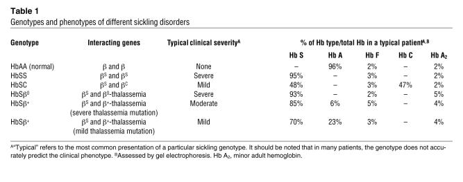
Homozygosity for the sickle mutation (i.e., HbSS disease) is responsible for the most common and most severe variant of SCD. Several other genetic variants of SCD result from the interaction of different mutations of the human β-globin genes (Table 1). When the βS gene interacts with the βC gene, the resulting sickling disorder known as HbSC disease is typically very mild (14). When a βS gene interacts with a β-thalassemia gene (a mutant β-globin gene that either fails to produce normal β-globin mRNA or produces it at markedly decreased levels), the severity of the resulting sickling disorder depends on the severity of the coinherited β-thalassemia mutation. When the coinherited β-thalassemia gene is completely inactive (i.e., β0-thalassemia), the resulting sickling disorder known as Sβ0-thalassemia tends to be of severity similar to that of homozygous HbSS disease (27). In contrast, when the coinherited β-thalassemia gene is partially active (i.e., β+-thalassemia), the resulting sickling disorder known as Sβ+-thalassemia can have a spectrum of clinical severity. If the β+-thalassemia mutation is mild, as is commonly the case in people of African descent, the resulting Sβ+-thalassemia tends to be clinically mild. In contrast, if the β+-thalassemia mutation is severe, as is commonly the case in the Mediterranean populations, the clinical sickling disorder tends to be moderate (28).
Table 1 .
Genotypes and phenotypes of different sickling disorders
Advances in the pathophysiology of SCD
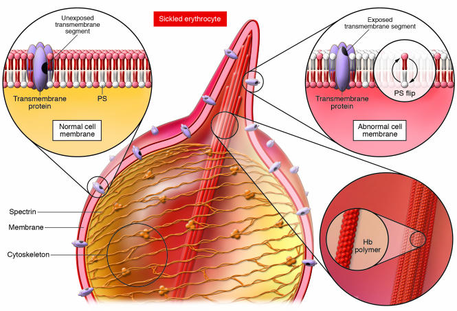
In addition to the obvious shape changes that result from the formation of intracellular hemoglobin polymers, the polymers can have a direct impact on the rbc plasma membrane, leading to the extracellular exposure of protein epitopes and glycolipids that are normally found inside the cell (Figure 3). These changes and the aberrant expression of adhesion molecules on stress reticulocytes likely explain the increased adherence of sickle rbc to vascular endothelium. Although the increased propensity of sickle rbc to stick to one another was noted many years before the field of cell adhesion was even conceptualized at the molecular level, pioneering independent studies by Hebbel, Hoover, and their colleagues demonstrated that sickle rbc were more apt than normal rbc to adhere to endothelial cells in vitro (29, 30). During the two decades that followed, multiple studies implicated virtually all major adhesion pathways in the interactions between sickle cells and endothelial cells. These pathways include those involving the integrins (α4β1, αVβ3) (31–33) and their receptors; immunoglobulin family members (VCAM-1, ICAM-4) (34, 35); the endothelial selectins (36, 37); soluble adhesion proteins such as thrombospondin (38), fibrinogen (39), fibronectin (40), von Willebrand factor (41, 42); and other exposed membrane components such as Band3 and sulfated glycolipids (43, 44). Thus, inasmuch as sickle adhesion to the endothelium plays a role in sickle cell vasoocclusion, the presence of such diverse mechanisms of adhesion presents an enormous challenge for delineating physiologically relevant therapeutic targets. Interestingly, recent studies have suggested that targeting a specific adhesion pathway may be sufficient to reduce vasoocclusion (33, 37, 45).
Figure 3. Alteration of the rbc membrane by polymers of sickle hemoglobin.
Deoxygenation of Hb S induces a change in conformation in which the mutant β chain binds to a complementary hydrophobic site resulting from a valine replacement, leading to the formation of a hemoglobin polymer (Hb polymer; lower right, inset). The hemoglobin polymers disrupt the rbc cytoskeleton and form protrusions, giving rise to the characteristic sickle appearance. Interruption of the attachment of the membrane to the protein cytoskeleton results in exposure of transmembrane protein epitopes and lipid exchanges, notably of phosphatidylserine (PS), between the inside and the outside of the cell (upper right, inset). Exposure of negatively charged glycolipids contributes to the proinflammatory and prothrombotic state of sickle cell blood. Adapted with permission from Blackwell Publishing (129).
The studies of the pathophysiology of SCD have been facilitated by the development of a number of mouse models that express either a mixture of mouse globins with Hb S, a super-sickling hemoglobin (e.g., SAD, NY1, S-Antilles mice), or human globin chains exclusively (e.g., Berkeley, NY1KO mice) (46). The severity of the phenotype of these transgenic mice depends on the presence of mouse globin chains, the mean corpuscular hemoglobin concentration (MCHC), and the presence of human Hb F. Although mice that express the human globin chains exclusively have a much more severe phenotype that closely mimics the major features of the human disease, all transgenic sickle mice exhibit pathological features of the disease, either spontaneously or in an inducible manner (46, 47).
As in the human disease, there is a clear role for inflammatory mediators in the pathogenesis of disease in murine models of SCD (48–52). Inflammation is likely triggered by the abnormal erythrocyte membrane and the presence of chronic hemolysis. Dense sickle cells that become dehydrated after several rounds of sickling expose their annexin V–binding phosphatidyl serine on the outer layer of the plasma membrane (53, 54). These negatively charged glycolipids can activate the coagulation cascade (55), leading to the generation of tissue factor and thrombin, which in turn promote the inflammatory response. Chronic hemolysis, on the other hand, leads to the release of plasma-free hemoglobin, which can scavenge NO and result in endothelial dysfunction (56). Moreover, the release of heme iron from lysed rbc is a major cause of oxidative stress that can induce redox-sensitive transcription factors such as NF-κB and activator protein–1. These transcription factors in turn induce the expression of E-selectin, VCAM-1, and ICAM-1 and the recruitment of adherent leukocytes in venules (51).
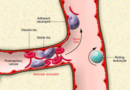
The presence of adherent leukocytes in small postcapillary venules is emerging as a key factor that contributes to vasoocclusion during SCD (57, 58) (Figure 4). Leukocytes are large cells that are rigid and not easily deformed as a result of a high viscoelastic coefficient (59). These physical properties endow leukocytes with a greater potential than discoid or sickle-shaped rbc (which would lie flat along the endothelium) to promote vascular obstruction. It has been known for many years, as a result of microdynamic measurements, that leukocyte recruitment in cat mesenteric venules can lead to a decrease in the effective diameter of the blood vessels and an increase in blood flow resistance due to obstruction of the lumen by wbc (60). In addition, sickle rbc have been shown to interact directly with adherent wbc in a mouse model of vasoocclusion induced by surgical trauma and TNF-α, leading to reduced blood flow and death of the mouse (49). Leukocyte adhesion was shown to be critical in this process, since flow reductions and death were prevented in mice deficient in both P- and E-selectins, key adhesion molecules mediating leukocyte rolling and adhesion (49, 61). Moreover, in vitro studies have revealed that human sickle cells can directly bind to activated neutrophils (62). Recent studies using high-speed fluorescence digital videomicroscopy suggest that most interactions between rbc and wbc in vivo are mediated by adherent neutrophils (63). As leukocyte adhesion plays a key role in vasoocclusion, it thus offers an attractive therapeutic target for this disease and is consistent with several clinical studies derived from the CSSCD cohort in which high leukocyte counts correlated with mortality (14), acute chest syndrome (16), stroke (17, 64, 65), and poor prognosis later in life when identified in infants with SCD (66).
Figure 4. Sickle cell vasoocclusion.
Abnormal, sickle rbc induce the expression of inflammatory and coagulation mediators, leading to the activation of the vascular endothelium. Sickle rbc themselves may also stimulate endothelial cells directly by adhesion. The stimulated endothelial cells are poised to recruit rolling and adherent leukocytes in venules by expressing chemokines and cell adhesion molecules such as the selectins and immunoglobulin family members. Activated, firmly adherent neutrophils capture circulating discoid and sickle-shaped rbc, leading to transient episodes of vascular occlusions that are initiated in the smallest postcapillary venules. Interactions between rbc and leukocytes tend to occur at vessel junctions, where leukocyte recruitment is the most active. In sickle mice, vasoocclusion can be prevented by the inhibition of leukocyte adhesion or the inflammatory response. The large arrow indicates the direction of blood flow.
Advances in the diagnosis of SCD
The diagnosis of SCD is usually simple and rarely poses a major challenge. Although the sickle-shaped rbc that gave the disease its name are not always present in the patient’s blood film, the characteristic migration of Hb S by gel electrophoresis is sufficient to make a diagnosis of a sickling disorder. Gel electrophoresis allows a definitive diagnosis of some but not all sickling genotypes. For example, when Hb A is present in the blood of a patient with SCD at a lower level than Hb S, this is indicative of HbSβ+-thalassemia (Table 1). However, the distinction between HbSβ0-thalassemia and homozygous HbSS can be much more challenging to make when no Hb A is detected by gel electrophoresis. In such situations, the diagnosis of HbSβ0-thalassemia is suggested by an elevated Hb A2 level and a low mean corpuscular volume. Fortunately, there are a number of excellent reference laboratories in the US where a definitive molecular diagnosis can be made in essentially every patient with a hemoglobin disorder.
The major challenge in the diagnosis of sickling disorders is to identify the disease during the prenatal period, at a time when such information would be critically important in enabling a couple at risk to make an informed decision about potential termination of pregnancy. The marked differences in the expected clinical severity of the different sickling disorders discussed above should be taken into consideration when making such decisions. Before the advent of molecular diagnostics, the only way to make a diagnosis prenatally was to obtain a fetal blood sample for analysis, which could only be performed after the 20th week of pregnancy. By that time, the pregnancy is already too advanced to make it possible to terminate safely. With the advent of DNA diagnostics, it has become possible to make definitive diagnoses of the different sickling disorders during the first trimester by analyzing fetal DNA obtained by chorionic villous biopsy (67). The molecular diagnostic technology is being pushed further to allow the diagnosis to be made from a small number of fetal cells that can be harvested from the maternal circulation (68).
Advances in the therapy of SCD
Induction of Hb F.
A large number of epidemiological, clinical, and laboratory observations have converged to support the notion that Hb F administration can ameliorate the clinical severity of SCD. Patients with SCD from the eastern provinces of Saudi Arabia (69) and from India (70) typically have a very mild sickling disorder associated with high levels of Hb F. Furthermore, the CSSCD identified Hb F as a prognostic factor for several sickle cell complications, including painful events (19), acute chest syndrome (16), and death (14). Elegant laboratory studies conducted many years earlier had demonstrated that Hb F interferes with the polymerization of deoxygenated Hb S in vitro (71). Based on all these observations, it was proposed that pharmacological induction of Hb F production may be an effective therapeutic strategy for ameliorating the severity of SCD. When the different globin genes were cloned in the late 1970s (72) and the mechanisms responsible for their regulation were elucidated during the 1980s (73, 74), it became clear that epigenetic factors such as DNA methylation and histone acetylation played important roles in the developmental regulation of globin gene expression (75–77). Thus, it was proposed that pharmacological agents that alter the epigenetic configuration of the γ-globin genes may provide a viable therapeutic approach to the induction of Hb F.
5-Azacytidine.
5-Azacytidine was the first agent to be used to induce Hb F expression via epigenetic silencing of the γ-globin genes in adult life. The rationale for this approach was based on the discovery that the actively transcribed adult β-globin genes are hypomethylated and the nontranscribed fetal γ-globin genes are hypermethylated in adult life. In contrast, the adult β-globin genes are hypermethylated and the γ-globin genes are hypomethylated in fetal life (75, 76). 5-Azacytidine was shown to induce very high levels of Hb F in anemic baboons (78). Its ability to stimulate Hb F production was also demonstrated in a small number of patients with SCD and β-thalassemia (79, 80). In spite of these promising results, this drug was never tested in large-scale clinical trials because of concerns about potential carcinogenicity.
Hydroxyurea.
The clinical development of hydroxyurea as an agent that induces Hb F production in SCD provides a vivid illustration of how scientific discoveries can be translated into therapies that improve the outlook for patients afflicted with a debilitating disease. Interestingly, controversy over the mechanism of induction of Hb F by 5-azacytidine provided the motivation for a study in which hydroxyurea, an S phase–specific chemotherapeutic agent that does not inhibit DNA methyltransferase, was shown to result in a marked increase in Hb F levels in baboons (81). Hydroxyurea is an inhibitor of ribonucleotide reductase that had been in use for many years in the treatment of myeloproliferative disorders. It is an orally available drug that is relatively well tolerated and simple to use. After the demonstration of its ability to induce Hb F production in baboons, hydroxyurea was tested in a number of small clinical trials in adults with SCD (82–84). A larger MSH study showed a marked decrease in the frequency of painful crises and acute chest syndrome and a reduction in transfusion requirements and hospitalizations in adults with moderate to severe SCD after hydroxyurea treatment (85). After 9 years of follow-up, patients with SCD treated with hydroxyurea were shown to have improved survival (86). Other studies demonstrated the clinical efficacy and short-term safety of hydroxyurea in children with SCD (87–89).
Although hydroxyurea was shown to have Hb F–inducing activity similar to that of 5-azacytidine in anemic baboons and patients with SCD, its molecular targets and mechanism(s) of action are still not fully elucidated. It was originally proposed that hydroxyurea may elevate Hb F levels by accelerating erythroid differentiation in the bone marrow, leading to the appearance of “fetal-like” cells in the peripheral blood (25). More recent studies have shown that hydroxyurea generates NO in vivo, which results in the activation of the NO/cGMP signaling pathway and the upregulation of γ-globin gene expression in patients with SCD (90). Hydroxyurea has other effects that may also benefit patients with SCD. For example, hydroxyurea was shown to decrease the adhesion of sickle cells to endothelium and to decrease the expression level of soluble VCAM-1 (91, 92). Owing to its myelosuppressive activity, hydroxyurea reduces circulating wbc counts and likely the number of adherent leukocytes recruited to the wall of small venules. The reduction of wbc counts was correlated with the clinical benefit from hydroxyurea (93). It is still not entirely clear how much of the clinical benefit from hydroxyurea could be attributed to its effect on Hb F levels compared with its other activities.
Butyrate.
Concerns over the potential for serious side effects of chemotherapeutic agents such as 5-azacytidine and hydroxyurea has stimulated the continuation of the search for safe and effective inducers of Hb F production. Butyrate, a short-chain fatty acid that inhibits histone deacetylase (HDAC), was shown to stimulate embryonic or fetal globin gene expression in chicken, mice, and baboons (94–96). When arginine butyrate was administered to patients with SCD intermittently (four days every four weeks), it resulted in sustained induction of Hb F production in a majority of patients (97). In spite of the considerable promise of this agent in the treatment of SCD, the difficulty of administrating large volumes of this drug through central venous catheters poses a major therapeutic challenge. It is unlikely that the full potential of butyrate and other HDAC inhibitors will be realized until an oral compound is identified that has the same efficacy as butyrate.
Decitabine.
The recent introduction of decitabine (5-aza-2-deoxycytidine), a new analog of 5-azacytidine that does not incorporate into RNA, has resulted in renewed interest in the use of DNA hypomethylation therapy for the induction of Hb F production in SCD. In recent small-scale clinical trials in patients with SCD, treatment with decitabine resulted in significant increases in mean γ-globin synthesis, Hb F levels, and the number of F cells (rbc that contain Hb F) (98–100). Interestingly, increased Hb F levels were observed in 100% of patients with SCD who received decitabine, including patients who had previously failed to respond to hydroxyurea. The increase in the levels of Hb F was associated with significant improvement in several factors that are important in the pathophysiology of vasoocclusion, including rbc adhesion, endothelial damage, and activation of the coagulation pathway (100). Larger and longer-term studies are needed to confirm the efficacy and safety of decitabine in the treatment of SCD.
Bone marrow transplantation
The idea of replacing the bone marrow, which is the source of the defective sickle cells, with bone marrow that produces normal rbc is an intuitive therapeutic approach in SCD. However, for many years, such an approach was considered too risky for a nonmalignant disorder such as SCD, since the mortality of the procedure itself was around 20%. The reduction in mortality following bone marrow transplantation (BMT), resulting from recent advances in immunosuppressive therapy and supportive care and the fact that long-term survival of patients with β-thalassemia after BMT was shown to be greater than 90% (101), resulted in renewed interest in this therapy for SCD. Clinical trials that were conducted in children with SCD in Europe and the US showed greater than 90% long-term survival (102–104). A major limitation in the use of BMT for the treatment of SCD is the fact that a matched sibling donor is available to less than 15% of patients who are suitable candidates for transplantation (105). In an effort to increase the availability of sources of hematopoietic stem cells for transplantation, clinical trials are being conducted to evaluate cord blood transplantation in the treatment of SCD (106). To date, very few transplantation procedures have been performed in adults with SCD because of concerns that the morbidity and mortality of BMT is higher in adults than in children. The use of nonmyeloablative BMT to reduce peritransplant morbidity and mortality has been associated with a very high graft rejection rate (107). BMT is the only curative therapy for SCD, and the major challenge is to make it more widely available to patients with a severe disease phenotype.
Impediments that limit the impact of the advances in SCD
In spite of the fact that hydroxyurea has been shown to improve both survival and the quality of life in patients with SCD, only a small fraction of eligible patients with SCD in the US are currently receiving hydroxyurea (108, 109). Although the reasons for the reluctance to use hydroxyurea are not entirely clear, there are many potential contributing factors. These include patient concerns about a drug that is used primarily to treat cancer, physician concerns about potential long-term mutagenic effects, lack of familiarity of primary care providers with the use of a chemotherapeutic agent, and resistance among patients with SCD to use therapies that are perceived to be experimental in nature. Careful investigation into the impediments to the use of hydroxyurea is necessary in order to realize the full potential of this important therapeutic advance.
Prenatal diagnosis is another area in which the development of important new technology has had a very limited impact in SCD. In spite of the fact that DNA diagnostics have made it possible to identify an affected fetus much earlier during pregnancy, the impact of these advances on the number of new births with SCD in the US has been extremely small. In contrast, the same technology has had a very large impact on the number of new births with β-thalassemia in Mediterranean regions including Greece, Cyprus, and Sardinia (110). Although the reasons for the differences in the impact of the same technology in these closely related disorders have not been investigated, it is conceivable that they are a reflection of the fact that a majority of patients with β-thalassemia die from iron overload before the third decade of life, while survival of patients with SCD into the fifth, sixth, and even seventh decades of life is not unusual. The reluctance to terminate an affected pregnancy may also be motivated by cultural and ethnic factors that have not received adequate attention. It is sobering to keep in mind that the one intervention that has had the largest impact on the natural history of SCD during the last few decades is the introduction of penicillin prophylaxis during childhood (13). This should serve as a reminder that important therapeutic advances are bound to have a very limited impact on the natural history of any human disease unless they are widely accepted by the patients they are intended to help.
Future directions
Future of diagnostics in SCD.
As discussed above, the molecular methods of identifying the sickle mutation in utero and after birth are well established and widely available. However, although the same sickle mutation in the β-globin gene is responsible for the spectrum of the pathophysiology of the sickling disorder, the clinical manifestations of the disease are extremely heterogeneous. Many factors that contribute to this heterogeneity, such as an interaction of the βC gene and the β-thalassemia gene, are well known. Other factors such as the Hb F levels and the coinheritance of α-thalassemia have also been known to modulate the clinical severity of sickling disorders for many years (69, 70, 111). Other genetic determinants that contribute to the variability of Hb F levels were identified outside the β-globin gene cluster and mapped to two different chromosomes (112, 113). More recent studies have demonstrated that the gene responsible for the variability in Hb F levels that was previously mapped to chromosome 6p23 (112) is cMYB (114). As the understanding of the pathophysiology of the disease evolves, the number of potential epistatic genes (i.e., modifying genes) increases. Thus, mutations or polymorphisms that have an impact on cell adhesion, thrombosis, rbc dehydration, and inflammation are likely in candidate epistatic genes in SCD. As is the case with the coinheritance of α-thalassemia, some of these genetic determinants might increase the risk of some complications and decrease the risks of others. A number of studies have investigated the potential effects of candidate modifying genes that are implicated in the pathophysiology of the disease (e.g., methylenetetrahydrofolate reductase and the pathogenesis of avascular necrosis [ref. 115], factor V R485K and the risk of venous thrombosis [ref. 116], and UDP glucuronosyltransferase-1 polymorphism and serum bilirubin levels [ref. 117]). Other investigators are using an unbiased genetic approach that consists of the analysis of hundreds of SNPs in a large number of patients to identify genes that increase the risk of a particular complication. Using such an approach, Sebastiani and colleagues recently identified 31 SNPs in 12 genes that interact with Hb F to modulate the risk of stroke (118). In the future, such biased and unbiased approaches may make it possible to identify a genetic blueprint in a particular patient that defines his/her risk of developing any of the known complications of SCD. It might even be possible at some point to use this information to implement therapeutic interventions before the complications develop.
Future of therapeutics in SCD.
Gene therapy offers enormous promise as a potential curative therapy for SCD, but concerns over the safety of random genomic insertion must first be resolved (119). Preclinical studies in mice have provided the proof of principle that transduction of bone marrow stem cells with lentiviral vectors that express a β-globin gene can prevent Hb S polymerization in vivo (120, 121). The wide range of abnormalities engendered by the sickle cell mutation offers several other opportunities for therapeutic interventions. For example, the NIH Road Map is supporting ongoing investigations in which high-throughput screening approaches are used to discover novel low-molecular-weight compounds that can alter key aspects of the disease, including hemoglobin polymerization, expression of Hb F, and leukocyte adhesion. Current clinical trials are evaluating the efficacy of Ca2+-sensitive Gardos channel inhibitors (e.g., ICA-17043), with or without hydroxyurea, in preventing dehydration of erythrocytes (122). Vasoactive drugs (e.g., NO, sildenafil, endothelin antagonists) are being evaluated for the treatment of pulmonary hypertension. Statins are of potentially great interest since they can increase NO production and reduce leukocyte adhesion (123, 124). Antiinflammatory drugs that inhibit NF-κB and the upregulation of adhesion molecules have shown promise in pilot clinical studies (125). Intravenous gammaglobulins are currently under clinical evaluation following a study demonstrating a dose-dependent reduction in leukocyte adhesion and in the number of interactions between rbc and wbc, accompanied by improvements in microcirculatory blood flow and survival of sickle transgenic mice (126). Furthermore, there is growing interest in the prevention and treatment of vasoocclusion by novel selectin antagonists since they appear to participate in multiple pathways involved in sickle vasoocclusion, including the adhesion of leukocytes, rbc, and platelets to the endothelium and to each other (127). Almost a century after SCD was first described, we may be at the dawn of a new era in which a physician might be able to use genetic information to select one or more drugs that target specific aspects of disease pathophysiology that are relevant to a particular patient with SCD.
Acknowledgments
The authors would like to acknowledge the support of the NIH for their research on SCD (HL69438 to P.S. Frenette and HL073438 to G.F. Atweh).
Footnotes
Nonstandard abbreviations used: BMT, bone marrow transplantation; CSSCD, Cooperative Study of Sickle Cell Disease; Hb F, fetal hemoglobin; Hb S, sickle hemoglobin; MSH, Multicenter Study of Hydroxyurea; SCD, sickle cell disease.
Conflict of interest: The authors have declared that no conflict of interest exists.
Citation for this article: J. Clin. Invest. 117:850–858 (2007). doi:10.1172/JCI30920.
References
- 1.Herrick J.B. Peculiar elongated and sickle-shaped red blood corpuscules in a case of severe anemia. Arch. Int. Med. 1910;6:517–521. [PMC free article] [PubMed] [Google Scholar]
- 2.Herrick J.B. Abstract of discussion. JAMA. 1924;83:16. [Google Scholar]
- 3.Sydenstricker V.P. Further observations on sickle cell anemia. JAMA. 1924;83:12–15. [Google Scholar]
- 4.Hanh E.V., Gillespie E.B. Sickle cell anemia. Arch. Int. Med. 1927;39:233. [Google Scholar]
- 5.Scriver J.R., Waugh T.R. Studies on a case of sickle cell anemia. Can. Med. Assoc. J. 1930;23:375–380. [PMC free article] [PubMed] [Google Scholar]
- 6.Pauling L. Molecular disease and evolution. Bull. N. Y. Acad. Med. 1964;40:334–342. [PMC free article] [PubMed] [Google Scholar]
- 7.Pauling L., Itano H.A., Singer S.J., Wells I.C. Sickle cell anemia, a molecular disease. . Science. 1949;110:543–548. doi: 10.1126/science.110.2865.543. [DOI] [PubMed] [Google Scholar]
- 8.Neel J.V. The inheritance of sickle cell anemia. . Science. 1949;110:64–66. doi: 10.1126/science.110.2846.64. [DOI] [PubMed] [Google Scholar]
- 9.Watson J., Stahman A.W., Bilello F.P. The significance of the paucity of sickle cells in newborn Negro infants. Am. J. Med. Sci. 1948;215:419–423. doi: 10.1097/00000441-194804000-00008. [DOI] [PubMed] [Google Scholar]
- 10.Ingram V.M. Abnormal human haemoglobins. I. The comparison of normal human and sickle-cell haemoglobins by fingerprinting. Biochim. Biophys. Acta. 1958;28:539–545. doi: 10.1016/0006-3002(58)90516-x. [DOI] [PubMed] [Google Scholar]
- 11.Ferrone F.A. Polymerization and sickle cell disease: a molecular view. Microcirculation. 2004;11:115–128. doi: 10.1080/10739680490278312. [DOI] [PubMed] [Google Scholar]
- 12.Gaston M., Rosse W.F. The cooperative study of sickle cell disease: review of study design and objectives. Am. J. Pediatr. Hematol. Oncol. 1982;4:197–201. [PubMed] [Google Scholar]
- 13.Gaston M.H., et al. Prophylaxis with oral penicillin in children with sickle cell anemia. A randomized trial. N. Engl. J. Med. 1986;314:1593–1599. doi: 10.1056/NEJM198606193142501. [DOI] [PubMed] [Google Scholar]
- 14.Platt O.S., et al. Mortality in sickle cell disease. Life expectancy and risk factors for early death. N. Engl. J. Med. 1994;330:1639–1644. doi: 10.1056/NEJM199406093302303. [DOI] [PubMed] [Google Scholar]
- 15.Vichinsky E.P., et al. Acute chest syndrome in sickle cell disease: clinical presentation and course. Cooperative Study of Sickle Cell Disease. Blood. 1997;89:1787–1792. [PubMed] [Google Scholar]
- 16.Castro O., et al. The acute chest syndrome in sickle cell disease: incidence and risk factors. The Cooperative Study of Sickle Cell Disease. Blood. 1994;84:643–649. [PubMed] [Google Scholar]
- 17.Ohene-Frempong K., et al. Cerebrovascular accidents in sickle cell disease: rates and risk factors. . Blood. 1998;91:288–294. [PubMed] [Google Scholar]
- 18.Adams R.J., et al. Prevention of a first stroke by transfusions in children with sickle cell anemia and abnormal results on transcranial Doppler ultrasonography. N. Engl. J. Med. 1998;339:5–11. doi: 10.1056/NEJM199807023390102. [DOI] [PubMed] [Google Scholar]
- 19.Platt O.S., et al. Pain in sickle cell disease. Rates and risk factors. N. Engl. J. Med. 1991;325:11–16. doi: 10.1056/NEJM199107043250103. [DOI] [PubMed] [Google Scholar]
- 20.Rosse W.F., et al. Transfusion and alloimmunization in sickle cell disease. The Cooperative Study of Sickle Cell Disease. Blood. 1990;76:1431–1437. [PubMed] [Google Scholar]
- 21.Koshy M., Burd L., Wallace D., Moawad A., Baron J. Prophylactic red-cell transfusions in pregnant patients with sickle cell disease. A randomized cooperative study. N. Engl. J. Med. 1988;319:1447–1452. doi: 10.1056/NEJM198812013192204. [DOI] [PubMed] [Google Scholar]
- 22.Koshy M., et al. Surgery and anesthesia in sickle cell disease. Cooperative Study of Sickle Cell Diseases. Blood. 1995;86:3676–3684. [PubMed] [Google Scholar]
- 23.Castro O., Gladwin M.T. Pulmonary hypertension in sickle cell disease: mechanisms, diagnosis, and management. Hematol. Oncol. Clin. North Am. 2005;19:881–896, vii. doi: 10.1016/j.hoc.2005.07.007. [DOI] [PubMed] [Google Scholar]
- 24.Ingram V.M. Abnormal human haemoglobins. III. The chemical difference between normal and sickle cell haemoglobins. Biochim. Biophys. Acta. 1959;36:402–411. doi: 10.1016/0006-3002(59)90183-0. [DOI] [PubMed] [Google Scholar]
- 25.Stamatoyannopoulos G. Control of globin gene expression during development and erythroid differentiation. Exp. Hematol. 2005;33:259–271. doi: 10.1016/j.exphem.2004.11.007. [DOI] [PMC free article] [PubMed] [Google Scholar]
- 26.Bank A. Regulation of human fetal hemoglobin: new players, new complexities. Blood. 2006;107:435–443. doi: 10.1182/blood-2005-05-2113. [DOI] [PMC free article] [PubMed] [Google Scholar]
- 27.Serjeant G.R., Sommereux A.M., Stevenson M., Mason K., Serjeant B.E. Comparison of sickle cell-beta0 thalassaemia with homozygous sickle cell disease. Br. J. Haematol. 1979;41:83–93. doi: 10.1111/j.1365-2141.1979.tb03684.x. [DOI] [PubMed] [Google Scholar]
- 28.Serjeant G.R., Ashcroft M.T., Serjeant B.E., Milner P.F. The clinical features of sickle-cell-thalassaemia in Jamaica. Br. J. Haematol. 1973;24:19–30. doi: 10.1111/j.1365-2141.1973.tb05723.x. [DOI] [PubMed] [Google Scholar]
- 29.Hebbel R.P., et al. Abnormal adherence of sickle erythrocytes to cultured vascular endothelium: possible mechanism for microvascular occlusion in sickle cell disease. J. Clin. Invest. 1980;65:154–160. doi: 10.1172/JCI109646. [DOI] [PMC free article] [PubMed] [Google Scholar]
- 30.Hoover R., Rubin R., Wise G., Warren R. Adhesion of normal and sickle erythrocytes to endothelial monolayer cultures. Blood. 1979;54:872–876. [PubMed] [Google Scholar]
- 31.Joneckis C.C., Ackley R.L., Orringer E.P., Wayner E.A., Parise L.V. Integrin alpha 4 beta 1 and glycoprotein IV (CD36) are expressed on circulating reticulocytes in sickle cell anemia. Blood. 1993;82:3548–3555. [PubMed] [Google Scholar]
- 32.Swerlick R.A., Eckman J.R., Kumar A., Jeitler M., Wick T.M. Alpha 4 beta 1-integrin expression on sickle reticulocytes: vascular cell adhesion molecule-1-dependent binding to endothelium. Blood. 1993;82:1891–1899. [PubMed] [Google Scholar]
- 33.Kaul D.K., et al. Monoclonal antibodies to alphaVbeta3 (7E3 and LM609) inhibit sickle red blood cell-endothelium interactions induced by platelet-activating factor. Blood. 2000;95:368–374. [PubMed] [Google Scholar]
- 34.Spring F.A., et al. Intercellular adhesion molecule-4 binds alpha(4)beta(1) and alpha(V)-family integrins through novel integrin-binding mechanisms. Blood. 2001;98:458–466. doi: 10.1182/blood.v98.2.458. [DOI] [PubMed] [Google Scholar]
- 35.Gee B.E., Platt O.S. Sickle reticulocytes adhere to VCAM-1. Blood. 1995;85:268–274. [PubMed] [Google Scholar]
- 36.Natarajan M., Udden M.M., McIntire L.V. Adhesion of sickle red blood cells and damage to interleukin-1 beta stimulated endothelial cells under flow in vitro. Blood. 1996;87:4845–4852. [PubMed] [Google Scholar]
- 37.Embury S.H., et al. The contribution of endothelial cell P-selectin to the microvascular flow of mouse sickle erythrocytes in vivo. Blood. 2004;104:3378–3385. doi: 10.1182/blood-2004-02-0713. [DOI] [PubMed] [Google Scholar]
- 38.Hillery C.A., Scott J.P., Du M.C. The carboxy-terminal cell-binding domain of thrombospondin is essential for sickle red blood cell adhesion. . Blood. 1999;94:302–309. [PubMed] [Google Scholar]
- 39.Wautier J.L., et al. Fibrinogen, a modulator of erythrocyte adhesion to vascular endothelium. . J. Lab. Clin. Med. 1983;101:911–920. [PubMed] [Google Scholar]
- 40.Kasschau M.R., Barabino G.A., Bridges K.R., Golan D.E. Adhesion of sickle neutrophils and erythrocytes to fibronectin. Blood. 1996;87:771–780. [PubMed] [Google Scholar]
- 41.Kaul D.K., Nagel R.L., Chen D., Tsai H.M. Sickle erythrocyte-endothelial interactions in microcirculation: the role of von Willebrand factor and implications for vasoocclusion. Blood. 1993;81:2429–2438. [PubMed] [Google Scholar]
- 42.Wick T.M., et al. Unusually large von Willebrand factor multimers increase adhesion of sickle erythrocytes to human endothelial cells under controlled flow. J. Clin. Invest. 1987;80:905–910. doi: 10.1172/JCI113151. [DOI] [PMC free article] [PubMed] [Google Scholar]
- 43.Thevenin B.J.M., Crandall I., Ballas S.K., Sherman I.W., Shohet S.B. Band 3 peptides block the adherence of sickle cells to endothelial cells in vitro. Blood. 1997;90:4172–4179. [PubMed] [Google Scholar]
- 44.Hillery C.A., Du M.C., Montgomery R.R., Scott J.P. Increased adhesion of erythrocytes to components of the extracellular matrix: isolation and characterization of a red blood cell lipid that binds thrombospondin and laminin. Blood. 1996;87:4879–4886. [PubMed] [Google Scholar]
- 45.Belcher J.D., et al. Critical role of endothelial cell activation in hypoxia-induced vasoocclusion in transgenic sickle mice. Am. J. Physiol. Heart Circ. Physiol. 2005;288:H2715–H2725. doi: 10.1152/ajpheart.00986.2004. [DOI] [PubMed] [Google Scholar]
- 46.Atweh G.F., et al. Hemoglobinopathies. Hematology Am. Soc. Hematol. Educ. Program. 2003;2003:14–39. doi: 10.1182/asheducation-2003.1.14. [DOI] [PubMed] [Google Scholar]
- 47.Manci E.A., et al. Pathology of Berkeley sickle cell mice: similarities and differences with human sickle cell disease. Blood. 2006;107:1651–1658. doi: 10.1182/blood-2005-07-2839. [DOI] [PMC free article] [PubMed] [Google Scholar]
- 48.Kaul D.K., Hebbel R.P. Hypoxia/reoxygenation causes inflammatory response in transgenic sickle mice but not in normal mice. J. Clin. Invest. 2000;106:411–420. doi: 10.1172/JCI9225. [DOI] [PMC free article] [PubMed] [Google Scholar]
- 49.Turhan A., Weiss L.A., Mohandas N., Coller B.S., Frenette P.S. Primary role for adherent leukocytes in sickle cell vascular occlusion: a new paradigm. Proc. Natl. Acad. Sci. U. S. A. 2002;99:3047–3051. doi: 10.1073/pnas.052522799. [DOI] [PMC free article] [PubMed] [Google Scholar]
- 50.Belcher J.D., et al. Transgenic sickle mice have vascular inflammation. Blood. 2003;101:3953–3959. doi: 10.1182/blood-2002-10-3313. [DOI] [PubMed] [Google Scholar]
- 51.Belcher J.D., et al. Heme oxygenase-1 is a modulator of inflammation and vaso-occlusion in transgenic sickle mice. J. Clin. Invest. 2006;116:808–816. doi: 10.1172/JCI26857. [DOI] [PMC free article] [PubMed] [Google Scholar]
- 52.Kaul D.K., et al. Anti-inflammatory therapy ameliorates leukocyte adhesion and microvascular flow abnormalities in transgenic sickle mice. Am. J. Physiol. Heart Circ. Physiol. 2004;287:H293–H301. doi: 10.1152/ajpheart.01150.2003. [DOI] [PubMed] [Google Scholar]
- 53.de Jong K., Larkin S.K., Styles L.A., Bookchin R.M., Kuypers F.A. Characterization of the phosphatidylserine-exposing subpopulation of sickle cells. Blood. 2001;98:860–867. doi: 10.1182/blood.v98.3.860. [DOI] [PubMed] [Google Scholar]
- 54.Yasin Z., et al. Phosphatidylserine externalization in sickle red blood cells: associations with cell age, density, and hemoglobin F. Blood. 2003;102:365–370. doi: 10.1182/blood-2002-11-3416. [DOI] [PubMed] [Google Scholar]
- 55.Chiu D., Lubin B., Roelofsen B., van Deenen L.L. Sickled erythrocytes accelerate clotting in vitro: an effect of abnormal membrane lipid asymmetry. Blood. 1981;58:398–401. [PubMed] [Google Scholar]
- 56.Reiter C.D., et al. Cell-free hemoglobin limits nitric oxide bioavailability in sickle-cell disease. Nat. Med. 2002;8:1383–1389. doi: 10.1038/nm1202-799. [DOI] [PubMed] [Google Scholar]
- 57.Okpala I. Leukocyte adhesion and the pathophysiology of sickle cell disease. Curr. Opin. Hematol. 2006;13:40–44. doi: 10.1097/01.moh.0000190108.62414.06. [DOI] [PubMed] [Google Scholar]
- 58.Frenette P.S. Sickle cell casoocclusion: heterotypic, multicellular aggregations driven by leukocyte adhesion. Microcirculation. 2004;11:167–177. [PubMed] [Google Scholar]
- 59.Schmid-Schonbein G.W. The damaging potential of leukocyte activation in the microcirculation. . Angiology. 1993;44:45–56. doi: 10.1177/000331979304400108. [DOI] [PubMed] [Google Scholar]
- 60.House S.D., Lipowsky H.H. Leukocyte-endothelium adhesion: microhemodynamics in mesentery of the cat. Microvasc. Res. 1987;34:363–379. doi: 10.1016/0026-2862(87)90068-9. [DOI] [PubMed] [Google Scholar]
- 61.Frenette P.S., Mayadas T.N., Rayburn H., Hynes R.O., Wagner D.D. Susceptibility to infection and altered hematopoiesis in mice deficient in both P- and E-selectins. Cell. 1996;84:563–574. doi: 10.1016/s0092-8674(00)81032-6. [DOI] [PubMed] [Google Scholar]
- 62.Hofstra T.C., Kalra V.K., Meiselman H.J., Coates T.D. Sickle erythrocytes adhere to polymorphonuclear neutrophils and activate the neutrophil respiratory burst. Blood. 1996;87:4440–4447. [PubMed] [Google Scholar]
- 63.Chiang E.Y., Hidalgo A., Chang J., Frenette P.S. Imaging receptor microdomains on leukocyte subsets in live mice. Nat. Methods. 2007;4:219–222. doi: 10.1038/nmeth1018. [DOI] [PubMed] [Google Scholar]
- 64.Kinney T.R., et al. Silent cerebral infarcts in sickle cell anemia: a risk factor analysis. The Cooperative Study of Sickle Cell Disease. Pediatrics. 1999;103:640–645. doi: 10.1542/peds.103.3.640. [DOI] [PubMed] [Google Scholar]
- 65.Balkaran B., et al. Stroke in a cohort of patients with homozygous sickle cell disease. . J. Pediatr. 1992;120:360–366. doi: 10.1016/s0022-3476(05)80897-2. [DOI] [PubMed] [Google Scholar]
- 66.Miller S.T., et al. Prediction of adverse outcomes in children with sickle cell disease. N. Engl. J. Med. 2000;342:83–89. doi: 10.1056/NEJM200001133420203. [DOI] [PubMed] [Google Scholar]
- 67.Orkin S.H., Little P.F., Kazazian H.H., Jr., Boehm C.D. Improved detection of the sickle mutation by DNA analysis: application to prenatal diagnosis. N. Engl. J. Med. 1982;307:32–36. doi: 10.1056/NEJM198207013070106. [DOI] [PubMed] [Google Scholar]
- 68.Cheung M.C., Goldberg J.D., Kan Y.W. Prenatal diagnosis of sickle cell anaemia and thalassaemia by analysis of fetal cells in maternal blood. Nat. Genet. 1996;14:264–268. doi: 10.1038/ng1196-264. [DOI] [PubMed] [Google Scholar]
- 69.Perrine R.P., Pembrey M.E., John P., Perrine S., Shoup F. Natural history of sickle cell anemia in Saudi Arabs. A study of 270 subjects. Ann. Intern. Med. 1978;88:1–6. doi: 10.7326/0003-4819-88-1-1. [DOI] [PubMed] [Google Scholar]
- 70.Kar B.C., et al. Sickle cell disease in Orissa State, India. Lancet. 1986;2:1198–1201. doi: 10.1016/s0140-6736(86)92205-1. [DOI] [PubMed] [Google Scholar]
- 71.Nagel R.L., et al. Structural bases of the inhibitory effects of hemoglobin F and hemoglobin A2 on the polymerization of hemoglobin S. Proc. Natl. Acad. Sci. U. S. A. 1979;76:670–672. doi: 10.1073/pnas.76.2.670. [DOI] [PMC free article] [PubMed] [Google Scholar]
- 72.Lawn R.M., Fritsch E.F., Parker R.C., Blake G., Maniatis T. The isolation and characterization of linked delta- and beta-globin genes from a cloned library of human DNA. Cell. 1978;15:1157–1174. doi: 10.1016/0092-8674(78)90043-0. [DOI] [PubMed] [Google Scholar]
- 73.Collins F.S., Weissman S.M. The molecular genetics of human hemoglobin. Prog. Nucleic Acid Res. Mol. Biol. 1984;31:315–462. doi: 10.1016/s0079-6603(08)60382-7. [DOI] [PubMed] [Google Scholar]
- 74.Orkin S.H. Globin gene regulation and switching: circa 1990. Cell. 1990;63:665–672. doi: 10.1016/0092-8674(90)90133-y. [DOI] [PubMed] [Google Scholar]
- 75.van der Ploeg L.H., Flavell R.A. DNA methylation in the human gamma delta beta-globin locus in erythroid and nonerythroid tissues. Cell. 1980;19:947–958. doi: 10.1016/0092-8674(80)90086-0. [DOI] [PubMed] [Google Scholar]
- 76.Mavilio F., et al. Molecular mechanisms of human hemoglobin switching: selective undermethylation and expression of globin genes in embryonic, fetal, and adult erythroblasts. Proc. Natl. Acad. Sci. U. S. A. 1983;80:6907–6911. doi: 10.1073/pnas.80.22.6907. [DOI] [PMC free article] [PubMed] [Google Scholar]
- 77.Groudine M., Weintraub H. Activation of globin genes during chicken development. Cell. 1981;24:393–401. doi: 10.1016/0092-8674(81)90329-9. [DOI] [PubMed] [Google Scholar]
- 78.DeSimone J., Heller P., Hall L., Zwiers D. 5-Azacytidine stimulates fetal hemoglobin synthesis in anemic baboons. Proc. Natl. Acad. Sci. U. S. A. 1982;79:4428–4431. doi: 10.1073/pnas.79.14.4428. [DOI] [PMC free article] [PubMed] [Google Scholar]
- 79.Ley T.J., et al. 5-Azacytidine selectively increases gamma-globin synthesis in a patient with beta+ thalassemia. N. Engl. J. Med. 1982;307:1469–1475. doi: 10.1056/NEJM198212093072401. [DOI] [PubMed] [Google Scholar]
- 80.Ley T.J., et al. 5-Azacytidine increases gamma-globin synthesis and reduces the proportion of dense cells in patients with sickle cell anemia. . Blood. 1983;62:370–380. [PubMed] [Google Scholar]
- 81.Letvin N.L., Linch D.C., Beardsley G.P., McIntyre K.W., Nathan D.G. Augmentation of fetal-hemoglobin production in anemic monkeys by hydroxyurea. N. Engl. J. Med. 1984;310:869–873. doi: 10.1056/NEJM198404053101401. [DOI] [PubMed] [Google Scholar]
- 82.Platt O.S., et al. Hydroxyurea enhances fetal hemoglobin production in sickle cell anemia. . J. Clin. Invest. 1984;74:652–656. doi: 10.1172/JCI111464. [DOI] [PMC free article] [PubMed] [Google Scholar]
- 83.Charache S., et al. Hydroxyurea: effects on hemoglobin F production in patients with sickle cell anemia. Blood. 1992;79:2555–2565. [PubMed] [Google Scholar]
- 84.Rodgers G.P., Dover G.J., Noguchi C.T., Schechter A.N., Nienhuis A.W. Hematologic responses of patients with sickle cell disease to treatment with hydroxyurea. N. Engl. J. Med. 1990;322:1037–1045. doi: 10.1056/NEJM199004123221504. [DOI] [PubMed] [Google Scholar]
- 85.Charache S., et al. Effect of hydroxyurea on the frequency of painful crises in sickle cell anemia. Investigators of the Multicenter Study of Hydroxyurea in Sickle Cell Anemia. N. Engl. J. Med. 1995;332:1317–1322. doi: 10.1056/NEJM199505183322001. [DOI] [PubMed] [Google Scholar]
- 86.Steinberg M.H., et al. Effect of hydroxyurea on mortality and morbidity in adult sickle cell anemia: risks and benefits up to 9 years of treatment. JAMA. 2003;289:1645–1651. doi: 10.1001/jama.289.13.1645. [DOI] [PubMed] [Google Scholar]
- 87.Hankins J.S., et al. Long-term hydroxyurea therapy for infants with sickle cell anemia: the Husoft extension study. Blood. 2005;106:2269–2275. doi: 10.1182/blood-2004-12-4973. [DOI] [PMC free article] [PubMed] [Google Scholar]
- 88.Kinney T.R., et al. Safety of hydroxyurea in children with sickle cell anemia: results of the HUG-KIDS study, a phase I/II trial. Pediatric Hydroxyurea Group. Blood. 1999;94:1550–1554. [PubMed] [Google Scholar]
- 89.Wang W.C., et al. A two-year pilot trial of hydroxyurea in very young children with sickle-cell anemia. J. Pediatr. 2001;139:790–796. doi: 10.1067/mpd.2001.119590. [DOI] [PubMed] [Google Scholar]
- 90.Cokic V.P., et al. Hydroxyurea induces fetal hemoglobin by the nitric oxide–dependent activation of soluble guanylyl cyclase. J. Clin. Invest. 2003;111:231–239. doi: 10.1172/JCI200316672. [DOI] [PMC free article] [PubMed] [Google Scholar]
- 91.Saleh A.W., Hillen H.F., Duits A.J. Levels of endothelial, neutrophil and platelet-specific factors in sickle cell anemia patients during hydroxyurea therapy. Acta Haematol. 1999;102:31–37. doi: 10.1159/000040964. [DOI] [PubMed] [Google Scholar]
- 92.Bridges K.R., et al. A multiparameter analysis of sickle erythrocytes in patients undergoing hydroxyurea therapy. Blood. 1996;88:4701–4710. [PubMed] [Google Scholar]
- 93.Charache S., et al. Hydroxyurea and sickle cell anemia. Clinical utility of a myelosuppressive “switching” agent. The Multicenter Study of Hydroxyurea in Sickle Cell Anemia. Medicine (Baltimore). 1996;75:300–326. doi: 10.1097/00005792-199611000-00002. [DOI] [PubMed] [Google Scholar]
- 94.Ginder G.D., Whitters M.J., Pohlman J.K. Activation of a chicken embryonic globin gene in adult erythroid cells by 5-azacytidine and sodium butyrate. Proc. Natl. Acad. Sci. U. S. A. 1984;81:3954–3958. doi: 10.1073/pnas.81.13.3954. [DOI] [PMC free article] [PubMed] [Google Scholar]
- 95.Constantoulakis P., et al. Locus control region-A gamma transgenic mice: a new model for studying the induction of fetal hemoglobin in the adult. Blood. 1991;77:1326–1333. [PubMed] [Google Scholar]
- 96.Constantoulakis P., Knitter G., Stamatoyannopoulos G. On the induction of fetal hemoglobin by butyrates: in vivo and in vitro studies with sodium butyrate and comparison of combination treatments with 5-AzaC and AraC. Blood. 1989;74:1963–1971. [PubMed] [Google Scholar]
- 97.Atweh G.F., et al. Sustained induction of fetal hemoglobin by pulse butyrate therapy in sickle cell disease. Blood. 1999;93:1790–1797. [PMC free article] [PubMed] [Google Scholar]
- 98.Koshy M., et al. 2-Deoxy 5-azacytidine and fetal hemoglobin induction in sickle cell anemia. Blood. 2000;96:2379–2384. [PubMed] [Google Scholar]
- 99.DeSimone J., et al. Maintenance of elevated fetal hemoglobin levels by decitabine during dose interval treatment of sickle cell anemia. Blood. 2002;99:3905–3908. doi: 10.1182/blood.v99.11.3905. [DOI] [PubMed] [Google Scholar]
- 100.Saunthararajah Y., et al. Effects of 5-aza-2’-deoxycytidine on fetal hemoglobin levels, red cell adhesion, and hematopoietic differentiation in patients with sickle cell disease. Blood. 2003;102:3865–3870. doi: 10.1182/blood-2003-05-1738. [DOI] [PubMed] [Google Scholar]
- 101.Lucarelli G., et al. Bone marrow transplantation in thalassemia. Hematol. Oncol. Clin. North Am. 1991;5:549–556. [PubMed] [Google Scholar]
- 102.Vermylen C., Cornu G., Ferster A., Ninane J., Sariban E. Bone marrow transplantation in sickle cell disease: the Belgian experience. Bone Marrow Transplant. 1993;12(Suppl. 1):116–117. [PubMed] [Google Scholar]
- 103.Bernaudin F., et al. Bone marrow transplantation (BMT) in 14 children with severe sickle cell disease (SCD): the French experience. GEGMO. Bone Marrow Transplant. 1993;12(Suppl. 1):118–121. [PubMed] [Google Scholar]
- 104.Walters M.C., et al. Bone marrow transplantation for sickle cell disease. N. Engl. J. Med. 1996;335:369–376. doi: 10.1056/NEJM199608083350601. [DOI] [PubMed] [Google Scholar]
- 105.Walters M.C., et al. Barriers to bone marrow transplantation for sickle cell anemia. Biol. Blood Marrow Transplant. 1996;2:100–104. [PubMed] [Google Scholar]
- 106.Locatelli F., et al. Related umbilical cord blood transplantation in patients with thalassemia and sickle cell disease. Blood. 2003;101:2137–2143. doi: 10.1182/blood-2002-07-2090. [DOI] [PubMed] [Google Scholar]
- 107.Iannone R., et al. Results of minimally toxic nonmyeloablative transplantation in patients with sickle cell anemia and beta-thalassemia. Biol. Blood Marrow Transplant. 2003;9:519–528. doi: 10.1016/s1083-8791(03)00192-7. [DOI] [PubMed] [Google Scholar]
- 108.Lanzkron S., Haywood C., Jr., Segal J.B., Dover G.J. Hospitalization rates and costs of care of patients with sickle-cell anemia in the state of Maryland in the era of hydroxyurea. Am. J. Hematol. 2006;81:927–932. doi: 10.1002/ajh.20703. [DOI] [PubMed] [Google Scholar]
- 109.Zumberg M.S., et al. Hydroxyurea therapy for sickle cell disease in community-based practices: a survey of Florida and North Carolina hematologists/oncologists. Am. J. Hematol. 2005;79:107–113. doi: 10.1002/ajh.20353. [DOI] [PubMed] [Google Scholar]
- 110.Cao A. Results of programmes for antenatal detection of thalassemia in reducing the incidence of the disorder. Blood Rev. 1987;1:169–176. doi: 10.1016/0268-960x(87)90032-4. [DOI] [PubMed] [Google Scholar]
- 111.Higgs D.R., et al. The interaction of alpha-thalassemia and homozygous sickle-cell disease. . N. Engl. J. Med. 1982;306:1441–1446. doi: 10.1056/NEJM198206173062402. [DOI] [PubMed] [Google Scholar]
- 112.Garner C., et al. Haplotype mapping of a major quantitative-trait locus for fetal hemoglobin production, on chromosome 6q23. Am. J. Hum. Genet. 1998;62:1468–1474. doi: 10.1086/301859. [DOI] [PMC free article] [PubMed] [Google Scholar]
- 113.Dover G.J., et al. Fetal hemoglobin levels in sickle cell disease and normal individuals are partially controlled by an X-linked gene located at Xp22.2. Blood. 1992;80:816–824. [PubMed] [Google Scholar]
- 114.Jiang J., et al. cMYB is involved in the regulation of fetal hemoglobin production in adults. Blood. 2006;108:1077–1083. doi: 10.1182/blood-2006-01-008912. [DOI] [PubMed] [Google Scholar]
- 115.Kutlar A., Kutlar F., Turker I., Tural C. The methylene tetrahydrofolate reductase (C677T) mutation as a potential risk factor for avascular necrosis in sickle cell disease. Hemoglobin. 2001;25:213–217. doi: 10.1081/hem-100104029. [DOI] [PubMed] [Google Scholar]
- 116.Helley D., et al. Polymorphism in exon 10 of the human coagulation factor V gene in a population at risk for sickle cell disease. Hum. Genet. 1997;100:245–248. doi: 10.1007/s004390050499. [DOI] [PubMed] [Google Scholar]
- 117.Kutlar A. Sickle cell disease: a multigenic perspective of a single gene disorder. Hematology. 2005;10(Suppl. 1):92–99. doi: 10.1080/10245330512331390069. [DOI] [PubMed] [Google Scholar]
- 118.Sebastiani P., Ramoni M.F., Nolan V., Baldwin C.T., Steinberg M.H. Genetic dissection and prognostic modeling of overt stroke in sickle cell anemia. Nat. Genet. 2005;37:435–440. doi: 10.1038/ng1533. [DOI] [PMC free article] [PubMed] [Google Scholar]
- 119.Sadelain M. Recent advances in globin gene transfer for the treatment of beta-thalassemia and sickle cell anemia. Curr. Opin. Hematol. 2006;13:142–148. doi: 10.1097/01.moh.0000219658.57915.d4. [DOI] [PubMed] [Google Scholar]
- 120.Pawliuk R., et al. Correction of sickle cell disease in transgenic mouse models by gene therapy. Science. 2001;294:2368–2371. doi: 10.1126/science.1065806. [DOI] [PubMed] [Google Scholar]
- 121.Levasseur D.N., Ryan T.M., Pawlik K.M., Townes T.M. Correction of a mouse model of sickle cell disease: lentiviral/antisickling beta-globin gene transduction of unmobilized, purified hematopoietic stem cells. Blood. 2003;102:4312–4319. doi: 10.1182/blood-2003-04-1251. [DOI] [PubMed] [Google Scholar]
- 122.Mueller B.U., Brugnara C. Prevention of red cell dehydration: a possible new treatment for sickle cell disease. Pediatr. Pathol. Mol. Med. 2001;20:15–25. [PubMed] [Google Scholar]
- 123.Solovey A., et al. Endothelial cell expression of tissue factor in sickle mice is augmented by hypoxia/reoxygenation and inhibited by lovastatin. Blood. 2004;104:840–846. doi: 10.1182/blood-2003-10-3719. [DOI] [PubMed] [Google Scholar]
- 124.Takemoto M., Liao J.K. Pleiotropic effects of 3-hydroxy-3-methylglutaryl coenzyme a reductase inhibitors. Arterioscler. Thromb. Vasc. Biol. 2001;21:1712–1719. doi: 10.1161/hq1101.098486. [DOI] [PubMed] [Google Scholar]
- 125.Solovey A.A., Solovey A.N., Harkness J., Hebbel R.P. Modulation of endothelial cell activation in sickle cell disease: a pilot study. Blood. 2001;97:1937–1941. doi: 10.1182/blood.v97.7.1937. [DOI] [PubMed] [Google Scholar]
- 126.Turhan A., et al. Intravenous immune globulin prevents venular vaso-occlusion in sickle cell mice by inhibiting leukocyte adhesion and the interactions between sickle erythrocytes and adherent leukocytes. Blood. 2004;103:2397–2400. doi: 10.1182/blood-2003-07-2209. [DOI] [PubMed] [Google Scholar]
- 127.Chiang E.Y., Frenette P.S. Sickle cell vaso-occlusion. Hematol. Oncol. Clin. North Am. 2005;19:771–784. doi: 10.1016/j.hoc.2005.08.002. [DOI] [PubMed] [Google Scholar]
- 128.Bank A. Understanding globin regulation in β-thalassemia: it’s as simple as α, β, γ, and δ. J. Clin. Invest. 2005;115:1470–1473. doi: 10.1172/JCI25398. [DOI] [PMC free article] [PubMed] [Google Scholar]
- 129. . Statius van Eps, L.W. 1999. Sickle cell disease. In Atlas of diseases of the kidney. Volume 4, chapter 4. Saulo Klahr, editor. Current Medicine LLC. Philadelphia, Pennsylvania, USA. http://www.kidneyatlas.org/toc.htm [Google Scholar]


