Abstract
The neurons of the locus ceruleus are responsible for most of the noradrenergic innervation in the brain and nicotine potentiates noradrenaline release from their terminals. Here we investigated the diversity and subcellular distribution of nicotinic acetylcholine receptors (nAChRs) in the locus ceruleus both somatically, by combining single-cell reverse transcription–PCR with electrophysiological characterization, and at the level of nerve terminals, by conducting noradrenaline efflux experiments. The proportion of neurons in the locus ceruleus expressing the nicotinic subunit mRNAs varied from 100% (β2) to 3% (α2). Yet, two populations of neurons could be distinguished on the basis of the pattern of expression of nAChR mRNAs and electrophysiological properties. One population (type A) of small cells systematically expressed α3 and β4 mRNAs (and often α6, β3, α5, α4), and nicotinic agonists elicited large currents with a potency order of cytisine > nicotine. Another population (type B) of cells with large soma did not contain α3 and β4 mRNAs but, systematically, α6 and β3 (and often α4) and responded to nicotinic agonists in the order of nicotine > cytisine. The nicotinic modulation of noradrenaline release in the hippocampus displayed an order of potency nicotine > cytisine, suggesting that noradrenergic terminals in the hippocampus originate largely from type B cells of the locus ceruleus. Accordingly, immunocytochemical labeling showed that β3 is present in hippocampal terminals. The α6β3β2(α4) heterooligomer thus behaves as the main nicotinic regulator of the ceruleo–hippocampal pathway.
Brain nicotinic acetylcholine receptors (nAChRs) are heteropentameric ligand-gated ion channels that result from the combinatorial assembly of up to nine different subunits (α2–7, β2–4) (1). These subunits are differentially expressed throughout the central nervous system (2–6) and contribute to a wide diversity of brain functions and pathologies (7, 8). The large number of possible subunit combinations allows the formation of many different subtypes of nAChRs. However, the small number of subunits expressed by a single neuron restrains the number of possible combinations of subunit. Furthermore, preferential association of subunits has been demonstrated in neurons containing a large number of nAChR subunits (9).
Immunoprecipitation studies have demonstrated the existence of two main subtypes of nAChRs in the mammalian brain: α4β2 heteromers (10) and α7 homomers (11). Several other associations have been demonstrated in chicken autonomic ganglia, such as α3β4(β2)α5 (12), and in chicken retina, such as α4β2α5 (13) and α6β4β3 (14). Furthermore, only a limited number of pharmacological tools is available to discriminate between various forms of nAChR in the nervous system. However, at least three pharmacological classes of nAChRs may be distinguished on the basis of a subunit composition including α7, β2, or β4 (15, 16). Cytisine is a partial agonist in β2-containing nAChRs but it is more efficient than nicotine in β4-containing nAChRs. These results, first established in oocytes experiments (17), have been confirmed for native nAChRs in mice lacking the β2 subunit (16, 18). nAChRs containing the α7 subunit produce fast-desensitizing currents that are sensitive to α-bungarotoxin and methyllicaconitine in oocyte experiments (5, 19), and these properties have been confirmed with mice lacking the α7 subunit (20).
The present study focuses on locus ceruleus (LC) neurons, which are the source of the ascending brain noradrenergic innervation and are involved in the control of vigilance and in emotional activation (see review in ref. 21). In this structure, most nicotinic subunits mRNAs are expressed. Functional responses to nicotine in LC neurons have been demonstrated in electrophysiological experiments (22), and nAChRs are also present on LC nerve terminals (23–25). However, there is still a large uncertainty in the subtypes of nAChRs present in the noradrenergic neurons.
We have studied the distribution of nAChR subunits in the noradrenergic neurons with the technique of single-cell reverse transcription–PCR (RT-PCR) (26) combined with electrophysiological characterization to investigate specific coexpression of subunits in single neurons and identify the corresponding nAChRs oligomers in the cell body. We also studied presynaptic and “preterminal” (27) nAChRs on LC noradrenergic terminals in the hippocampus.
Methods
Preparation of the Slices and Solutions.
Young Sprague–Dawley rats (16–22 days old) were anesthetized with pentobarbital and then decapitated, and their brain was removed rapidly and placed in ice-cold Krebs solution: 126 mM NaCl/26 mM NaHCO3/25 mM glucose/1.25 mM NaH2PO4/2.5 mM KCl/2 mM CaCl2/1 mM MgCl2 bubbled with 95% O2 and 5% CO2. Slices (300-μm thick) were obtained as described previously (28). The drugs were applied locally in a solution containing 150 mM NaCl, 10 mM Hepes, 2 mM CaCl2, 1 mM MgCl2, and 2.5 mM KCl (pH 7.3 with NaOH). In fast-application experiments, the drugs were pressure-applied through a patch pipette during 5–20 ms (29).
Electrophysiological Recordings.
The patch pipettes were pulled from thin, hard glass tubes (Hilgenberg, Malsfeld, Germany) with a P-87 Sutter Instruments (Novato, CA) puller and filled with 135 mM CsCl/10 mM 1,2-bis(2-aminophenoxy)ethane-N,N,N′,N′-tetraacetate (BAPTA)/10 mM Hepes/5 mM MgCl2/4 mM Na2ATP (pH 7.3) yielding a 2- to 3-MΩ resistance. When RT-PCR was performed on the neurons, the intracellular medium was 140 mM KCl/3 mM MgCl2/10 mM BAPTA/10 mM Hepes, pH 7.2. Voltage clamp experiments were performed with an Axopatch1D amplifier (Axon Instruments). The LC neurons were easily identified under the microscope (Axioscope; Zeiss). All neurons exhibited a multiphasic capacitance transient, indicating the presence of multiple electric compartments. Capacitance measurement was performed by correcting the first exponential component at the beginning of recordings. Membrane resistance was measured by using the asymptotic current elicited by a 1- to 10-mV negative voltage jump. The holding membrane potential was usually set to −70 mV. The duration of the recordings was limited to 5–10 min per cell when RT-PCR was performed, because longer duration reduced the success of RT-PCR in preliminary experiments.
Drugs and Chemicals.
All drugs were purchased from Sigma.
Cytoplasm Harvest and RT.
At the end of the recording, the content of the neuron was aspired in the recording pipette by application of a negative pressure under visual control. The content of the pipette was expelled into a test tube, RT was performed in a final volume of 10 μl as in Lambolez et al. (26), and the mix was incubated overnight at 37°C in an air incubator.
Multiplex PCR.
The following set of primers were used (from 5′ to 3′, position 1 being the first base of the start codon), which were located in different exons. Specific endonuclease for the different cDNAs and their cut position are also given.
α2 For: (426) CTTCTTCACGGGCACTGTGCACTGGGTG, α2α4 Rev (908) GGGATGACCAGCGAGGTGGACGGGATGAT NdeI (546); α3 For: (345) CGTACTGTACAACAACGCTGATGGGG, α3 Rev (870) GGAAGGAATGGTCTCGGTGATCACCAGG AvaI (619); α4 For: (435) GTTCTATGACGGAAGGGTGCAGTGGACA, α2α4 Rev (917) AatII (510); α5 For: (322) GTTCCTTCGGACTCCCTGTGGATCCC, α5 Rev (1005) GGGCGCCATAGCGTTGTGTGTGGAGG SalI (574); α6 For: (417) TCTTAAGTACGATGGGGTGATAACC, α6 Rev (687) AAGATGGTCTTCACCCACTTG EcoRV (1034); α7 For: (264) GTGGAACATGTCTGAGTACCCCGGAGTGAA, α7 Rev (773) GAGTCTGCAGGCAGCAAGAATACCAGCA HaeII (363); β2 For: (399) CTTCTATTCCAATGCTGTGGTCTCCTATG, β2β4 Rev (896) AGCGGTACGTCGAGGGAGGTGGGAGG HinFI (661) (30); β3 For: (334) CCATCAGAATCGCTCTGGCTGCCGGAC, β3 Rev (799) CGTCCGAGGGTAGGTAGAACACCAGGAC NcoI (479, 554); β4 For: (381) TGTCTACACCAACGTGATTGTGCGTTCCA, β2β4 Rev (875) AflII (547); GAD65 For: (713) TCTTTTCTCCTGGTGGTGCC (31), GAD65–67 Rev (1103) CCCCAAGCAGCATCCACAT (31); GAD67 For: (529) TACGGGGTTCGCACAGGTC (31), GAD65–67 Rev (1127); TH For: (36) GGGCTTCAGAAGGGCCGTCTCAGAGCAGGA, TH Rev (610) GACGCTGGCGATACACCTGGTCAGAGAAGC BanI (408).
The resulting cDNAs of nAChR subunits α2–7, β2–4, GAD65–67, and TH contained in a 10-μl RT reaction first were amplified simultaneously. Taq polymerase (2.5 units) (Qiagen) and 10 pmol of each of the 21 primers were added in the buffer supplied by the manufacturer (final volume, 100 μl), and 20 cycles (94°C, 1 min; 60°C, 1 min; 72°C, 1 min) of PCR were run. Second rounds of PCR then were performed by using 2 μl of the first PCR as template (final volume, 50 μl). In this second PCR, each cDNA was amplified individually by using its specific primer pair (excepted for GAD65 and GAD67, which are performed together) by performing 40 PCR cycles as described above.
Then 20 μl of each PCR were run on a 2% agarose gel. To confirm the specificity of PCR product, the last 30 μl was purified on a QIAquick spin column (Qiagen) and was restricted with specific endonucleases (30). The products digested yielded uniquely identifying fragments.
The possibility of contamination by nAChR subunit cDNAs used in the laboratory was ruled out by inclusion of a template minus negative control (by routinely aspirating extracellular solution near harvested neurons) in our experiments. To limit the number of cells with poor retrieval and/or RT-PCR, we used only the results from neurons that were found to express the tyrosine hydroxylase gene (involved in the synthesis of noradrenaline) and the subunit β2 (normally present in all neurons; ref. 32). All cells found to express glutamic acid decarboxylase (GAD) were removed from the set of neurons taken into account (n = 7).
Neurotransmitter Superfusion Release.
Sprague–Dawley rats (200–300 g) were decapitated and the brain was dissected rapidly on ice. The dorsal third of the hippocampus was cut transversely (300 μm) in a DSK-1000 slicer (Dosaka, Kyoto). The slices were incubated in the presence of 100 nM [3H]norepinephrine (48 Ci/mmol; Amersham) and 10 μM pargyline for 30 min at 36°C in Krebs buffer containing 120 mM NaCl, 2.5 mM KCl, 1.2 mM NaH2PO4, 2 mM CaCl2, 1.2 mM MgSO4, 25 mM NaHCO3, and 10 mM glucose, gassed with 95%/5% O2/CO2 mixture. The slices were rinsed several times, transferred to homemade Plexiglas chambers, and then continuously superfused with the oxygenated buffer at a rate of 0.5 ml/min. The perfusate collected during the first 30 min was discarded, and subsequent superfusion fluid was collected in 1.25-ml (2.5-min) fractions. Antagonist/agonists were added at the fifth collection period.
Immunocytochemistry.
A rabbit polyclonal antibody specific for the rat β3 subunit was generated. The peptide sequence, AVRGKVSGKRKQTPASDGER, beginning at residue 373 of the preprotein, was selected in the variable portion of the rat β3 subunit cytoplasmic loop. This sequence shows no similarities with any of the other nicotinic subunits. Rabbit immunizations were performed by Eurogentec (Seraing, Belgium). Antibodies were purified from serum on β3-peptides coupled to active ester agarose.
Adult rats (350 g) were killed by intracardiac perfusion of saline solution, followed by a solution of 4% paraformaldehyde. The brains were dissected out and postfixed overnight. Sections of 50 μm were cut with a vibratome. Free-floating brain sections were prepared and used in immunocytochemical experiments as described elsewhere (33). The localization of anti-β3 immunoreactivity was revealed by a reaction of peroxidase/diaminobenzidine.
Statistical Analysis.
The probability of all the possible association of pairs of subunits was calculated with the χ2 test of association. The associations not mentioned in the text are not statistically significant.
Values are the mean ± SEM. Values in brackets are [min, max]. Unless otherwise specified, statistics correspond to Mann–Whitney (nonparametric) test. Seven neurons have been excluded from analysis but their inclusion in statistical analysis changed only mildly the average/SEM values and probability in comparative tests.
Results
Expression of nAChR Subunits in Single Neurons.
Patch-clamp recordings were performed in 92 neurons of the LC. The presence of transcripts coding for individual nAChR subunits was examined by single-cell RT-PCR in 35 of these neurons. The probes for the nicotinic subunit mRNAs were directed against portions of the gene separated by an intron to avoid signal contamination by genomic DNA. These probes detected all subunits in RT-PCR from 0.5–1 ng of whole-brain total RNA (34) and could detect tens of cDNA copies in dilutions from rat plasmid cDNA (data not shown). The PCR-generated fragments were confirmed by restriction analysis (see Methods).
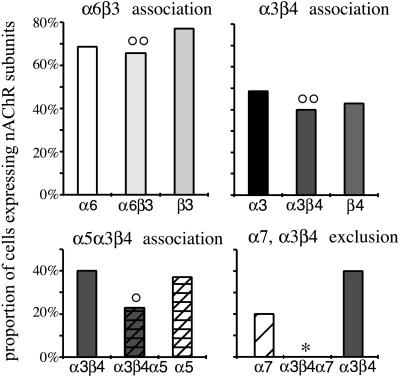
The β2 subunit was present in all neurons, and the β3, α6, and α4 subunits were present in more than 50% of the neurons sampled (77%, n = 27; 69%, n = 24; 60%, n = 21, respectively). The subunits α3, β4, and α5 were found in less than 50% of the sample (49%, n = 17; 42%, n = 15; 39%, n = 13, respectively). α7 was found in only seven neurons (20%) and α2 was found in a single neuron.
The subunits were not distributed randomly in the population of neurons (Fig. 1). A significant association was found between the subunits α6 and β3 (P = 1.0 × 10⁻⁴). α3 and β4 also were found significantly associated (P = 4.5 × 10⁻⁶) and the pair α3β4 often was associated with α5 (P = 1.5 × 10⁻²). The α7 subunit was found to be excluded from neurons expressing α3 and β4 (P = 4.5 × 10⁻²).
Figure 1.
Coexpression of nAChR subunits in noradrenergic neurons in the LC. Each bar represents the fraction of cells in the sample that express the subunit or the association of subunits indicated in the label of x axis. The subunit β2 is expressed in all neurons. Significant association of subunits is observed for α6 and β3, α3 and β4, and α3β4 and α5. Significant exclusion of subunits is observed for α7 and α3β4. Significant association (○, P < 5%; ○○, P < 1%) and significant exclusion (∗, P < 5%) are given.
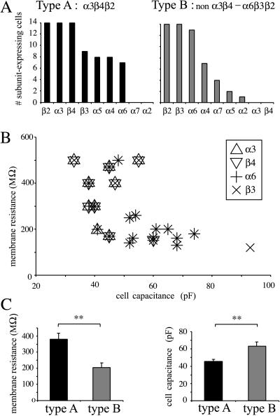
Examination of the expression pattern allowed us to distinguish two populations of neurons. The neurons expressing α3 and β4 were referred as type A (n = 14). In four neurons, only α3 (n = 3) or only β4 (n = 1) was detected; these cells were not included in the rest of the analysis. Type A neurons expressed frequently α4, α5, α6, and β3 (Fig. 2A). The neurons containing α6 or β3, but neither α3 nor β4, were named type B (n = 14). These type B neurons also sometimes contain α4 and α7 (Fig. 2A). A fraction of neurons expressing α5 did not express α3 nor β4 but always expressed α4 (n = 3). Three cells did not contain α3, β4, α6, or β3 but contained only α4β2 (with α2, α7, or α5; n = 1 for each) and were not included in the rest of the analysis.
Figure 2.
Difference in electrical characteristics of LC neurons in two populations distinguished by their expression pattern. (A) Pattern of expression of the nAChR subunits in the two populations of LC neurons. (B) Pattern of expression of nAChR subunits in neurons when both the cell capacitance and the membrane resistance have been measured. Note the frequent association of α6 and β3 (star symbol) and of α3 and β4 (superimposition of large, empty symbols), as well as the segregation of these last subunits in neurons with large membrane resistance and small capacitance. (C) Average membrane resistance and capacitance in types A and B neurons (∗∗, P < 0.01).
Electrophysiological Properties of LC Neurons.
Electrophysiological experiments in the LC neurons revealed a large diversity in cell size and nicotinic responses. Neurons of types A and B displayed different electrical characteristics (Fig. 2 B and C): the membrane resistance of type A neurons was significantly higher (380 ± 41 [170–500], n = 8, vs. 205 ± 30 [120–500] MΩ, n = 12, P = 5.3 × 10⁻³). Furthermore, the cell capacitance was significantly lower (44.5 ± 2.2 pF [30–60], n = 14, vs. 63.0 ± 3.6 pF [48–93], n = 14, P = 2.0 × 10⁻⁴) compared with type B neurons, indicating a smaller size of the electrical compartment of cell soma and proximal dendrites. This was consistent with the visual examination of the recorded neurons: type A neurons tended to be of smaller size with a round or multipolar soma whereas type B neurons presented generally a larger size and elongated shape.
The pharmacology of the nicotinic responses was tested with consecutive applications of 10 μM nicotine and cytisine, which permit β2- and β4-containing nAChRs to be discriminated (16). Only two consecutive applications per cell were used for optimal cytoplasm retrieval and RT-PCR (see Methods).
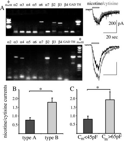
The type A neurons presented larger responses to nicotine than the type B neurons (336 ± 76 [110–801] pA, n = 8, vs. 137 ± 16 [38–206] pA, n = 9, P = 5.3 × 10⁻³), and nicotine elicited smaller currents than cytisine (Fig. 3 A and B). On the other hand, cytisine was less efficient than nicotine in type B neurons (Fig. 3 A and B). Therefore, the difference in subunit expression was also reflected in the pharmacology of the nicotinic responses recorded at the level of the cell soma.
Figure 3.
Difference in the pharmacological profile of LC neurons in relation with the subunits they express. (A) Agarose gel electrophoresis of the RT-PCR products from two cells with different expression patterns (Left) and electrophysiological responses in the same cells to the application of 10 μM nicotine or cytisine (Right). Transcripts for subunits α6, β2, and β3 are detected in the first cell (type B), and nicotine elicits larger currents than cytisine. Transcripts coding for subunits α3, α4, β2, and β4 are detected in the second cell (type A), and nicotine elicits smaller responses than cytisine. The faint band in the TH lanes corresponds to nonspecific amplification. (B) Ratio of current amplitude elicited by 10 μM nicotine and cytisine in types A and B neurons (see text); P = 1.85 × 10⁻². Nicotine elicits larger currents than cytisine in type A neurons whereas the reverse is true in type B neurons. (C) Ratio of current amplitude elicited by 10 μM nicotine and cytisine in a separate set of cells (for which the expression pattern is unknown) divided in two groups with respectively small and large membrane capacitance. The same difference as types A and B neurons is observed between small and large capacitance neurons (P = 1.0 × 10⁻²); ∗, P < 0.05.
We then confirmed the link between cell capacitance and pharmacology in another set of neurons without RT-PCR. All the cells with a capacitance below 45 pF are expected to express α3β4 (see results above), with cytisine being a more efficient agonist than nicotine, whereas the reverse should be true in cells with a capacitance above 65 pF. Neurons with a capacitance below 45 pF presented a nicotine/cytisine ratio of 0.81 ± 0.16 (n = 6), to be compared with the ratio of 0.77 ± 0.13 (n = 5) in the set of type A neurons. Neurons with a capacitance higher than 65 pF presented a significantly higher nicotine/cytisine ratio of 1.92 ± 0.46 (n = 6), to be compared with the ratio 1.79 ± 0.26 (n = 7) in type B neurons (Fig. 3 B and C).
All the nicotinic responses tested were inhibited by the nicotinic antagonists dihydro-β-erythroidine (dose, 1–10 μM, n = 9) or mecamylamine (dose, 10–30 μM, n = 5). The presence of α7-nAChR-mediated nicotinic responses (29) was examined with fast (5- to 20-ms) application of acetylcholine (1 mM, n = 8) and choline (10 mM, n = 4). These compounds only elicited responses with a slow onset (not shown). None of these responses was sensitive to 20 nM methyllycaconitine, an antagonist of α7-containing nAChRs. This is consistent with the observation that no α7 mRNA could be detected in these cells.
Properties of nAChRs Modulating Noradrenaline Release.
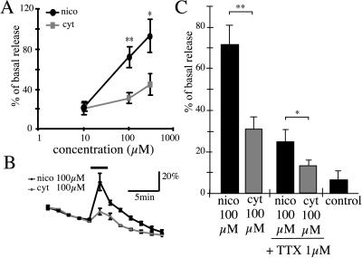
The pharmacological profile of nAChRs in the cells then was compared with that of nAChRs modulating the release of noradrenaline from nerve terminals. The rat hippocampus receives a large, noradrenergic innervation that originates exclusively in the LC. Slices from the hippocampus were loaded with tritiated noradrenaline, and the efflux of radioactivity elicited by nicotinic-agonist perfusion was monitored (Fig. 4). Nicotine or cytisine (10 μM) elicited a small efflux of tritium whereas, at higher concentrations, nicotine (100–300 μM) elicited a larger efflux of tritium than cytisine (Fig. 4). Tetrodotoxin (TTX, 1 μM) blocked 70% of the neurotransmitter release, indicating that the largest fraction of tritium release was due to the activation of preterminal nAChRs (27). Nicotine (100 μM) also was found to be more potent than cytisine in the presence of 1 μM TTX, indicating that presynaptic nAChRs on noradrenergic terminals presented the same pharmacology as preterminal nAChRs (see Discussion). No effect of nicotine could be observed when extracellular calcium was omitted or when slices were preincubated with nisoxetine (300 nM), a blocker of noradrenaline uptake (not shown). The effect of nicotine was blocked by 30 μM mecamylamine (n = 5, not shown).
Figure 4.
Low efficacy of cytisine in eliciting efflux of tritiated noradrenaline from hippocampal slices. (A) Dose–response curve for nicotine and cytisine (10 μM nicotine or cytisine, n = 5; 100–300 μM nicotine or cytisine, n = 10–15). The amplitude of each response was measured toward the linear extrapolation of the baseline during the period of drug application. (B) Averaged traces for noradrenaline efflux elicited by 100 μM nicotine and cytisine. Traces were normalized to the average release in the baseline period and plotted as a percentage of baseline release. (C) Relative potency of 100 μM nicotine and cytisine in the absence and presence of TTX; ∗, P < 0.05; ∗∗, P < 0.01.
β3 Immunoreactivity in the Hippocampus.
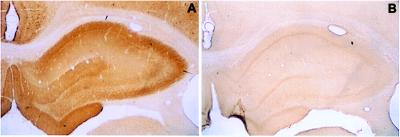
Because α6 and β3 are the prevalent subunits in the LC, they might be components of the nAChRs in noradrenergic terminals in the hippocampus. The distribution of the β3 subunit in hippocampus was assessed by immunocytochemistry. Whereas preimmune serum gave no detectable signal above background (see Fig. 5), the purified specific antibody largely stained the hippocampus. No somatodendritic labeling was detected, indicating that the signal is mainly due to afferent projections. The stratum oriens and stratum lucidum were strongly labeled, the stratum radiatum presented a medium labeling, and the pyramidal cell layer was almost devoid of staining. The staining of the dentate gyrus was similar. The stratum moleculare and stratum lacunosum moleculare presented a strong labeling, whereas the polymorph and granular layers exhibited a weaker labeling. This pattern of labeling was strongly reminiscent of the density of noradrenergic terminals (35), suggesting that β3 protein is located on the LC afferents.
Figure 5.
Immunocytochemical detection of the β3 subunit in the neuropil in the hippocampus. (A) Experiment is performed with the purified anti-β3 antibody. (B) Experiment is performed with the preimmune serum of the rabbit that produced the anti-β3 antibody afterward. Note that the cell nuclei are nonspecifically labeled (particularly in the pyramidal cell layer), as demonstrated by the labeling also present with the preimmune serum.
Discussion
The present study shows that nAChRs are differentially distributed in LC neurons. At least two different types of neurons (types A and B) of different size, different nAChR subunit mRNAs expressed, and different nicotinic pharmacology have been distinguished. In contrast, one class of nAChRs was detected in the noradrenergic terminals in the hippocampus.
The composition of nAChRs in noradrenergic neurons may not be inferred directly from the presence of nAChR subunit transcripts, and because these neurons expressed, in general, more than one pair of subunits, it is likely that multiple subtypes of nAChRs coexist in single cells. However, regularities in the coexpression of subunits coupled with simple pharmacological characterization suggests the predominance of specific subtypes of nAChRs oligomers.
Predominance of β2, α6, and β3 nAChRs Subunits.
The presence of α6, β3, and β2 mRNAs in the LC is in agreement with previous in situ hybridization studies that detected a high level for these mRNAs in the LC (6). These mRNAs generally were found to be coexpressed in various brain structures (catecholaminergic nuclei, reticularis thalamus, medial habenula, and mesencephalic V nucleus). With the use of single-cell RT-PCR, we show here that these subunits are coexpressed in single neurons that are sensitive to nicotine. This provides a new argument in favor of the assembly of these subunits into functional nAChRs. Each of the subunits, α6, β2, and β3, may participate in functional nAChRs (14, 36–38). Moreover α6 and β3 have been shown to form functional oligomers (together with β2 and β4) in the chicken retina (14). The expression of these subunits in most of the LC neurons tested suggests the frequent occurence of nAChRs containing these subunits in the noradrenergic neurons.
The subunit α4 mRNA is also expressed in a large number of neurons. However, the small amount of mRNA for this subunit detected in the LC by in situ hybridization (2, 6) suggests a minor role of this subunit in the LC, though there may be a difference between the abundance of mRNA and the abundance of protein.
Two Principal Populations of Noradrenergic Neurons in the LC.
The single-cell RT-PCR experiment demonstrates the existence of at least two populations of noradrenergic neurons in the LC, characterized by the presence (type A) or the absence (type B) of transcripts coding for the nAChR subunits α3 and β4.
The first population, type A, contains neurons that express α3 and β4 subunits. These neurons generally presented a smaller size and multipolar shape. They have, on average, a higher membrane resistance and smaller cell capacitance than the other cells. The nAChRs in these neurons are activated by cytisine, a property typical of β4-containing nAChRs (16, 39); because the subunit β2 is also expressed in these neurons, this indicates a minor contribution of β2-containing (non-β4) nAChRs to the nicotinic currents recorded at the level of the cell soma. The currents resulting from the activation of β2-containing nAChRs could be hidden by the large currents resulting from the activation of β4-containing nAChRs. Accordingly, the nicotinic currents are larger in these cells than in the type B neurons. Finally, the systematic coexpression of α3 and β4 and the frequent expression of α5 in the same population of neurons suggest that these three subunits assemble together in the LC, as observed in other neuronal populations (9, 12, 13).
The LC neurons belonging to the type B population do not express α3 or β4 mRNAs but generally express the subunits α6, β3, and β2. These neurons generally display a large fusiform shape and, on average, have a larger cell capacitance and a smaller membrane resistance. Cytisine is a weak agonist of the nAChRs activated by local application, indicating that most, if not all, of the nAChRs in these cells contain the β2 subunit (16, 17). We propose, in addition, that the main nAChR isotype of these neurons contains the subunits α6 and β3. However, the frequent expression of α4 mRNA in this population also suggests the participation of this subunit in the pool of nAChRs in a fraction of cells.
The nAChRs in the Noradrenergic Terminals in the Hippocampus.
We have compared the pharmacological profile of nAChRs recorded in the LC with nAChRs in noradrenergic nerve terminals. Previous work on GABAergic nerve terminals has led to the distinction of presynaptic and preterminal nAChRs: preterminal nAChRs increase the release of neurotransmitter, and their effect is sensitive to the blockade of sodium channels by TTX. Preterminal receptors are located upstream to the release sites (27, 40, 41). Presynaptic nAChRs increase the release of neurotransmitter but this effect is resistant to the blockade of sodium channels (28, 42, 43). Presynaptic nAChRs thus are likely to reside near the release site and correspond to the nAChRs recorded in synaptosomes (see review in ref. 44).
Both presynaptic and preterminal nAChRs are present in noradrenergic nerve terminals. In agreement with previous studies (24, 45), we found that TTX blocks 70% of the release induced by nicotine from slices, indicating that activation of preterminal nAChRs accounts for the largest fraction of the release in this preparation. The TTX might also block the recruitment of hippocampal interneurons (46). However, nAChRs in these neurons are more likely to be α7-containing nAChRs (29, 46), and the contribution of such nAChRs on noradrenaline release is not visible in the slice preparation (24, 45). Cytisine and nicotine at the concentration used in patch-clamp experiments (10 μM) only elicited a small response in efflux experiments. However, because the whole slice is likely to contribute to noradrenaline release, the efficacy of the drug may not be compared with the patch-clamp experiments performed in superficial neurons. Cytisine (100–300 μM) is less potent than nicotine in the presence or in the absence of TTX, indicating the prevalence of β2-containing nAChRs at preterminal and presynaptic levels. β4-Containing nAChRs also could be present in some noradrenergic nerve terminals in a synaptosomal preparation (23), which is likely to be enriched in presynaptic vs. preterminal receptors. However, in a similar preparation, the α3β4-subtype-specific toxin AuIB blocked only 30% of the release of noradrenaline (47).
The potent blockade of nicotine-evoked noradrenaline release by the subtype-specific neuronal bungarotoxin has led to the proposal that α3β2 nAChRs might account for most of the hippocampal nAChRs in noradrenergic terminals (24). However, given the high-sequence identity of the α3 and α6 nAChR subunit (48), α6β2-containing nAChRs also could be sensitive to the neuronal bungarotoxin.
The presence of β3 immunoreactivity in the hippocampus suggests that this subunit may also contribute to form nAChRs in noradrenergic terminals. Neurons in the hippocampus do not express β3 (6, 49), and the LC is the only nucleus that expresses β3 and provides a dense projection to the hippocampus. Moreover, the pattern of β3-like immunoreactivity matches with the density of noradrenergic terminals visualized with tritiated noradrenaline (35). Sequence resemblance (38, 48, 50) indicate that β3 could play within the α6β2β3 oligomer, a role analogous to that of β1 in the muscle α1γδβ1 oligomer (6). Therefore, we propose that β3, together with other subunits like α6β2, contributes to the formation of nAChRs in noradrenergic nerve terminals in the hippocampus.
Relevance of nAChR Distribution.
The morphology of LC neurons may be related to their area of projection. The “core cells” are medium-sized multipolar neurons that innervate multiple brain regions (hippocampus, cortex, cerebellum, hypothalamus, etc.), whereas larger neurons innervate more restricted areas in the brain; notably, the large fusiform neurons generally project to the hippocampus and to the cortex (51). The analogy in cell morphology suggests that the type A neurons are “core cells.” Conversely, most of the type B neurons would be the large fusiform cells that project to the hippocampus or the cortex.
Using local injection of nicotinic antagonists in the cerebral aqueduct in vivo, Fu et al. (46) suggested that systemic nicotine raises hippocampal noradrenaline levels by activating mostly β2-containing nAChRs in the LC, with only a minor contribution of β4-containing nAChR subtypes. In light of our results, the nicotinic activation of the ceruleo–hippocampal pathway thus is likely to involve mostly activation of nAChRs in type B neurons. Together with the results of tritiated noradrenaline efflux in hippocampal slices, this also suggests that β4-containing nAChRs only play a minor role in the modulation of the hippocampal noradrenergic innervation.
The physiological relevance of the existence of two types of LC neurons with different nAChR expression patterns is unclear. These cells could receive a cholinergic innervation from different sources and be differentially activated depending on the state of animal’s arousal. Alternatively, the difference could be physiologically relevant at the level of nerve terminals, where regional variations in the relative abundance of terminals arising from α3β4-expressing neurons and non-α3β4-expressing neurons could provide a region-specific presynaptic regulation of noradrenaline release.
Conclusion
In summary, our study has allowed us to discriminate two populations of LC neurons with distinct patterns of expression of nAChR subunits and distinct electrophysiological and pharmacological characteristics. This study notably stresses the predominance of putative α6β3β2-nAChRs in the LC and the limited role of (α3)β4-containing nAChRs in the regulation of noradrenaline release in the hippocampus. The presence of multiple subtypes of nAChRs in the LC may reflect the complexity and possibly the physiological importance of the cholinergic control of the noradrenergic system in the brain. Nicotine acts as a cognitive enhancer by several manners, including the potentiation of hippocampal synaptic transmission (43). The elucidation of nAChR diversity in the ceruleotelencephalic systems paves the way toward a targeted pharmacology, able to handle the nicotinic properties in cognitive enhancement.
Acknowledgments
We thank Drs. M. Zoli, R. Klink, and L. Rondi for critical reading of the manuscript. This work was supported by the Collége de France, the Centre National de la Recherche Scientifique, the Association Française contre la Myopathie, the Council for Tobacco Research, the Commission of the European Communities (BIOTECH program, BIOTECH fellowship to A.d.K.dE.). C.L. was supported by a grant from the Association France Alzheimer.
Abbreviations
- nAChRs
nicotinic acetylcholine receptors
- TTX
tetrodotoxin
- LC
locus ceruleus
- RT
reverse transcription
References
- 1.Changeux J-P, Bessis A, Bourgeois J-P, Corringer P-J, Devillers-Thiéry A, Eiselé J-L, Kerszberg M, Léna C, Le Novère N, Picciotto M, et al. Cold Spring Harbor Symp Quant Biol. 1996;LXI:343–362. [PubMed] [Google Scholar]
- 2.Wada K, Dechesne C, Shimasaki S, King R, Kusano K, Buonanno A, Hampson D, Banner C, Wenthold R, Nakatani Y. Nature (London) 1989;342:684–689. doi: 10.1038/342684a0. [DOI] [PubMed] [Google Scholar]
- 3.Wada E, McKinnon D, Heinemann S, Patrick J W, Swanson L W. Brain Res. 1990;526:45–53. doi: 10.1016/0006-8993(90)90248-a. [DOI] [PubMed] [Google Scholar]
- 4.Dineley-Miller K, Patrick J W. Mol Brain Res. 1992;16:339–344. doi: 10.1016/0169-328x(92)90244-6. [DOI] [PubMed] [Google Scholar]
- 5.Séguéla P, Wadiche J, Dineley M K, Dani J A, Patrick J W. J Neurosci. 1993;13:596–604. doi: 10.1523/JNEUROSCI.13-02-00596.1993. [DOI] [PMC free article] [PubMed] [Google Scholar]
- 6.Le Novère N, Zoli M, Changeux J. Eur J Neurosci. 1996;8:2428–2439. doi: 10.1111/j.1460-9568.1996.tb01206.x. [DOI] [PubMed] [Google Scholar]
- 7.Léna C, Changeux J-P. Curr Opin Neurobiol. 1997;7:674–682. doi: 10.1016/s0959-4388(97)80088-8. [DOI] [PubMed] [Google Scholar]
- 8.Lindstrom J M. Mol Neurobiol. 1997;15:193–222. doi: 10.1007/BF02740634. [DOI] [PubMed] [Google Scholar]
- 9.Vernallis A B, Conroy W G, Berg D K. Neuron. 1993;10:451–464. doi: 10.1016/0896-6273(93)90333-m. [DOI] [PubMed] [Google Scholar]
- 10.Whiting P J, Liu R S, Morley B J, Lindstrom J M. J Neurosci. 1987;7:4005–4016. doi: 10.1523/JNEUROSCI.07-12-04005.1987. [DOI] [PMC free article] [PubMed] [Google Scholar]
- 11.Chen D, Patrick J W. J Biol Chem. 1997;272:24024–24029. doi: 10.1074/jbc.272.38.24024. [DOI] [PubMed] [Google Scholar]
- 12.Conroy W G, Berg D K. J Biol Chem. 1995;270:4424–4431. doi: 10.1074/jbc.270.9.4424. [DOI] [PubMed] [Google Scholar]
- 13.Conroy W G, Vernallis A B, Berg D K. Neuron. 1992;9:679–691. doi: 10.1016/0896-6273(92)90031-8. [DOI] [PubMed] [Google Scholar]
- 14.Vailati S, Hanke W, Bejan A, Barabino B, Longhi B, Balestra B, Moretti M, Clementi F, Gotti C. Mol Pharmacol. 1999;56:11–19. doi: 10.1124/mol.56.1.11. [DOI] [PubMed] [Google Scholar]
- 15.Alkondon M, Albuquerque E X. J Pharmacol Exp Ther. 1993;265:1455–1473. [PubMed] [Google Scholar]
- 16.Zoli M, Léna C, Picciotto M R, Changeux J P. J Neurosci. 1998;18:4461–4472. doi: 10.1523/JNEUROSCI.18-12-04461.1998. [DOI] [PMC free article] [PubMed] [Google Scholar]
- 17.Papke R L, Heinemann S F. Mol Pharmacol. 1994;45:142–149. [PubMed] [Google Scholar]
- 18.Picciotto M R, Zoli M, Léna C, Bessis A, Lallemand Y, Le Novère N, Vincent P, Pich E M, Brûlet P, Changeux J P. Nature (London) 1995;374:65–67. doi: 10.1038/374065a0. [DOI] [PubMed] [Google Scholar]
- 19.Palma E, Bertrand S, Binzoni T, Bertrand D. J Physiol (London) 1996;491:151–161. doi: 10.1113/jphysiol.1996.sp021203. [DOI] [PMC free article] [PubMed] [Google Scholar]
- 20.Orr-Urtreger A, Goldner F M, Saeki M, Lorenzo I, Goldberg L, De Biasi M, Dani J A, Patrick J W, Beaudet A L. J Neurosci. 1997;17:9165–9171. doi: 10.1523/JNEUROSCI.17-23-09165.1997. [DOI] [PMC free article] [PubMed] [Google Scholar]
- 21.Aston-Jones G, Rajkowski J, Kubiak P, Valentino R J, Shipley M T. Prog Brain Res. 1996;107:379–402. doi: 10.1016/s0079-6123(08)61877-4. [DOI] [PubMed] [Google Scholar]
- 22.Egan T M, North R A. Neuroscience. 1986;19:565–571. doi: 10.1016/0306-4522(86)90281-2. [DOI] [PubMed] [Google Scholar]
- 23.Clarke P B, Reuben M. Br J Pharmacol. 1996;117:595–606. doi: 10.1111/j.1476-5381.1996.tb15232.x. [DOI] [PMC free article] [PubMed] [Google Scholar]
- 24.Sershen H, Balla A, Lajtha A, Vizi E S. Neuroscience. 1997;77:121–130. doi: 10.1016/s0306-4522(96)00425-3. [DOI] [PubMed] [Google Scholar]
- 25.Li X, Rainnie D G, McCarley R W, Greene R W. J Neurosci. 1998;18:1904–1912. doi: 10.1523/JNEUROSCI.18-05-01904.1998. [DOI] [PMC free article] [PubMed] [Google Scholar]
- 26.Lambolez B, Audinat E, Bochet P, Crepel F, Rossier J. Neuron. 1992;9:247–258. doi: 10.1016/0896-6273(92)90164-9. [DOI] [PubMed] [Google Scholar]
- 27.Léna C, Changeux J P, Mulle C. J Neurosci. 1993;13:2680–2688. doi: 10.1523/JNEUROSCI.13-06-02680.1993. [DOI] [PMC free article] [PubMed] [Google Scholar]
- 28.Léna C, Changeux J P. J Neurosci. 1997;17:576–585. doi: 10.1523/JNEUROSCI.17-02-00576.1997. [DOI] [PMC free article] [PubMed] [Google Scholar]
- 29.Frazier C J, Rollins Y D, Breese C R, Leonard S, Freedman R, Dunwiddie T V. J Neurosci. 1998;18:1187–1195. doi: 10.1523/JNEUROSCI.18-04-01187.1998. [DOI] [PMC free article] [PubMed] [Google Scholar]
- 30.Poth K, Nutter T J, Cuevas J, Parker M J, Adams D J, Luetje C W. J Neurosci. 1997;17:586–596. doi: 10.1523/JNEUROSCI.17-02-00586.1997. [DOI] [PMC free article] [PubMed] [Google Scholar]
- 31.Cauli B, Audinat E, Lambolez B, Angulo M C, Ropert N, Tsuzuki K, Hestrin S, Rossier J. J Neurosci. 1997;17:3894–3906. doi: 10.1523/JNEUROSCI.17-10-03894.1997. [DOI] [PMC free article] [PubMed] [Google Scholar]
- 32.Hill J A, Zoli M, Bourgeois J P, Changeux J P. J Neurosci. 1993;13:1551–1568. doi: 10.1523/JNEUROSCI.13-04-01551.1993. [DOI] [PMC free article] [PubMed] [Google Scholar]
- 33.Arroyo-Jiménez M d M, Bourgeois J P, Marubio L M, Le Sourd A M, Ottersen O P, Rinvik E, Fairén A, Changeux J P. J Neurosci. 1999;19:6475–6487. doi: 10.1523/JNEUROSCI.19-15-06475.1999. [DOI] [PMC free article] [PubMed] [Google Scholar]
- 34.Chomczynski P, Sacchi N. Anal Biochem. 1987;162:156–159. doi: 10.1006/abio.1987.9999. [DOI] [PubMed] [Google Scholar]
- 35.Oleskevich S, Descarries L, Lacaille J C. J Neurosci. 1989;9:3803–3815. doi: 10.1523/JNEUROSCI.09-11-03803.1989. [DOI] [PMC free article] [PubMed] [Google Scholar]
- 36.Gerzanich V, Kuryatov A, Anand R, Lindstrom J M. Mol Pharmacol. 1997;51:320–327. [PubMed] [Google Scholar]
- 37.Fucile S, Matter J M, Erkman L, Ragozzino D, Barabino B, Grassi F, Alema S, Ballivet M, Eusebi F. Eur J Neurosci. 1998;10:172–178. doi: 10.1046/j.1460-9568.1998.00001.x. [DOI] [PubMed] [Google Scholar]
- 38.Groot-Kormelink P J, Luyten W H, Colquhoun D, Sivilotti L G. J Biol Chem. 1998;273:15317–15320. doi: 10.1074/jbc.273.25.15317. [DOI] [PubMed] [Google Scholar]
- 39.Luetje C W, Patrick J W. J Neurosci. 1991;11:837–845. doi: 10.1523/JNEUROSCI.11-03-00837.1991. [DOI] [PMC free article] [PubMed] [Google Scholar]
- 40.McMahon L L, Yoon K W, Chiappinelli V A. Neuroscience. 1994;59:689–698. doi: 10.1016/0306-4522(94)90187-2. [DOI] [PubMed] [Google Scholar]
- 41.Kristufek D, Stocker E, Boehm S, Huck S. J Physiol (London) 1999;516:739–756. doi: 10.1111/j.1469-7793.1999.0739u.x. [DOI] [PMC free article] [PubMed] [Google Scholar]
- 42.McMahon L L, Yoon K W, Chiappinelli V A. J Neurophysiol. 1994;71:826–829. doi: 10.1152/jn.1994.71.2.826. [DOI] [PubMed] [Google Scholar]
- 43.Gray R, Rajan A S, Radcliffe K A, Yakehiro M, Dani J A. Nature (London) 1996;383:670–671. doi: 10.1038/383713a0. [DOI] [PubMed] [Google Scholar]
- 44.Wonnacott S. Trends Neurosci. 1997;20:92–98. doi: 10.1016/s0166-2236(96)10073-4. [DOI] [PubMed] [Google Scholar]
- 45.Sacaan A, Dunlop J, Lloyd G. J Pharm Exp Ther. 1995;274:224–230. [PubMed] [Google Scholar]
- 46.Fu Y, Matta S G, James T J, Sharp B M. J Pharmacol Exp Ther. 1998;284:1188–1196. [PubMed] [Google Scholar]
- 47.Luo S, Kulak J M, Cartier G E, Jacobsen R B, Yoshikami D, Olivera B M, McIntosh J M. J Neurosci. 1998;18:8571–8579. doi: 10.1523/JNEUROSCI.18-21-08571.1998. [DOI] [PMC free article] [PubMed] [Google Scholar]
- 48.Le Novère N, Changeux J P. J Mol Evol. 1995;40:155–172. doi: 10.1007/BF00167110. [DOI] [PubMed] [Google Scholar]
- 49.Deneris E S, Boulter J, Swanson L W, Patrick J W, Heinemann S. J Biol Chem. 1989;264:6268–6272. [PubMed] [Google Scholar]
- 50.Wang F, Gerzanich V, Wells G B, Anand R, Peng X. J Biol Chem. 1996;271:17656–17665. doi: 10.1074/jbc.271.30.17656. [DOI] [PubMed] [Google Scholar]
- 51.Loughlin S E, Foote S L, Grzanna R. Neuroscience. 1986;18:307–319. doi: 10.1016/0306-4522(86)90156-9. [DOI] [PubMed] [Google Scholar]


