Abstract
The production of several virulence factors in Vibrio cholerae O1, including cholera toxin and the pilus colonization factor TCP (toxin-coregulated pilus), is strongly influenced by environmental conditions. To specifically identify membrane proteins involved in these signal transduction events, we examined a transposon library of V. cholerae generated by Tnbla mutagenesis for cells that produce TCP when grown under various nonpermissive conditions. To select for TCP-producing cells we used the recently described bacteriophage CTXΦ-Kan, which uses TCP as its receptor and carries a gene encoding resistance to kanamycin. Among the isolated mutants was a transposon insertion in a gene homologous to nqrB from Vibrio alginolyticus, which encodes a subunit of a Na+-translocating NADH:ubiquinone oxidoreductase, and tcpI, encoding a chemoreceptor previously implicated in the negative regulation of TCP production. A third transposon mutant had an insertion in tcpP, which is in an operon with tcpH, a known positive regulator of TCP production. However, TcpP was shown to be essential for TCP production in V. cholerae, as a tcpP-deletion strain was deficient in pili production. The amino-terminal region of TcpP shows sequence homology to the DNA-binding domains of several regulatory proteins, including ToxR from V. cholerae and PsaE from Yersinia pestis. Like ToxR, TcpP activates transcription of the toxT gene, an essential activator of tcp operon transcription. Furthermore, TcpH, with its large periplasmic domain and inner membrane anchor, has a structure similar to that of ToxS and was shown to enhance the activity of TcpP. We propose that TcpP/TcpH constitute a pair of regulatory proteins functionally similar to ToxR/ToxS and PsaE/PsaF that are required for toxT transcription in V. cholerae.
Keywords: transcription activation, fimbriae, pathogenesis, toxin-coregulated pilus
Vibrio cholerae is a Gram-negative bacterium that causes the diarrheal disease cholera. To establish infection and cause disease, V. cholerae must express a variety of virulence factors, including cholera toxin (CT) and colonization factors such as the toxin-coregulated pilus (TCP). Expression of CT and TCP is strongly influenced by environmental stimuli, including temperature, pH, osmolarity, and composition of the growth medium (1–3). The mechanisms by which environmental conditions affect the coordinate expression of virulence factors by V. cholerae remain poorly understood but certainly involve transcriptional regulation mediated by several regulatory proteins (4, 5). The current model for virulence regulation is that of a cascade in which ToxR controls expression of ToxT, which itself is a transcriptional activator and directly controls expression of several virulence genes (5). ToxR is an inner membrane protein that contains a cytoplasmic DNA-binding domain with homology to the OmpR subfamily of the regulator proteins of the two-component signal transduction family (6). The periplasmic domain of ToxR is thought to interact with another transmembrane regulatory protein, ToxS, that stimulates its activity (7). ToxR, in conjunction with ToxS, regulates transcription of the toxT gene as well as a set of outer membrane proteins. ToxT is an AraC-like transcriptional activator that activates transcription of several genes, including ctx and tcpA, the latter encoding the structural subunit of TCP (8).
We aimed to identify proteins that are involved in the regulation of virulence factors by environmental stimuli in V. cholerae. We used the transposon Tnbla (9) to concentrate on the subset of those proteins that are membrane associated or secreted. Mutant bacteria that express TCP under noninducing conditions were selected for by using a kanamycin (Kan) resistance-encoding derivative of the recently described bacteriophage CTXΦ, which uses TCP as its receptor (10). The results presented here indicate that TcpP, a protein encoded by a previously identified ORF in the tcp cluster, is an important regulator of virulence gene transcription in V. cholerae.
MATERIALS AND METHODS
Bacterial Strains and Growth Media.
Escherichia coli DH5α (BRL) was used for cloning experiments, and E. coli SM10λpir (3) was used to transfer plasmids to V. cholerae by conjugation. V. cholerae strain O395-N1 (11) was used as the host for tansposon mutagenesis. E. coli and V. cholerae strains were maintained at −70°C in Luria–Bertani (LB) medium containing 20% (vol/vol) glycerol. Antibiotics were used at the following concentrations: ampicillin (Amp), 50 μg/ml; streptomycin (Sm), 100 μg/ml; Kan, 50 μg/ml; chloramphenicol (Cam), 50 μg/ml for E. coli and 5 μg/ml for V. cholerae. For TCP-producing conditions, V. cholerae strains were grown in LB broth (pH 6.5) at 30°C for 18 hr with aeration. For conditions not favoring pili production, cells were grown in LB at pH 8.5 at 30°C, in LB containing 20 mM procaine at 30°C, or in LB at 37°C.
Phage Transduction.
Phage particles of a derivative of CTXΦ (10) that carries a gene encoding resistance to Kan were prepared from the culture supernatants of V. cholerae strain Peru-2 (pCtx-Kan) after overnight growth at 30°C by filtration through a 0.2-μm-pore filter unit. Recipient TCP-producing V. cholerae strains were grown under various conditions and incubated with an equal volume of phage at room temperature for 30 min and plated on LB plates containing the appropriate antibiotics.
Genetic Methods.
Random insertions of Tnbla into the chromosome of V. cholerae were accomplished through the use of pWL10, a derivative of pGP704 (3) carrying a copy of Tnbla. Mutant strains were screened for single transposon insertions by Southern blotting (12). Fragments carrying the insertion sequences as well as adjacent chromosomal junctions were amplified by inverse PCR using primers homologous to bla sequences. The PCR products were then isolated from agarose gels and used directly in sequencing reactions (DNA Core Facility, Harvard Medical School) using the same primers.
Isolation of V. cholerae Mutants Derepressed for Pili Production.
CTXΦ phage particles (10) carrying a gene encoding resistance to Kan (CTXΦ-Kan) were used to select for V. cholerae mutants derepressed for the expression of TCP at normally nonpermissive conditions. Pools of V. cholerae colonies carrying inserts of Tnbla were grown in LB medium with a starting pH of 8.5 or in LB supplemented with 20 mM procaine. TCP-producing cells were isolated on Kan-containing plates after phage transduction. Kan-resistant colonies isolated after the initial selection were pooled, grown in the absence of Kan overnight, and streaked to obtain single colonies. Loss of the phage was screened for by replica plating colonies onto Kan-containing media. Individual Kan-sensitive colonies were then retested for TCP production under the noninducing conditions by phage transduction.
Generation of V. cholerae Strains Carrying Specific Chromosomal Mutations.
The desired mutation were introduced into the chromosome of V. cholerae strains by using the suicide plasmid vectors pCVD442 (13) or pKAS32 or pKAS46 (14). Mutant alleles were first cloned in E. coli strain SM10λpir and then conjugated into V. cholerae. The appropriate counter-selections were used to isolate V. cholerae strains that had lost the vector DNA followed by screening for the mutation by PCR. To generated the ΔtcpP strain, a PCR product carrying the entire tcpP gene as well as flanking sequences was inserted into pCR2.1 (Invitrogen). After passage through a dam− E. coli strain, a 426-bp BclI restriction fragment internal to tcpP was deleted, resulting in an in-frame deletion in tcpP. The resulting DNA fragment was then inserted into pCVD442 and introduced into the V. cholerae chromosome by homologous recombination. The Tnbla mutation was reintroduced into a wild-type V. cholerae strain by cloning the PCR product from the initial transposon mutant into pKAS46 followed by conjugation into V. cholerae.
Construction of toxT∷lacZ Reporter Strains.
The toxT gene and flanking sequences were cloned from pGJ40 (15) as a SnaBI–SpeI fragment into pKAS32 digested with EcoRV and XbaI (pKAS32-T). The lacZ gene was amplified from pCH110 (Pharmacia) by using primers with added HindIII sites and inserted into the HindIII site of toxT after a partial digestion of pKAS32-T leading to a ToxT-LacZ fusion protein under the control of the toxT promoter. This construct was introduced into the chromosome of several V. cholerae strains.
β-Galactosidase Assays.
V. cholerae cells carrying various plasmid constructs were grown overnight in LB medium at 30°C in the presence or absence of arabinose and assayed for β-galactosidase by using a modified version of the method described by Miller (16).
RESULTS
Isolation and Characterization of Mutant V. cholerae Strains.
To identify membrane or secreted proteins that, when mutated, allow expression of TCP in V. cholerae under noninducing conditions, random mutagenesis was performed with Tnbla. Tnbla carries a promoterless and signal sequence-less β-lactamase gene that will result in ampicillin-resistant cells only if it is fused to a gene for an expressed membrane or secreted protein (9). A Kan-marked derivative of the CTXΦ phage (10) was used to select for TCP-producing cells after growth under noninducing conditions, such as elevated pH or addition of procaine. We had previously observed that the addition of local anesthetics such as procaine to the growth medium strongly inhibited transcription of genes controlled by the ToxR/ToxT regulatory cascade (J.J.M. and R. Taylor, unpublished results), including tcpA.
Three mutant strains that had single Tnbla insertions and were derepressed for TCP production were isolated. The chromosomal genes interrupted by the Tnbla insertions were determined by cloning insertion junctions and DNA sequencing. One mutant strain had a transposon insertion in a gene highly homologous to nqrB from Vibrio alginolyticus (17, 18). This gene is believed to encode a subunit of an enzyme involved in production of an electrochemical gradient of sodium ions during aerobic respiration. The other mutant strains had insertions in two genes, tcpI and tcpP, that were previously implicated in the regulation of TCP and lie within the tcp gene cluster (Fig. 1A). A tcpI mutation has previously been described, and its gene product was implicated in the negative regulation of TCP production in V. cholerae (19). No mutations in tcpP had previously been reported, although the gene immediately downstream of tcpP is tcpH, whose gene product was suggested to be a positive regulator of TCP production (19). Furthermore, recent data suggested that spontaneous CT− TCP− mutants of V. cholerae have frameshift mutations in tcpH, also supporting a role for TcpH in the regulation of the ToxR regulon (27).
Figure 1.
(A) Genomic map of the region in the tcp gene cluster affected by two independent transposon insertions (arrowheads). The BclI sites show the extent of the deletion in the ΔtcpP strain. (B) Comparison of the tcpP and tcpH genes to psaE and psaF from Yersinia pestis (20) and toxR and toxS from V. cholerae (6, 7). Arrows indicate the direction and sizes of the ORFs. Sequences encoding hydrophobic regions representing potential transmembrane domains are shown in black. (C) Proposed membrane topology of ToxR, ToxS, TcpP, and TcpH. Carboxyl (C) and amino (N) termini of the proteins are indicated.
Characterization of V. cholerae Strains Carrying Mutations in tcpP.
To verify that the deregulated TCP-producing phenotype is linked to the transposon insertion, the tcpP∷Tnbla mutation was reintroduced into a wild-type strain, where it showed a pattern of elevated TCP production in the presence of procaine similar to that of the original mutant (data not shown). Because we could not distinguish whether the TcpP-Bla fusion protein was active, inactive, or constitutive for TcpP activity, we decided to construct a null mutation in tcpP. A V. cholerae strain carrying an in-frame deletion mutant in tcpP was constructed (ΔtcpP) and was found to be resistant to the CTXΦ-Kan phage, indicating that no functional pili are produced (Table 1). However, this strain was fully complemented by introducing a plasmid containing the tcpP gene under an arabinose-inducible promoter (pBAD-P) only in the presence of arabinose (Table 1). As the tcpP∷Tnbla insertion occurred in the very carboxyl-terminal region of TcpP (Fig. 1), it is possible that the TcpP-Bla fusion molecule remains functional. In fact, the cloned tcpP∷Tnbla gene can complement the ΔtcpP mutation (Table 1). Additionally, the ΔtcpP mutant strain was complemented for TCP production by a toxT-expressing plasmid (pBAD-T) (Table 1).
Table 1.
Gene complementation
| Strain | Growth in arabinose
|
|
|---|---|---|
| 0% | 0.04% | |
| ΔtcpP | — | |
| ΔtcpP (pBAD-P) | — | ++ |
| ΔtcpP (PBAD-P:bla) | — | ++ |
| ΔtcpP (pBAD-T) | — | ++ |
| ΔtcpP (pBAD-R) | — | — |
| ΔtoxR | — | |
| ΔtoxR (pBAD-R) | — | ++ |
| ΔtoxR (pBAD-T) | — | ++ |
| ΔtoxR (pBAD-P) | — | + |
| ΔtoxT | — | |
| ΔtoxT (pBAD-T) | — | ++ |
| ΔtoxT (pBAD-R) | — | — |
| ΔtoxT (pBAD-P) | — | — |
V. cholerae strains with chromosomal deletions in tcpP, toxR, or toxT carrying plasmids with these genes under an arabinose-inducible promoter were grown overnight in LB medium at 30°C without or with 0.04% arabinose, followed by transduction. A + indicates at least 50 Kan-resistant colonies, whereas a ++ indicates a confluent lawn of Kan-resistant colonies in 25 μl of plated culture.
TcpP Is a Positive Regulator.
Sequence alignment of TcpP with proteins in the databases revealed similarities between the amino-terminal region of TcpP and the DNA-binding domains of several regulatory proteins (Fig. 2), including PsaE from Y. pestis (20), HilA from S. typhimurium (21), and ToxR from V. cholerae (6). Furthermore, like ToxR, TcpP has a hydrophobic segment (from Ile-141 to Gln-169) that likely defines a transmembrane helix, suggesting it may have a topology similar to that of ToxR. These similarities suggested that TcpP was likely to be a transcriptional regulator. To investigate at which level in the regulatory cascade of TCP production in V. cholerae TcpP exerts its effect, plasmids expressing toxT, toxR, or tcpP were introduced into various V. cholerae deletion strains and assayed for TCP production by phage transduction (Table 1). As expected, all genes complemented their respective chromosomal mutations. In addition, toxT, when expressed from an inducible promoter, led to pili production regardless of which regulatory gene was deleted in the chromosome, consistent with previous results indicating that ToxT is hierarchically the last regulator in the ToxR–ToxT cascade (22). Furthermore, the tcpP-expressing plasmid weakly complemented a toxR-deletion strain but not a toxT-deletion strain for TCP production (Table 1). In contrast, the toxR-expressing plasmid did not complement a tcpP-deletion or a toxT-deletion strain (Table 1).
Figure 2.
Amino acid sequence alignment of the amino-terminal region of the V. cholerae TcpP protein and the proteins PsaE from Y. pestis (20), HilA from Salmonella typhimurium (21), and ToxR from V. cholerae (6). Identical amino acids are shown in black boxes, whereas gray boxes indicate similar amino acid residues.
TcpP Is Involved in Transcription of toxT.
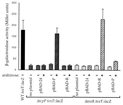
To investigate whether TcpP is involved in the transcription of toxT, a toxT∷lacZ fusion reporter construct was generated and introduced into the chromosome of various V. cholerae strains, and then plasmids carrying different complementing genes were introduced. The β-galactosidase expression levels of these strains are summarized in Fig. 3. As expected, the ΔtoxR strain expressed very low β-galactosidase activity, but was complemented by the toxR-carrying plasmid. Similarly, the ΔtcpP strain expressed almost no β-galactosidase activity but was fully complemented by a tcpP-carrying plasmid. As in a ΔtoxR strain, when the plasmid pRSI-2, carrying the toxT promoter (−172 to +45 relative to the ToxR-dependent start site) in front of a promoterless lacZ gene (23), was introduced into the ΔtcpP strain, β-galactosidase expression compared with the parental strain was reduced (data not shown). This finding suggests that TcpP acts directly upstream of the toxT gene. Analogous to the phage transduction results, the plasmid encoding the tcpP gene partially complemented a toxR deletion strain for toxT transcription (Fig. 3). These data indicate that while both ToxR ad TcpP can partially activate the toxT promoter, both are required to get efficient activation.
Figure 3.
β-Galactosidase activities of V. cholerae strains carrying a toxT∷lacZ reporter construct in the chromosome with plasmids carrying the tcpP or toxR gene under arabinose-inducible promoters. Arabinose was added to a final concentration of 0.04%.
TcpH Enhances the Activity of TcpP.
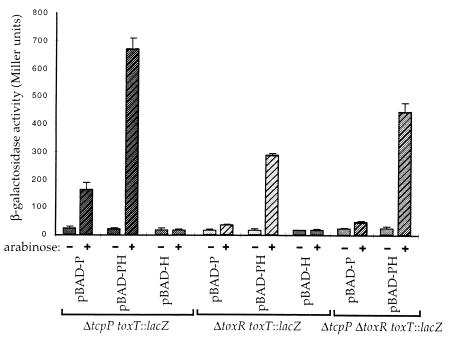
Sequence comparison of TcpH, encoded by the gene downstream and possibly in an operon with tcpP, did not reveal any significant homology with other known proteins. However, TcpH shows an amino-terminal hydrophobic region that might represent a membrane anchor; consistent with its putative membrane protein structure, an active PhoA fusion to TcpH has been previously reported (19). A similar structure is found in ToxS from V. cholerae as well as PsaF from Y. pestis, and these proteins have been shown to enhance the activities of the transcriptional activators ToxR and PsaE, respectively. Both toxS and psaF are located immediately downstream and in an operon with their respective regulators (Fig. 1B). This similarity prompted us to investigate whether TcpH can enhance the activity of TcpP. Comparison of the two constructs pBAD-P and pBAD-PH showed that the presence of tcpH leads to higher β-galactosidase activities when the toxT∷lacZ reporter construct is assayed in several V. cholerae deletion strains (Fig. 4). However, pBAD-H, which was constructed by deleting the BclI DNA fragment internal to tcpP from the pBAD-PH construct, did not activate the toxT∷lacZ reporter construct (Fig. 4). Furthermore, less arabinose was required for complementation of a ΔtcpP mutant strain for TCP production by the plasmid expressing tcpP and tcpH compared with the plasmid expressing only tcpP (data not shown).
Figure 4.
β-Galactosidase activities of V. cholerae strains carrying a toxT∷lacZ reporter construct in the chromosome with plasmids carrying the tcpP and/or tcpH genes under arabinose-inducible promoters. Arabinose was added to a final concentration of 0.04%.
ToxR/S and TcpP/H Act Synergistically.
The plasmid pVJ21 (24), expressing toxR and toxS constitutively, weakly increased the β-galactosidase activities in a ΔtcpP (data not shown) and a ΔtcpP ΔtoxR toxT∷lacZ reporter strain (Table 2). Cells carrying pVJ21 and pBAD-PH show very high β-galactosidase activities when arabinose is added, even at a low arabinose concentration that is not sufficient to activate the toxT∷lacZ reporter construct by pBAD-PH alone (Table 2), suggesting a synergistic effect of ToxR/S and TcpP/H in activating the toxT promoter.
Table 2.
ToxR/S and TcpP/H activation of toxT
| Strain | β-Galactosidase activity (Miller units) after growth in arabinose
|
||
|---|---|---|---|
| 0% | 0.002% | 0.02% | |
| ΔtcpP ΔtoxR toxT::lacZ | 21.47 ± 3.95 | 26.16 ± 2.53 | 25.60 ± 2.15 |
| ΔtcpP ΔtoxR toxT::lacZ (pVJ21) | 39.59 ± 4.87 | 55.11 ± 3.52 | 53.09 ± 2.53 |
| ΔtcpP ΔtoxR toxT::lacZ (PBAD-PH) | 22.66 ± 6.95 | 20.32 ± 0.76 | 368.22 ± 31.38 |
| ΔtcpP ΔtoxR toxT::lacZ (pBAD-PH, pVJ21) | 36.94 ± 2.56 | 770.66 ± 50.52 | 2,346.27 ± 429.23 |
β-Galactosidase activities are shown for a ΔtcpP ΔtoxR double mutant V. cholerae strain carrying a toxT::lacZ reporter construct in the chromosome with plasmids expressing the toxR and toxS genes constitutively (pVJ21) and/or the tcpP and tcpH genes from an arabinose-inducible promoter (pBAD-PH). Results are mean ± SD.
DISCUSSION
In the present study, we used a novel approach to select V. cholerae strains that are at least partially constitutive for production of TCP pili. We used the recently described phage derivative CTXΦ-Kan, which employs TCP as its receptor (10), to identify genes in V. cholerae that, when mutated, allow pili production during growth under nonpermissive environmental conditions (elevated pH or the presence of the drug procaine). After transduction, piliated cells become antibiotic resistant and can be selected from a large background of nonpiliated wild-type cells. In conjunction with the use of Tnbla (9) as the mutagenizing agent, this method allows the identification of membrane proteins that are involved in the regulation of pili production in response to environmental stimuli. The production of the type IV pilus, TCP, in V. cholerae is strongly influenced by growth conditions such as temperature, pH, and osmolarity (3), and several regulatory proteins have been identified that are involved in pilus expression (5). Our selection technique for mutants that constitutively express TCP has identified several known as well as previously unknown genes involved in TCP regulation.
Three mutant V. cholerae strains were characterized and the transposon insertions were mapped. One mutant strain had a Tnbla insertion in a gene homologous to nqrB from V. alginolyticus, a gene believed to encode a subunit of a Na+-translocating NADH:ubiquinone oxidoreductase (17, 18). In V. alginolyticus this enzyme is induced under alkaline pH and produces an electrochemical gradient of sodium ions during aerobic respiration that is used to drive ATP synthesis, flagellar rotation, and solute transport. This alternative to H+ coupling in membrane energetics allows the bacteria to maintain a neutral intracellular pH in an alkaline environment. The V. cholerae nqrB mutant strain showed much reduced motility compared with wild-type cells when assayed in soft agar (data not shown); however, it is not clear how this mutation is connected to TCP production. Nonmotile mutants of V. cholerae have previously been reported to be partially constitutive for ToxR-regulated gene expression (25).
Another transposon mutant was found to have an insertion in tcpI, whose product was previously implicated in the negative regulation of TCP production (19). tcpI was identified as a ToxR-activated gene that encodes an inner membrane protein with extensive sequence similarity to the highly conserved signaling domain in methyl-accepting membrane chemoreceptors (26). Interestingly, the tcpI∷Tnbla mutant isolated in the present study produces pili even after growth at pH 8.5; however, as in the parental strain, pili production is still inhibited by elevated temperature and procaine (data not shown). This suggests that TcpI is mainly involved in the down-regulation of TCP in response to alkaline environments.
A third mutant carried a transposon insertion in tcpP. This gene has previously been thought to have some role in regulation, as it lies in an operon with tcpH, a known positive regulator of TCP. A TnphoA insertion in tcpH resulted in cells with sporadic, but visually normal, pili (19). More recently, evidence suggested that spontaneous null mutations in tcpH produce little or no TCP or cholera toxin (27). Similarly, we expected a transposon insertion in tcpP to lead to reduced rather than deregulated pili production. Indeed, an in-frame tcpP deletion mutant strain did not produce TCP when assayed by phage transduction but was complemented for pili production by a plasmid carrying the tcpP gene. As the Tnbla insertion in this strain occurred in the very carboxyl-terminal region of TcpP, it was possible that the resulting fusion protein remains functional. In fact, the cloned tcpP∷bla hybrid gene did complement a ΔtcpP strain, indicating that this TcpP-Bla fusion protein is active.
The ΔtcpP strain was complemented by a toxT-expressing plasmid, indicating that the mutation results in a defect in regulatory rather than structural aspects of pilus assemble. A blast search of TcpP against proteins in the data banks revealed homology of the amino-terminal region of TcpP to several regulatory proteins, including PsaE from Y. pestis and ToxR from V. cholerae. PsaE, in conjunction with PsaF, is involved in the regulation of pH 6 antigen, a fimbrial surface structure found on Y. pestis and Yersinia pseudotuberculosis (28) in response to changes in temperature and pH. A similar pilus structure is found in Yersinia enterolytica, called Myf, in which myfE and myfF appear to be homologues of psaE and psaF. PsaE, like ToxR of V. cholerae, is an integral inner membrane protein with a amino-terminal cytoplasmic region with sequence similarity to transcriptional regulators and a periplasmic carboxyl-terminal region. PsaF as well as MyfF resemble ToxS from V. cholerae, as they are predicted to be located mostly in the periplasm with the hydrophobic amino terminus being either inserted in the inner membrane or cleaved as a signal peptide (28, 29). TcpP and TcpH of V. cholerae appear to have structures similar to these pairs of transcription activators (Fig. 1). This led us to investigate whether TcpP alone or in conjunction with TcpH is a transcriptional activator. Indeed, when tcpP was expressed in V. cholerae it activated a chromosomal toxT∷lacZ reporter construct. This activity was markedly enhanced by the simultaneous expression of tcpH. Additionally, tcpP as well as tcpPH, when strongly expressed from an independent promoter, complemented a toxR-deletion strain for phage transduction and for β-galactosidase activity of the toxT∷lacZ reporter construct. It was recently shown that the promoter upstream of tcpPH is transcribed independently of ToxR and ToxT in classical V. cholerae strains and is regulated by growth conditions such as temperature and pH (27, 30). Furthermore, there is some rather weak evidence that indicates that TcpP is involved in the regulation of the tcpP and tcpI promoters (30, 31). We propose a model where both ToxR/S and TcpP/H are involved in sensing various environmental and internal stimuli and are required for the production of TCP in V. cholerae by cooperating in the activation of the toxT promoter. Both TcpP and ToxR appear to be homologous membrane regulatory proteins; similarly, although TcpH and ToxS are not homologous, their predicated topology suggests that they too reside within the inner membrane and have similar topologies (Fig. 1C). Thus, it is conceivable that our observed requirement for both TcpP/H and ToxR/S for optimal expression of the toxT gene may reflect the interaction of these membrane regulatory proteins with each other in a complex. Alternatively, these pairs of regulatory proteins may activate different promoters upstream of toxT in a manner that produces the observed cooperativity between TcpP/H and ToxR/S. The membrane localization of these four regulatory proteins also suggests their interaction with membrane components of the motility and chemotaxis systems as well as other membrane proteins involved in the energizing of the cytoplasmic membrane.
Acknowledgments
We thank Victor DiRita for the generous gift of the plasmid pRSI-2 and the ΔtoxT strain, Karl Klose for kindly providing the ΔtoxR strain and for the plasmids pBAD-R and pBAD-T, and Wei Lin for the plasmid pWL10. We also thank Eric Rubin for many helpful discussions and for critically reading this manuscript. We are grateful to John W. Tobias and Nicholas Judson for assistance with the graphic programs. This study was supported by National Institutes of Health Grant AI 18045-13.
Footnotes
This paper was submitted directly (Track II) to the Proceedings Office.
Abbreviations: CT, cholera toxin; TCP, toxin-coregulated pilus; Kan, kanamycin.
References
- 1.Taylor R K, Miller V L, Furlong D B, Mekalanos J J. Proc Natl Acad Sci USA. 1987;84:2833–2837. doi: 10.1073/pnas.84.9.2833. [DOI] [PMC free article] [PubMed] [Google Scholar]
- 2.Peterson K M, Mekalanos J J. Infect Immun. 1988;56:2822–2829. doi: 10.1128/iai.56.11.2822-2829.1988. [DOI] [PMC free article] [PubMed] [Google Scholar]
- 3.Miller V L, Mekalanos J J. J Bacteriol. 1988;170:2575–2583. doi: 10.1128/jb.170.6.2575-2583.1988. [DOI] [PMC free article] [PubMed] [Google Scholar]
- 4.Skorupski K, Taylor R K. Proc Natl Acad Sci USA. 1997;94:265–270. doi: 10.1073/pnas.94.1.265. [DOI] [PMC free article] [PubMed] [Google Scholar]
- 5.DiRita V J. In: Two-component Signal Transduction. Hoch J A, Silhavy T J, editors. Washington, DC: Am. Soc. Microbiol.; 1995. pp. 351–365. [Google Scholar]
- 6.Miller V L, Taylor R K, Mekalanos J J. Cell. 1987;48:271–279. doi: 10.1016/0092-8674(87)90430-2. [DOI] [PubMed] [Google Scholar]
- 7.DiRita V J, Mekalanos J J. Cell. 1991;64:29–37. doi: 10.1016/0092-8674(91)90206-e. [DOI] [PubMed] [Google Scholar]
- 8.DiRita V J. Mol Microbiol. 1992;6:451–458. doi: 10.1111/j.1365-2958.1992.tb01489.x. [DOI] [PubMed] [Google Scholar]
- 9.Reidl J, Mekalanos J J. Mol Microbiol. 1995;18:685–701. doi: 10.1111/j.1365-2958.1995.mmi_18040685.x. [DOI] [PubMed] [Google Scholar]
- 10.Waldor M K, Mekalanos J J. Science. 1996;272:1910–1914. doi: 10.1126/science.272.5270.1910. [DOI] [PubMed] [Google Scholar]
- 11.Mekalanos J J, Swartz D J, Pearson G D, Harford N, Groyne F, de Wilde M. Nature (London) 1983;306:551–557. doi: 10.1038/306551a0. [DOI] [PubMed] [Google Scholar]
- 12.Ausubel F M, Brent R, Kingston R E, Moore D E, Seidman J G, Smith J A, Struhl K. Current Protocols in Molecular Biology. New York: Wiley; 1991. [Google Scholar]
- 13.Donnenberg M S, Kaper J B. Infect Immun. 1991;59:4310–4317. doi: 10.1128/iai.59.12.4310-4317.1991. [DOI] [PMC free article] [PubMed] [Google Scholar]
- 14.Skorupski K, Taylor R K. Gene. 1996;169:47–52. doi: 10.1016/0378-1119(95)00793-8. [DOI] [PubMed] [Google Scholar]
- 15.DiRita V J, Parsot C, Jander G, Mekalanos J J. Proc Natl Acad Sci USA. 1991;88:5403–5407. doi: 10.1073/pnas.88.12.5403. [DOI] [PMC free article] [PubMed] [Google Scholar]
- 16.Miller J H. Experiments in Molecular Genetics. Plainview, NY: Cold Spring Harbor Lab. Press; 1972. [Google Scholar]
- 17.Beattie P, Tan K, Bourne R M, Leach D, Rich P R, Ward F B. FEBS Lett. 1994;356:333–338. doi: 10.1016/0014-5793(94)01275-x. [DOI] [PubMed] [Google Scholar]
- 18.Hayashi M, Hirai K, Unemoto T. FEBS Lett. 1995;363:75–77. doi: 10.1016/0014-5793(95)00283-f. [DOI] [PubMed] [Google Scholar]
- 19.Shaw C, Peterson K M, Mekalanos J J, Taylor R K. In: Advances in Research on Cholera and Related Diarrheas. Takeda Y, Sack R B, editors. Tokyo: KTK Science Publishers; 1988. pp. 5–17. [Google Scholar]
- 20.Lindler L E, Tall B D. Mol Microbiol. 1993;8:311–324. doi: 10.1111/j.1365-2958.1993.tb01575.x. [DOI] [PubMed] [Google Scholar]
- 21.Bajaj V, Hwang C, Lee C A. Mol Microbiol. 1995;18:715–727. doi: 10.1111/j.1365-2958.1995.mmi_18040715.x. [DOI] [PubMed] [Google Scholar]
- 22.DiRita V J, Neely M, Taylor R K, Bruss P M. Proc Natl Acad Sci USA. 1996;93:7991–7995. doi: 10.1073/pnas.93.15.7991. [DOI] [PMC free article] [PubMed] [Google Scholar]
- 23.Higgins D E, DiRita V J. Mol Microbiol. 1994;14:17–29. doi: 10.1111/j.1365-2958.1994.tb01263.x. [DOI] [PubMed] [Google Scholar]
- 24.Miller V L, DiRita V J, Mekalanos J J. J Bacteriol. 1989;171:1288–1293. doi: 10.1128/jb.171.3.1288-1293.1989. [DOI] [PMC free article] [PubMed] [Google Scholar]
- 25.Gardel C, Mekalanos J J. Infect Immun. 1995;64:2246–2255. doi: 10.1128/iai.64.6.2246-2255.1996. [DOI] [PMC free article] [PubMed] [Google Scholar]
- 26.Harkey C W, Everiss K D, Peterson K M. Infect Immun. 1994;62:2669–2678. doi: 10.1128/iai.62.7.2669-2678.1994. [DOI] [PMC free article] [PubMed] [Google Scholar]
- 27.Carroll P A, Tashima K T, Rogers M B, DiRita V J, Calderwood S B. Mol Microbiol. 1997;25:1099–1111. doi: 10.1046/j.1365-2958.1997.5371901.x. [DOI] [PubMed] [Google Scholar]
- 28.Yang Y, Isberg R. Mol Microbiol. 1997;24:499–510. doi: 10.1046/j.1365-2958.1997.3511719.x. [DOI] [PubMed] [Google Scholar]
- 29.Iriarte M, Cornelis G R. J Bacteriol. 1995;177:738–744. doi: 10.1128/jb.177.3.738-744.1995. [DOI] [PMC free article] [PubMed] [Google Scholar]
- 30.Thomas S, Williams S G, Manning P. Gene. 1995;166:43–48. doi: 10.1016/0378-1119(95)00610-x. [DOI] [PubMed] [Google Scholar]
- 31.Manning P. Gene. 1997;192:63–70. doi: 10.1016/s0378-1119(97)00036-x. [DOI] [PubMed] [Google Scholar]


