To the Editor-in-Chief:
In a recent issue of The American Journal of Pathology, Lawrence et al 1 reported their immunohistochemical studies on ALK in inflammatory myofibroblastic tumors (IMT). They found cytoplasmic ALK expression in association with TPM3-ALK or TPM4-ALK chimeric transcript, which is a well-known feature of anaplastic large cell lymphoma (ALCL), in 6 of 11 IMT cases and nuclear ALK staining in one case. In addition, ALK-positive IMT showed a noticeably younger age distribution and had the same characteristics with ALK-positive ALCL. Their diagnosis of IMT was based only on morphological characteristics; however, the differential diagnostic examinations from T cell lymphomas such as T cell receptor gene rearrangements or expression of cytotoxic molecules were not mentioned.
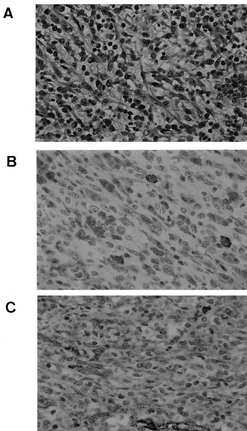
It is well recognized that the morphological spectrum of ALCL is broad 2,3 and includes sarcomatoid variant ALCL, which shows spindle shaped lymphoma cells and has a fibroblastic appearance. 4 Here, we report a case of sarcomatoid variant of ALCL with cytoplasmic ALK expression. A six-year-old girl presented at Aichi Medical University Hospital with axillary and supraclavicular lymphadenopathy in May, 2000. The biopsy specimen of the axillary lymph node showed a diffuse infiltration of spindle-shaped tumor cells with a storiform pattern (Figure 1A) ▶ . Although the histological appearance resembled that for IMT, 5 the restriction of the tumor to the lymph node and occasional mimicry of ALCL in the form of histiocytosis or carcinoma 2,3 prompted us to search for markers of lymphoid malignancies. The tumor cells were positive for ALK (cytoplasmic, Figure 1B ▶ ), CD30, CD4, EMA, TIA-1, and granzyme B, and negative for CD2, CD3, CD5, CD8, CD15, CD20, CD45RO, CD56, and EBV. The patient was consequently diagnosed with sarcomatoid variant of ALCL with null cell phenotype. However, it was of great interest that the lymphoma cells were positive for α-smooth muscle actin (Figure 1C) ▶ . Staining examination of the malignant lymphoma including bone marrow biopsy showed no extranodal involvement of the tumor. The patient was successfully treated with a chemotherapeutic regimen including doxorubicin, vincristine, cyclophosphamide, and prednisone, and is alive without disease, 10 months after the initial diagnosis.
Figure 1.

Lymph node biopsies of the patient. A: Diffuse infiltration of spindle-shaped tumor cells show a storiform pattern (hematoxylin and eosin staining, original magnification ×250). B and C: The lymphoma cells are positive for cytoplasmic ALK (B, original magnification ×375) and α-smooth muscle actin (C, original magnification ×375).
α-Smooth muscle actin has been reported to be expressed in IMTs 6 and also in several cases of malignant lymphoma, 7,8 indicating that the α-smooth muscle actin is not a golden marker for soft tissue tumors. The positive reaction of cytotoxic molecules on the tumor cells and the disease localization of lymph nodes strongly suggest that the neoplasm is derived from T cells. We therefore propose that differential diagnosis of lymphoid malignancies including ALCL is needed for the precise diagnosis of IMT, particularly in cases with ALK expression.
References
- 1.Lawrence B, Perez-Atayde A, Hibbard MK, Rubin BP, Dal Cin P, Pinkus JL, Pinkus GS, Xiao S, Yi ES, Fletcher CD, Fletcher JA: TPM3-ALK and TPM4-ALK oncogenes in inflammatory myofibroblastic tumors. Am J Pathol 2000, 157:377-384 [DOI] [PMC free article] [PubMed] [Google Scholar]
- 2.Stein H, Foss HD, Durkop H, Marafioti T, Delsol G, Pulford K, Pileri S, Falini B: CD30(+) anaplastic large cell lymphoma: a review of its histopathologic, genetic, and clinical features. Blood 2000, 96:3681-3695 [PubMed] [Google Scholar]
- 3.Drexler HG, Gignac SM, von Wasielewski R, Werner M, Dirks WG: Pathobiology of NPM-ALK and variant fusion genes in anaplastic large cell lymphoma and other lymphomas. Leukemia 2000, 14:1533-1559 [DOI] [PubMed] [Google Scholar]
- 4.Chan JK, Buchanan R, Fletcher CD: Sarcomatoid variant of anaplastic large-cell Ki-1 lymphoma. Am J Surg Pathol 1990, 14:983-988 [DOI] [PubMed] [Google Scholar]
- 5.Meis JM, Enzinger FM: Inflammatory fibrosarcoma of the mesentery and retroperitoneum. A tumor closely simulating inflammatory pseudotumor. Am J Surg Pathol 1991, 15:1146-1156 [DOI] [PubMed] [Google Scholar]
- 6.Griffin CA, Hawkins AL, Dvorak C, Henkle C, Ellingham T, Perlman EJ: Recurrent involvement of 2p23 in inflammatory myofibroblastic tumors. Cancer Res 1999, 59:2776-2780 [PubMed] [Google Scholar]
- 7.Fung CHK, Anter S, Yonan T, Lo JW: Actin-positive spindle cell lymphoma. Arch Pathol Lab Med 1993, 117:1053-1055 [PubMed] [Google Scholar]
- 8.Nozawa Y, Wang J, Weiss LM, Kikuchi S, Hakozaki H, Abe M: Diffuse large b-cell lymphoma with spindle cell features. Histopathology 2001, 38:117-178 [DOI] [PubMed] [Google Scholar]


