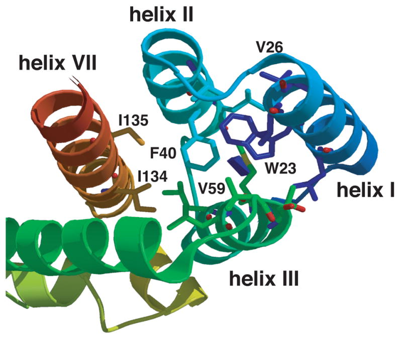
Fig. 1.

Ribbon diagram highlighting helices I, II, III, and VII in the N-terminal domain of HIV-1 CA. The ribbon diagram shows a top view of helices I, II, III, and VII in the N-terminal domain structure. The positions of two conserved hydrophobic residues Trp23 (helix I) and Phe40 (helix II) are shown. The positions of Val26 (helix I), Val59 (helix III), as well as I134 and I135 (helix VII) are also illustrated in the figure.
