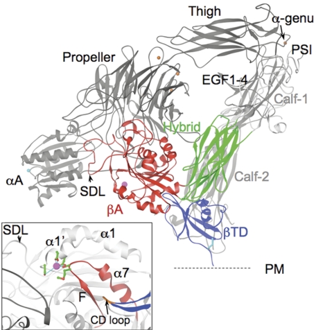
Figure 1.
Spatial relationships of the βTD domain. A ribbon diagram of a model of CD11b/CD18 based on the crystal structure of unliganded αVβ3 ectodomain.2 The 8 β2-subunit domains are labeled. The 5 CD11b-subunit domains αA (MIDAS ion in cyan), propeller (with 4 metal ions, orange circles), thigh, and calf-1 and -2 (light gray) are labeled. The metal ion in the α-genu (arrow) is in orange. The specificity-determining loop (SDL) in βA is indicated by an arrowhead. The C663-C687 disulfide bridge found at the bottom of the CD loop is shown in cyan. The position of the plasma membrane (PM) is indicated by a dotted line for orientation. (Inset) An enlarged image of the βTD's CD loop (brown, arrow) and F/α7 region (red) in the unliganded αVβ3 ectodomain.2 The ADMIDAS (adjacent to MIDAS) ion (magenta) links the α1′-α1 helix and F/α7 loop through D126D127 and M335 (equivalent to D119D120, E325 in the β2 subunit, respectively) (Cα is in green; oxygens, red). S123 in β3 (and S116 in β2) completes the metal coordination sphere.

