Abstract
Severe and malignant hyperthermia is a frequently reported factor in Emergency Department (ED) visits and fatalities in which use of amphetamine drugs, such as (±)3,4-methylenedioxymethamphetamine (MDMA), (±)3,4-methylenedioxyamphetamine (MDA) and (+)methamphetamine (METH), is confirmed. Individuals who use “Ecstasy“ are also often exposed, intentionally or otherwise, to several of these structurally-related compounds alone or in combination. In animal studies the degree of (subcritical) hyperthermia is often related to the severity of amphetamine-induced neurotoxicity, suggesting health risks to the human user even when emergency medical services are not invoked. A clear distinction of thermoregulatory risks posed by different amphetamines is therefore critical to understand factors that may produce medical emergency related to hyperthermia. The objective of this study was therefore to determine the relative thermoregulatory disruption produced by recreational doses of MDMA, MDA and METH in nonhuman primates. Body temperature and spontaneous home cage activity were monitored continuously in six male rhesus monkeys via radiotelemetric devices. The subjects were challenged intramuscularly with 0.56–2.4 mg/kg MDMA, 0.56–2.4 mg/kg MDA and 0.1–1.0 mg/kg METH. All three amphetamines significantly elevated temperature; however the timecourse of effects differed. The acute effect of METH lasted hours longer than MDA or MDMA and a disruption of nighttime circadian cooling was observed as long as 18 hours after 1.0 mg/kg METH and 1.78–2.4 mg/kg MDA, but not after MDMA. Activity levels were only reliably increased by 0.32 mg/kg METH. It is concluded that while all three substituted amphetamines produce hyperthermia in rhesus monkeys, the effects do not depend on elevated locomotor activity and exhibit differences between compounds. The results highlight physiological risks posed both by recreational use of the amphetamines and by current trials for clinical MDMA use.
Keywords: MDMA, drug abuse, neurotoxicity, Macaca mulatta, circadian, thermoregulation, serotonin, amphetamine
Introduction
Survey data from the United States show that in 2004 annual prevalence rates for 12th grade students’ illicit use of amphetamines was 10% with 4.6% reporting use in the past 30 days. It is important to establish the thermoregulatory impact of structurally distinct amphetamine compounds from a public health perspective since amphetamine related fatalities and Emergency Department (ED) admissions appear to feature significant and malignant elevations in body temperature (Dams et al. 2003; Gillman 1997; Green et al. 2003; Kamijo et al. 2002; Kojima et al. 1984; Mallick and Bodenham 1997; Wallace and Squires 2000). The Drug Abuse Warning Network estimates 4,000 annual ED visits in the US in which 3,4-methylenedioxymethamphetamine (MDMA or “Ecstasy”) is involved and at least 40,000 where methamphetamine or amphetamine are involved (Ball et al. 2003; Ball et al. 2004). The hyperthermic response may be a critical determinant of medical emergencies and deaths since many of the toxicological problems that are seen, such as rhabdomyolysis, disseminated intravenous coagulation and acute renal failure (Henry et al. 1992) can result from hyperthermia. The acute thermoregulatory disruption produced by amphetamines is also important beyond acute medical emergency. For example hyperthermia can markedly influence amphetamine-induced neurotoxicity in rodents and non-human primates (Bowyer et al. 1994; Bowyer et al. 1992; Malberg and Seiden 1998; Melega et al. 1998; Miller and O'Callaghan 1994).
It is difficult to determine the relative impact of each of these amphetamines on thermoregulation in humans because recreational users are frequently poly-drug abusers and are often positive for multiple drugs in ED medical situations. Therefore, nonhuman laboratory models are necessary to establish the relative thermoregulatory impact of different amphetamines in order to better understand the clinical implications of amphetamine use and abuse. The present study is focused on the acute effects of MDMA, 3,4-methylenedioxyamphetamine (MDA) and methamphetamine (METH), which are all commonly used in an intermittent pattern in the nightclub/rave party population.
All three of these amphetamines can produce an acute increase in body temperature. MDMA results in an acute elevation of body temperature in human laboratory studies at doses (1.5–2.0 mg/kg, p.o.) within the range of common recreational doses (Freedman et al. 2005; Liechti et al. 2000), but not reliably so at lower doses (Grob et al. 1996; Mas et al. 1999) suggesting a dose-related effect. MDMA (racemic or the S(+) enantiomer) also produces acute hyperthermia in rats (Brown and Kiyatkin 2004; Dafters 1994; Malberg and Seiden 1998), mice (Carvalho et al. 2002; Fantegrossi et al. 2003), guinea pigs (Saadat et al. 2004), pigs (Fiege et al. 2003; Rosa-Neto et al. 2004), rabbits (Pedersen and Blessing 2001) and non-human primates (Taffe et al. 2006). METH also increases body temperature in rodents (Bowyer et al. 1994; Brown et al. 2003). Recent studies also suggest that repeated dosing with METH can cause fatal/threatening hyperthermia in at least three nonhuman primate species (Madden et al. 2005; Ricaurte et al. 2002; 2003), and MDA can cause hyperthermia and death in canines (Davis et al. 1987).
However, given the wide variability of species and the type and doses of amphetamines administered in prior studies, the relative contribution to the thermodysregulatory effects of MDMA, METH and MDA is unclear. It is likely that significant thermoregulatory differences between related amphetamines exist. MDMA, MDA and METH are potent indirect monoaminergic agonists, acting to inhibit reuptake mechanisms and to enhance transmitter release, although their relative potencies for releasing serotonin, norepinephrine and dopamine differ from each other within species and these relationships may differ significantly across species (Battaglia and De Souza 1989; Han and Gu 2006; Verrico et al. 2005). With respect to human monoamine transporters, MDMA has greater affinity for noradrenergic and serotonin transporters compared with dopamine transporters whereas METH has greater affinity for dopamine transporters. Interpretation of the pharmacology can be complex; for example MDMA has greater affinity for noradrenergic transporters compared with serotonergic transporters and yet is more potent in stimulating serotonin release in a cell transfection model (Verrico et al. 2005). Such results support the need to compare systemic effects of the amphetamines in the intact organism, ideally one more closely related to humans such as nonhuman primates.
To date, there are few studies which have systematically measured the thermoregulatory impact of these amphetamines in non-human primates. Recent studies from this laboratory have established that unrestrained rhesus monkeys develop hyperthermia following administration of MDMA without any stimulation of locomotor activity (Taffe et al. 2006; Von Huben et al. 2006). Although much work has been done in rodent species, careful comparisons of the thermoregulatory effects of MDMA, MDA and METH have not been reported within a consistent model. Therefore, the present study was designed to directly compare the acute thermoregulatory effects of (±)MDMA with the effects of the related amphetamines (±)MDA and (+)METH within the same subjects. The goals were to 1) confirm our preliminary finding by testing a wider range of doses of (±)MDMA, 2) determine the relative thermoregulatory disruption of the closely-related (±)MDA (also a metabolite of MDMA) and 3) compare the effects of the more “empathogenic” amphetamines to those of (+)METH as a substituted amphetamine with typically a more classic psychomotor stimulant behavioral profile.
Experimental Procedures
Animals
Six male rhesus monkeys (Macaca mulatta; Chinese origin) participated in this study. Animals were 6–10 years of age, weighed 9.0–12.7 kg at the start of the study and exhibited body condition scores (Clingerman and Summers 2005) of 2.25–3.25 out of 5 at the nearest quarterly exam. Daily chow (Lab Diet 5038, PMI Nutrition International; 3.22 kcal of metabolizable energy (ME) per gram) allocations were determined by a power function (Taffe 2004a; b) fit to data provided in a National Research Council recommendation (NRC/NAS 2003) and modified individually by the veterinary weight management plan. Daily chow ranged from 160 to 230 g per day for the animals in this study. The animals' normal diet was supplemented with fruit or vegetables seven days per week and water was available ad libitum in the home cage at all times. All animals were individually housed throughout the study. Animals on this study had previously been immobilized with ketamine (5–20 mg/kg) no less than semiannually for purposes of routine care and some experimental procedures. Animals also had various acute exposure to scopolamine, raclopride, methylphenidate, SCH23390, Δ9-THC, nicotine and mecamylamine in behavioral pharmacological studies and 4 had been exposed to an oral ethanol induction procedure (Katner et al. 2004). No experimental drug treatments had been administered for a minimum of one year prior to the start of telemetry studies and thus were not anticipated to have any bearing on the results of the current study. The United States National Institutes of Health guidelines for laboratory animal care (Clark et al. 1996) were followed and all protocols were approved by the Institutional Animal Care and Use Committee of The Scripps Research Institute (La Jolla).
The monkeys’ body temperature and activity patterns naturally followed a diurnal pattern of daytime high and nighttime low; however temperature was consistent across the selected injection times (either 1030 or 1300). These times were carefully selected based on pilot data which indicated both stability of the normal body temperature and a lack of any differential effect of drug challenges across this interval. Activity also follows a circadian cycle as animals do not make many movements across the cage (triggering an activity “count”) when lights are off; however they engage in relatively variable levels of activity, both between animal and across the light period. Subcutaneous body temperature varies about 1.5–2 °C from nighttime low to daytime high which is consistent with reports for intraperitoneal temperature in Japanese (Takasu et al. 2002) macaques or subcutaneous temperature in cynomolgus (Almirall et al. 2001) or rhesus (Horn et al. 1998) macaques. The subcutaneous temperature values from Almirall (Almirall et al. 2001), Horn et al (Horn et al. 1998) and the present study all differ from the intraperitoneal values reported by Takasu et al (Takasu et al. 2002) by about −1 to −1.5 °C. In our recent studies we have compared rectal temperatures (obtained under light ketamine anesthesia) with concurrent telemetric temperature values in 17 monkeys; multiple determinations are available for most animals. The subcutaneous values vary from ~1–3 °C lower than rectal temperature across animals but the temperature differential is consistent across determinations within animals.
Apparatus
Radio telemetric transmitters (TA10TA-D70; Transoma / Data Sciences International) were implanted subcutaneously in the flank. The surgical protocol was adapted from the manufacturer’s surgical manual and implantation was conducted by, or under supervision of, the TSRI veterinary staff using sterile techniques under isoflurane anesthesia. Temperature and gross locomotor activity recordings were obtained continuously from the telemetric transmitters implanted in the monkey via an in-cage receiver (RMC-1; Transoma / Data Sciences International). Data were recorded on a 5 minute sample interval basis by the controlling computer and represented as a moving average of three samples (−5 min, current, + 5 min) for each 10 minutes. Occasional missing data points were replaced with a linear interpolation of adjacent points. Ambient room temperature was also recorded by the system via a thermometer mounted near the top of the housing room.
Drug challenge studies
For these studies doses of (±)3,4-methylenedioxymethamphetamine HCl (0.56, 1.0, 1.78, 2.4 mg/kg), (±)3,4-methylenedioxyamphetamine HCl (0.56, 1.0, 1.78, 2.4 mg/kg) and (+)methamphetamine HCl (0.1, 0.32, 0.56, 1.0 mg/kg) were administered intramuscularly in a volume of 0.1 ml/kg saline. Drugs were provided by the National Institute on Drug Abuse. Treatment order was pseudorandomized within compound to the extent possible with the small sample size to minimize the impact of any potential order effects. Generally, the MDMA studies were conducted first, MDA second and the METH last; however, there was some degree of overlap of the schedule across compounds. Dose ranges were originally based on pill-content analyses suggesting ~75–125 mg MDMA per “Ecstasy” pill, thus 1–1.78 mg/kg MDMA for a single pill taken by the standard 70kg person, but as much as 2.5 mg/kg in a 50 kg woman or as little as 0.83 mg/kg in a 90 kg man. Relevant dose ranges for MDA and METH were determined initially by reference to MDMA:MDA and MDMA:METH ratios in the pills analyzed by Ecstasydata.org. These ranges were further refined based on pilot studies conducted for this and other projects (Madden et al. 2005; Taffe et al. 2006), and taking in to consideration the minimum dose thresholds for lasting or neurotoxic effects (Ali et al. 1993; Melega et al. 1998; Ricaurte et al. 1988). All challenges were administered in the middle of the light cycle, either at 1030 (N=2) or 1300 (N=4) hours, with active doses separated by 1–2 weeks. The ambient room temperature averaged either 23°C (N=2) or 27°C (N=4) for these studies. Even larger differences in ambient room temperature (18°C–30°C) have not been shown to have a significant effect on MDMA-induced hyperthermia in humans (Freedman et al. 2005) or monkeys (Von Huben et al. 2006) and the latter study illustrates clearly that individual differences in response are greater than any effects associated with ambient temperature. Animals were visually observed for a period of two hours following injections and efforts were made to minimize noise and excitement in the rooms during these intervals. Normal daily activity such as afternoon feedings and interactions with other animals not on the study resumed after the two hour interval.
Data Analysis
Two way randomized block analysis of variance (ANOVA) was employed to evaluate acute treatment related effects starting with the sample collected immediately prior to injection (referred to as “baseline”). The first analysis was conducted on the 10 minutes sample data over the interval −10 to 240 min post injection as this was the designated interval in which room disruption was minimized; see (Taffe et al. 2006; Von Huben et al. 2006). A second analysis was conducted to evaluate potential effects lasting for 18 hours after injection; this interval is the time between the latest injection time (1300 hrs) and the time that the lights were turned on the following morning (0600 hrs). Thus the repeated measures factors for ANOVA were time relative to injection (−10 to 240 min; −1 to 18 hours) and drug dose (Vehicle, four active doses) for each compound. Significant main effects in the two-way ANOVAs were followed up with the Tukey-Kramer post hoc procedure to evaluate all pair-wise comparisons. All statistical analyses were conducted using GB-STAT v7.0 for Windows (Dynamic Microsystems, Inc., Silver Spring MD) and the criterion for significance in all tests was p < 0.05.
Results
3,4-Methylenedioxymethamphetamine (MDMA)
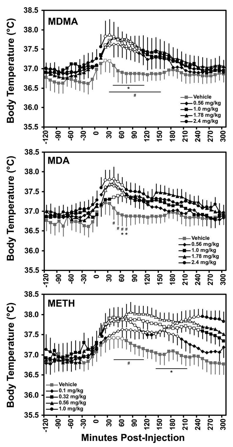
MDMA significantly increased body temperature within 10–15 mins of drug administration (Figure 1). The 10-min sample analysis confirmed significant main effects of drug condition [F4,20 = 4.33; p < 0.05], time post-injection [F25,125 = 11.78; p < 0.0001] and an interaction of factors [F100,500 = 1.80; p < 0.0001]. The post hoc test confirmed a significant temperature increase over baseline 20–30 minutes after vehicle, 20–90 min after the 0.56 dose, 20–100 minutes after the 1.0 dose, 20–100 and 140 min after the 1.78 mg/kg dose and 20–60 min after the 2.4 mg/kg dose. The post hoc test also confirmed that temperature was significantly higher than the respective vehicle time points 40–110 and 140 min after 0.56 mg/kg, 30–150 after 1.0 mg/kg, 30–160 min after 1.78 mg/kg and 10–160 min after 2.4 mg/kg.
Figure 1.

The mean (N=6, bars indicate SEM) subcutaneous temperature values following acute challenge with doses of (±)3,4-methylenedioxymethamphetamine (MDMA), (±)3,4-methylenedioxyamphetamine (MDA) and (+)methamphetamine (METH) are presented. Breaks in the series indicate the time of injection. The statistical analysis included the interval −10 to 240 minutes after injection and a significant change from the −10 min time point is indicated by the open symbol for each treatment condition. The * and # indicate timepoints in which all four (*) or three of four (#) active dose conditions differed significantly from the vehicle temperature; see text for additional effects determined to be statistically reliable.
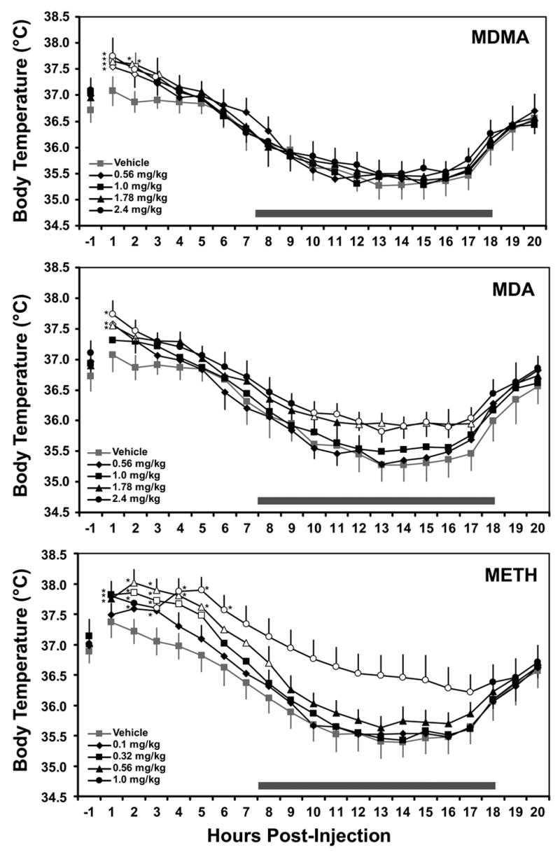
Effects of MDMA on temperature did not last beyond the first three hours after dosing (Figure 2). The analysis of hourly timepoints after dosing confirmed a significant main effect of time post-injection [F18,90 = 41.93; p < 0.0001] and an interaction of factors [F72,360 = 1.81; p < 0.001]. The post hoc test confirmed that temperature was significantly elevated for 1 hr after 0.56 and 2.4 mg/kg, and for 2 hrs after 1.0 or 1.78 mg/kg of MDMA. In addition, temperature was significantly elevated above vehicle for two hours after 0.56 and 1.0 mg/kg and for three hours after 1.78 or 2.4 mg/kg. Consistent with the usual nighttime cooling, temperature was significantly below baseline 6–18 hrs after 1.0 and 2.4 mg/kg doses, 7–18 hrs after the 1.78 mg/kg dose and 8–18 hrs after vehicle or 0.56 mg/kg MDMA.
Figure 2.

The mean (N=6, bars indicate SEM) subcutaneous temperature values in the 20 hours following acute challenge with doses of (±)MDMA, (±)MDA and (+)METH are presented. Error bars (SEM) are selectively presented for visual clarity. The statistical analysis included the interval −1 to 18 hours after injection and the open symbols indicate a significant difference from the vehicle condition at a given timepoint. A significant increase from the time point preceding injection is indicated by *, however significant decreases from baseline are not depicted, see Results.
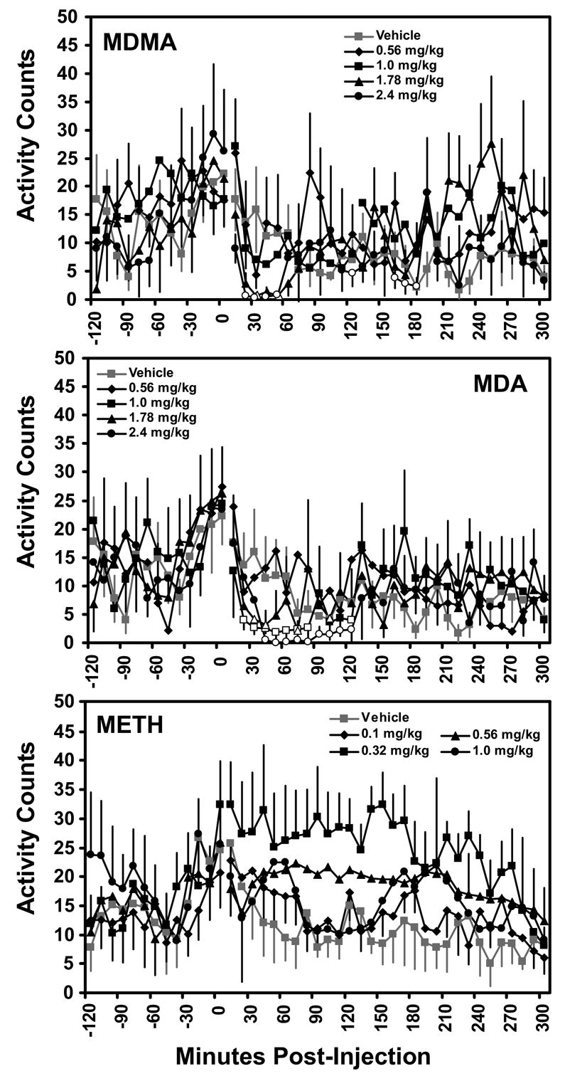
Activity was also significantly reduced in the few hours after injection as confirmed by significant effects of time post-injection [F25,125 = 2.11; p < 0.01] and an interaction of factors [F100,500 = 1.54; p < 0.01] ; see Figure 3. The post hoc test confirmed that activity was significantly reduced from the baseline 30 and 50 min after 1.7 mg/kg dose and 20–50, 120, 160–180 and 220 min after 2.4 mg/kg MDMA.
Figure 3.

The mean (N=6) activity values following acute challenge with doses of (±)MDMA, (±)MDA and (+)METH are presented. Breaks in the series indicate the time of injection. Error bars (SEM) are selectively presented for visual clarity. Statistical conventions are as in Figure 1.
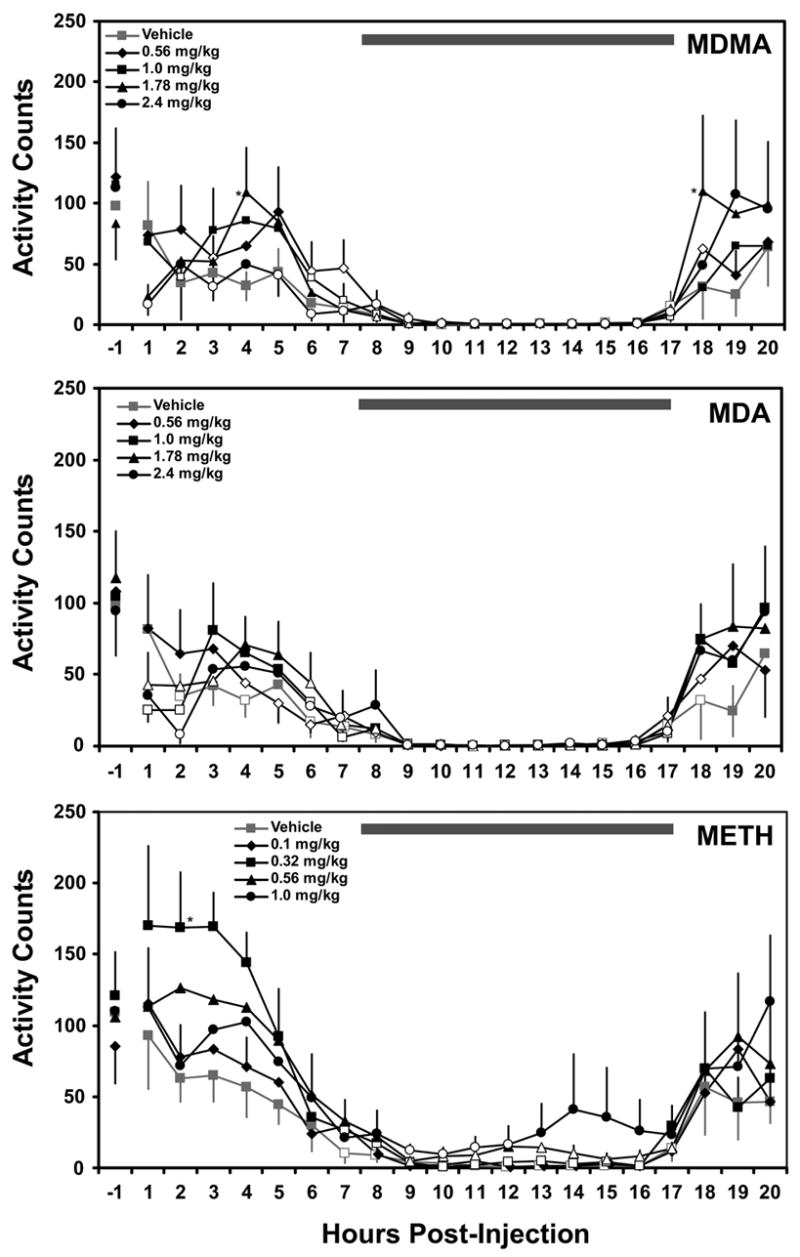
The hourly activity counts (Figure 4) were also significantly affected by time post-injection [F18,90 = 7.19; p < 0.0001] and an interaction of factors [F72,360 = 1.38; p < 0.05]. The post hoc test confirmed that activity was significantly lower than baseline 3 hrs after 0.56 mg/kg, 2 hrs after 1 mg/kg and 1 and 3 hrs after 2.4 mg/kg of MDMA. Activity was also consistent with the usual circadian pattern as it was significantly lower than baseline 6–18 hours after vehicle or 1.0 mg/kg, 6–17 hours after 0.56 mg/kg, 7–17 hours after 1.78 mg/kg and 5–17 hours after 2.4 mg/kg MDMA. Finally, activity was significantly higher than corresponding vehicle timepoints 4 and 18 hours after 1.78 mg/kg MDMA.
Figure 4.

The mean (N=6, bars indicate SEM) activity values in the 20 hours following acute challenge with doses of (±)MDMA, (±)MDA and (+)METH are presented. Error bars (SEM) are selectively presented for visual clarity. The statistical analysis included the interval −1 to 18 hours after injection and the open symbols indicate a significant difference from the pre-injection baseline and a significant increase from the vehicle conditions is indicated by *.
3,4-Methylenedioxyamphetamine (MDA)
MDA also significantly increased body temperature (Figure 1). The analysis confirmed significant main effects of drug condition [F4,20 = 5.60; p < 0.01] and time post-injection [F25,125 = 5.00; p < 0.0001]; however, a trend for an interaction of factors was not statistically reliable [F100,500 = 1.24; p = 0.077]. The post hoc test confirmed a significant temperature increase over baseline 20–60 minutes after the 0.56, 1.78 and 2.4 mg/kg doses and 40–70 minutes after the 1.0 dose. The post hoc test also confirmed that temperature was significantly higher than the respective vehicle time points 40–70 after 0.56 mg/kg, 60–70 after 1.0 mg/kg, 40–70, 90–100 and 140–160 min after 1.78 mg/kg and 20–120 min after 2.4 mg/kg of MDA. An apparent difference in temperature after the 1.0 mg/kg dose compared with other active doses was not statistically reliable.
The analysis of hourly timepoints after dosing (Figure 2) confirmed a significant main effect of drug condition [F4,20 = 11.39; p < 0.0001], time post-injection [F18,90 = 33.77; p < 0.0001] and an interaction of factors [F72,360 = 1.50; p < 0.01]. The post hoc test confirmed that temperature was significantly elevated from baseline 1 hr after 0.56 or 2.4 mg/kg, and for 2 hrs after 1.78 mg/kg of MDA. In addition, temperature was significantly elevated above vehicle 1 hr after 0.56 mg/kg and 1–2 hrs after 1.78 or 2.4 mg/kg of MDA. Consistent with the usual nighttime cooling, temperature was significantly below baseline 6–18 hrs after 0.56 mg/kg, 7–18 hrs after the 1.0 mg/kg dose and 8–18 hrs after vehicle and 1.78 or 2.4 mg/kg MDA. The effects of the higher doses lasted overnight as temperature was significantly higher than the respective vehicle time points 10, 12–17 hrs after 1.78 mg/kg and 10–17 hrs after 2.4 mg/kg MDA. The 1.78mg/kg dose also significantly elevated temperature compared with similar timepoints after 0.56 mg/kg (10–11, 13–15 hrs post-injection) and 1.0 mg/kg (13 hrs post-injection) doses of MDA. Similarly, the 2.4 mg/kg dose also significantly elevated temperature compared with similar timepoints after 0.56 mg/kg (7, 10–15 hrs post-injection) and 1.0 mg/kg (11 hrs post-injection) doses of MDA.
Activity was decreased in the few hours after injection (Figure 3) as confirmed by a significant main effect of time post-injection [F25,125 = 7.32; p < 0.0001]. The post hoc test confirmed that activity was significantly lower than baseline 20–80 and 120 min after 1.0 mg/kg MDA, 30, 40, 70 and 100 min after 1.78 mg/kg and 40–120 min after 2.4 mg/kg MDA.
The hourly activity counts (Figure 4) were also significantly reduced as was confirmed by a significant main effect of time post-injection [F18,90 = 7.90; p < 0.0001]. The post hoc test confirmed that activity counts were lower than baseline after vehicle (2,4, 6–18 hrs) as well as the 0.56 mg/kg (4–18 hrs), 1.0 mg/kg (1–2, 6–17 hrs), 1.78 mg/kg (1–3, 6–17 hrs) and 2.4 mg/kg (2, 6–17 hrs) doses of MDA.
(+)Methamphetamine (METH)
METH also significantly increased body temperature in the first few hours after administration, however the timecourse differed notably (Figure 1). The analysis confirmed significant main effects of drug condition [F4,20 = 15.71; p < 0.0001], time post-injection [F25,125 = 6.44; p < 0.0001] and of the interaction of factors [F100,500 = 2.82; p < 0.0001]. The post hoc test confirmed a significant temperature increase over baseline 30–60 min after vehicle, 30–190 minutes after the 0.1 mg/kg dose, 20–240 min after the 0.32 mg/kg dose, 10–240 min after the 0.56 mg/kg dose and 20–110 and 140–240 minutes after 1.0 mg/kg of METH. The post hoc test also confirmed that temperature was significantly higher than the respective vehicle time points 110–200 min after 0.1 mg/kg, 20–240 after 0.32 mg/kg, 40–240 min after 0.56 mg/kg and 20–100 and 130–240 min after 2.4 mg/kg of METH. No significant effects on activity were confirmed for the first four hours after administration in this analysis.
METH also significantly disrupted temperature during nighttime hours (Figure 2). Analysis of the hourly time averages confirmed a main effect of drug condition [F4,20 = 16.73; p < 0.0001], time post-injection [F18,90 = 69.19; p < 0.0001] and of the interaction of factors [F72,360 = 3.28; p < 0.0001]. The post hoc test confirmed that temperature was significantly elevated over baseline 2–3 hrs after 0.1 mg/kg, 1–3 hrs after 0.32 mg/kg, 1–5 hrs after 0.56 mg/kg and 1–6 hrs after 1.0 mg/kg METH. Similarly, temperature was significantly higher than the respective vehicle timepoints 2–5 hrs after 0.32 mg/kg, 2–8 hrs after 0.56 mg/kg and 3–17 hrs after 1.0 mg/kg METH. The 1.0 mg/kg dose also significantly elevated temperature 4–17 hrs post-injection relative to the 0.1 mg/kg dos, 6–17 hrs post-injection relative to the 0.32 mg/kg dose and 9–16 hrs post-injection relative to the 0.56 mg/kg dose. The posthoc test also confirmed significant circadian cooling with temperatures reliably lower than baseline 8–18 hrs after Vehicle, 0.1 or 0.32 mg/kg conditions, 9–18 hrs after 0.56 mg/kg METH and 14–18 hrs after 1.0 mg/kg METH.
The hourly activity counts (Figure 4) were also significantly lower as was confirmed by a significant main effect of time post-injection [F18,90 = 7.19; p < 0.0001]. The post hoc test confirmed that activity was significantly lower than baseline 7–17 hrs after vehicle, 7–16 hrs after 0.32 mg/kg, 9–11 and 13–17 hrs after 0.56 mg/kg and 9–12 hrs after the 1.0 mg/kg dose of METH. Activity was increased over the vehicle condition 2 hrs after 0.32 mg/kg METH.
Discussion
The results of the present study establish that rhesus monkeys develop elevated body temperature following an intramuscular injection of a range of doses of each of three substituted amphetamines. These data support and extend our initial report (Taffe et al. 2006) in confirming that monkeys’ hyperthermic responses to these compounds are similar to humans and not hypothermic, which contrasts with one prior report on the effects of (+)MDMA in rhesus monkeys (Bowyer et al. 2003). The study also shows that the immediate temperature response in ~4 hours after the administration of (±)MDA is quite similar to the (±)MDMA response at identical doses and that doses of METH elevate temperature over a more protracted time course. The immediate temperature response to all three amphetamines was not strongly dose-dependent across the tested ranges although dose-dependent elevations of nighttime temperature were observed after MDA and METH. The temperature responses did not appear to depend on significant increases in locomotor activity following any of the compounds. This latter finding may indicate important differences between nonhuman primate and rodent responses to the amphetamines.
The magnitude of the acute hyperthermic response subsequent to amphetamine exposure (maximum change in the 4 hr post-injection: 2.4 mg/kg MDMA 0.71°C; 2.4 mg/kg MDA 0.65°C; 1.0 mg/kg METH 0.94°C) is consistent with prior reports of drug-induced hyperthermia. For example, Freedman and colleagues (2005) reported that an oral dose of 2.0 mg/kg (±)MDMA elevates human temperature by ~0.3–0.6 °C. Yuan and colleagues (Yuan et al. 2006) found that the highest mean temperature increase observed in squirrel monkeys after a single oral 1.25 mg/kg dose of METH reached ~0.6 °C over the pre-injection baseline and ~0.8 °C over the vehicle condition at a similar time point. Furthermore racemic MDMA results in significant hyperthermia of ~ 0.7–1.0°C in rhesus monkeys under normal ambient temperature conditions (Taffe et al. 2006; Von Huben et al. 2006). The lack of a strong dose dependency of the immediate hyperthermic response confirmed a prior observation on the effects of MDMA (Von Huben et al. 2006) and was consistent across compounds in this study. This outcome suggests perhaps that thermoregulatory mechanisms in the rhesus monkey which are triggered upon temperature elevations of about 0.7–1.0°C may not be affected by the amphetamines at these doses.
Major differences emerged in the duration of the amphetamine-induced hyperthermia. The temperature responses to MDMA and MDA peaked sharply ~60–90 minutes post-injection but thereafter declined steadily. This pattern is consistent with reports that plasma MDMA levels peak within 60 minutes after administration in rhesus and squirrel monkeys (Bowyer et al. 2003; Mechan et al. 2006). In contrast, the temperature response to METH initially peaked ~60 minutes post-injection but was sustained at high levels for 180–300 minutes post-injection depending on the dose. This finding is consistent with a report that while plasma METH levels reach a peak within 60 minutes after administration and then rapidly decline in squirrel monkeys, the metabolite amphetamine reaches plasma levels which approximate the early METH peak and persists 60–180 minutes post administration (Yuan et al. 2006). In total then, the acute temperature responses in the present study correspond quite well to reported pharmacokinetic data assuming that amphetamine is an active metabolite of METH with respect to thermoregulation.
The highest dose of METH resulted in a disruption of temperature regulation that lasted overnight until the following morning. Similar effects of a smaller magnitude were observed following the highest two doses of MDA; however, MDMA did not result in overnight disruption at these doses. The mechanism of this extended elevated temperature response is unknown but might theoretically be related to pharmacokinetics since MDA has been reported to have a significantly longer half life than MDMA in humans and monkeys (Bowyer et al. 2003; de la Torre et al. 2004; Kraemer and Maurer 2002). Still it should be appreciated that most published reports on the pharmacokinetics of MDA derive from investigations of MDA as a metabolite of administered MDMA. MDA is, however, only a minor metabolite of MDMA with peak plasma levels of about 10% of the administered MDMA dose reported (ibid). Given that MDA levels rise slowly to a peak some 7 hrs after an MDMA injection in monkeys (Bowyer et al. 2003), it seems unlikely that (metabolite) MDA contributed much to the effects of MDMA observed in this study. In contrast the plasma levels of the METH metabolite amphetamine are equivalent to the administered METH dose at peak and remain significantly elevated at least 6 hours after a 1.25 mg/kg oral dose of METH in squirrel monkeys (Yuan et al. 2006). In addition, the current locomotor activity results suggest that some significant disruption of sleep may have occurred following METH (Figure 4). These results also suggest that it is important to consider drug effects that may be related to the timing of administration within the diurnal cycle and/or qualitative effects on the sleep cycle.
Although not directly addressed in this report, the data may also highlight different thermoregulatory patterns produced by amphetamines which differ in effect on serotonin, dopamine and noradrenaline signaling. The central nervous system mechanisms involved in amphetamine-induced thermodysregulation include all three of these monoamines, which all interact with all three monoamine transporters and release transmitters, albeit with varying potencies. Administration of 5-HT2A and 5-HT2C receptor antagonists can block MDMA hyperthermia in rodents (Fantegrossi et al. 2003; Herin et al. 2005; Mechan et al. 2002), as can depletion of pre-synaptic serotonin stores (Fantegrossi et al. 2005; Saadat et al. 2005). Conversely, administration of an MAO-A inhibitor (Freezer et al. 2005) or 5-HT1A receptor antagonist can prolong MDMA-induced hyperthermia (Saadat et al. 2004). Dopaminergic contributions to hyperthermia appear to be primarily mediated by the D1-like receptors since the D1-like antagonist SCH23390 blocks MDMA or METH hyperthermia where D2-like antagonists are less effective (Broening et al. 2005; Mechan et al. 2002). The α1- and β-adrenergic receptors also contribute to these effects, see (Sprague et al. 2005) for review. Significant differences are reported for relative potency of a given amphetamine to interact with SERT, DAT and NET derived from humans and rodents (Han and Gu 2006; Verrico et al. 2005). These findings suggest that additional exploration of specific monoaminergic contributions to amphetamine-induced hyperthermia in nonhuman primate species is warranted.
The activity data also support and extend our prior finding that MDMA, administered in doses similar to human recreational use, does not stimulate significant locomotor activity in rhesus monkeys under normal laboratory housing conditions in the first few hours after dosing (Taffe et al. 2006; Von Huben et al. 2006). Animals were observed by either direct observation or via video feed for 2.5 hrs after dosing in each condition and the activity data generated from the radiotelemetry devices are highly consistent with direct observation. The present data show that this effect is consistent across a range of relevant doses and MDA appears to have a similar profile. This finding is important because it demonstrates that the immediate hyperthermic effects of MDMA and MDA are not exclusively due to increased activity. In fact, in the case of MDMA and MDA the higher doses produced a marked reduction in activity 1–2 hrs after dosing and a slight increase in activity (over vehicle) 4–5 hrs after dosing, a pattern which contrasts with the temperature response. Importantly, all animals were consistently immobile in the period 1–2 hrs after MDMA or MDA without evidence of repetitive movements (stereotypy); however they tended to react with appropriate, if blunted, ocular/head movements and facial expressions to the behavior of other animals in the room and/or investigators entering briefly for direct observation. The effect of METH was different in that it did not consistently decrease locomotor activity in the first few hours. The effect of METH appeared to conform somewhat to a classic “inverted U” dose effect pattern common with many behavioral effects of stimulants. That is, individual animals tended to exhibit increased activity at one of the middle doses and lowered activity at the higher doses; individual differences in this pattern led to effects where are apparent but did not reach statistical significance, save for 2 hrs after 0.32 mg/kg (Figure 4). A modest amount of repetitive movement was observed in some animals after METH however these effects were not consistent across all individuals. Thus, our current findings provide further support that the nonhuman primate may be a closely matched analog of human laboratory findings, and even fatalities, in which individuals did not engage in substantial locomotor activity (Freedman et al. 2005; Liechti et al. 2000; Patel et al. 2005). These findings may also point to particular differences in the response of primates versus rodents to the substituted amphetamines of “empathogenic” character which appear to produce typical psychomotor-stimulant patterns of increased locomotion in rodents.
Lethal thresholds for amphetamines have not been well described in nonhuman primate models; however, evidence from studies suggests that lethality involving hyperthermia is indeed possible. One available report shows that the 24-hr LD50 in rhesus monkeys is 22 mg/kg (95% C.I. 17–28) MDMA, i.v., and 6 mg/kg (95% C.I. 5–9) MDA, i.v. (Hardman et al. 1973), although little information on correlates of fatality such as hyperthermia were described. More recent studies suggest that fatal hyperthermia can result from repeated dosing with METH in three species of nonhuman primates (Madden et al. 2005; Ricaurte et al. 2002; 2003). The effects of MDA on thermoregulation have been less studied in nonhuman primates but it can cause fatal hyperthermia (~4.5°C) in canines (Davis et al. 1987). Davis and colleagues also reviewed available information on human fatalities associated with MDA (Davis et al. 1987) in which the pathology appears to be quite similar to recent MDMA-associated fatality reports (Dams et al. 2003; Gillman 1997; Greene et al. 2003; Mallick and Bodenham 1997). Lethality has also been reported after single day repeated MDMA dosing (cumulative dose of 25.8 mg/kg, i.g.) in squirrel monkeys; however, the role of hyperthermia was not discussed (Mechan et al. 2006). Finally, we have recently observed two cases in which rhesus monkeys required emergency intervention following 10 mg/kg racemic MDMA, i.m., and exhibited a peak colonic temperature of 42.2°C and 43.2°C prior to emergency cooling and stabilization.
The findings from this study are also relevant to evidence that three of the more common constituents of street Ecstasy pills (Baggott et al. 2000; Tanner-Smith 2005) all disrupt thermoregulation, producing hyperthermia in monkeys under normal ambient temperatures. Over the past decade, studies have increasingly shown that hyperthermia can markedly influence the severity of neurotoxicity observed after MDMA and related amphetamines in rodent models. The relative thermodysregulatory contribution of each agent is therefore important to identify to determine risks posed by real world Ecstasy use, given that many Ecstasy pills are contaminated with non-MDMA compounds and that some Ecstasy consumers may explicitly seek non-MDMA pill constituents (i.e., of reputed “speedy” vs “dopey” subjective properties; (Levy et al. 2005). The behavioral and cognitive implications of the hyperthermic and neurotoxic effects are not fully known; however, cognitive deficits in Ecstasy users have been described repeatedly; see (Gouzoulis-Mayfrank and Daumann 2006; Morgan 2000; Parrott 2000; 2001) for review. An initial series of nonhuman primate behavioral studies failed to establish a clear relationship between MDMA-induced serotonin depletions and behavioral disruption (Frederick et al. 1998; Taffe et al. 2002; Taffe et al. 2001; Winsauer et al. 2002). Such studies were quite limited in size (treatment groups N=3); however, there was an indication in one of the studies that a treated individual with the most severe serotonin depletion and post-treatment behavioral impairment was the only one to become clearly hyperthermic (Bowyer et al. 2003). The broader importance of the present work is the establishment of a reliable and repeatable monkey model of amphetamine hyperthermia with which to investigate critical factors, individual, environmental or dose-related, which may contribute to unregulated and threatening temperature disruption.
Acknowledgments
The authors are grateful for helpful comments on an earlier draft supplied by Michael R. Weed, Ph.D. This work was supported by USPHS grant DA018418. This is publication #17890-MIND from The Scripps Research Institute.
Abbreviations Used
- MDMA
(±)3,4-Methylenedioxymethamphetamine
- MDA
(±)3,4-Methylenedioxyamphetamine
- METH
(+)Methamphetamine
- ED
Emergency Department
Footnotes
Suggested section editor: Dr Yoland Smith, Yerkes Regional Primate Center, Emory University, 954 Gatewood Rd NE, Atlanta, GA 30329, USA
References
- Ali SF, Newport GD, Scallet AC, Binienda Z, Ferguson SA, Bailey JR, Paule MG, Slikker W., Jr Oral administration of 3,4-methylenedioxymethamphetamine (MDMA) produces selective serotonergic depletion in the nonhuman primate. Neurotoxicol Teratol. 1993;15:91–6. doi: 10.1016/0892-0362(93)90067-x. [DOI] [PubMed] [Google Scholar]
- Almirall H, Bautista V, Sanchez-Bahillo A, Trinidad-Herrero M. Ultradian and circadian body temperature and activity rhythms in chronic MPTP treated monkeys. Neurophysiol Clin. 2001;31:161–70. doi: 10.1016/s0987-7053(01)00256-8. [DOI] [PubMed] [Google Scholar]
- Baggott M, Heifets B, Jones RT, Mendelson J, Sferios E, Zehnder J. Chemical analysis of ecstasy pills. Jama. 2000;284:2190. doi: 10.1001/jama.284.17.2190. [DOI] [PubMed] [Google Scholar]
- Ball J, Garfield T, Morin C, Steele D. Emergency Department Trends From the Drug Abuse Warning Network, Final Estimates 1995–2002. DAWN Series D-24, DHHS Publication No. (SMA) 03-3780. Substance Abuse and Mental Health Services Administration, Office of Applied Studies; Rockville, MD: 2003. [Google Scholar]
- Ball J, Morin C, Cover E, Green J, Sonnefeld J, Steele D, Williams T, Mallonee E. DAWN Series D-26, DHHS Publication No. (SMA) 04-3972. Substance Abuse and Mental Health Services Administration, Office of Applied Studies; Rockville, MD: 2004. Drug Abuse Warning Network, 2003: Interim National Estimates of Drug-Related Emergency Department Visits. [Google Scholar]
- Battaglia G, De Souza EB. Pharmacologic profile of amphetamine derivatives at various brain recognition sites: selective effects on serotonergic systems. NIDA Res Monogr. 1989;94:240–58. [PubMed] [Google Scholar]
- Bowyer JF, Davies DL, Schmued L, Broening HW, Newport GD, Slikker W, Jr, Holson RR. Further studies of the role of hyperthermia in methamphetamine neurotoxicity. J Pharmacol Exp Ther. 1994;268:1571–80. [PubMed] [Google Scholar]
- Bowyer JF, Tank AW, Newport GD, Slikker W, Jr, Ali SF, Holson RR. The influence of environmental temperature on the transient effects of methamphetamine on dopamine levels and dopamine release in rat striatum. J Pharmacol Exp Ther. 1992;260:817–24. [PubMed] [Google Scholar]
- Bowyer JF, Young JF, Slikker W, Itzak Y, Mayorga AJ, Newport GD, Ali SF, Frederick DL, Paule MG. Plasma levels of parent compound and metabolites after doses of either d-fenfluramine or d-3,4-methylenedioxymethamphetamine (MDMA) that produce long-term serotonergic alterations. Neurotoxicology. 2003;24:379–90. doi: 10.1016/S0161-813X(03)00030-5. [DOI] [PubMed] [Google Scholar]
- Broening HW, Morford LL, Vorhees CV. Interactions of dopamine D1 and D2 receptor antagonists with D-methamphetamine-induced hyperthermia and striatal dopamine and serotonin reductions. Synapse. 2005;56:84–93. doi: 10.1002/syn.20130. [DOI] [PubMed] [Google Scholar]
- Brown PL, Kiyatkin EA. Brain hyperthermia induced by MDMA (ecstasy): modulation by environmental conditions. Eur J Neurosci. 2004;20:51–8. doi: 10.1111/j.0953-816X.2004.03453.x. [DOI] [PubMed] [Google Scholar]
- Brown PL, Wise RA, Kiyatkin EA. Brain hyperthermia is induced by methamphetamine and exacerbated by social interaction. J Neurosci. 2003;23:3924–9. doi: 10.1523/JNEUROSCI.23-09-03924.2003. [DOI] [PMC free article] [PubMed] [Google Scholar]
- Carvalho M, Carvalho F, Remiao F, de Lourdes Pereira M, Pires-das-Neves R, de Lourdes Bastos M. Effect of 3,4-methylenedioxymethamphetamine ("ecstasy") on body temperature and liver antioxidant status in mice: influence of ambient temperature. Arch Toxicol. 2002;76:166–72. doi: 10.1007/s00204-002-0324-z. [DOI] [PubMed] [Google Scholar]
- Clark JD, Baldwin RL, Bayne KA, Brown MJ, Gebhart GF, Gonder JC, Gwathmey JK, Keeling ME, Kohn DF, Robb JW, Smith OA, Steggarda J-AD, Vandenbergh JG, White WJ, Williams-Blangero S, VandeBerg JL. Guide for the Care and Use of Laboratory Animals. Institute of Laboratory Animal Resources, National Research Council; Washington D.C: 1996. p. 125. [Google Scholar]
- Clingerman KJ, Summers L. Development of a body condition scoring system for nonhuman primates using Macaca mulatta as a model. Lab Anim (NY) 2005;34:31–6. doi: 10.1038/laban0505-31. [DOI] [PubMed] [Google Scholar]
- Dafters RI. Effect of ambient temperature on hyperthermia and hyperkinesis induced by 3,4-methylenedioxymethamphetamine (MDMA or "ecstasy") in rats. Psychopharmacology (Berl) 1994;114:505–8. doi: 10.1007/BF02249342. [DOI] [PubMed] [Google Scholar]
- Dams R, De Letter EA, Mortier KA, Cordonnier JA, Lambert WE, Piette MH, Van Calenbergh S, De Leenheer AP. Fatality due to combined use of the designer drugs MDMA and PMA: a distribution study. J Anal Toxicol. 2003;27:318–22. doi: 10.1093/jat/27.5.318. [DOI] [PubMed] [Google Scholar]
- Davis WM, Hatoum HT, Waters IW. Toxicity of MDA (3,4-methylenedioxyamphetamine) considered for relevance to hazards of MDMA (Ecstasy) abuse. Alcohol Drug Res. 1987;7:123–34. [PubMed] [Google Scholar]
- de la Torre R, Farre M, Roset PN, Pizarro N, Abanades S, Segura M, Segura J, Cami J. Human pharmacology of MDMA: pharmacokinetics, metabolism, and disposition. Ther Drug Monit. 2004;26:137–44. doi: 10.1097/00007691-200404000-00009. [DOI] [PubMed] [Google Scholar]
- Fantegrossi WE, Godlewski T, Karabenick RL, Stephens JM, Ullrich T, Rice KC, Woods JH. Pharmacological characterization of the effects of 3,4-methylenedioxymethamphetamine ("ecstasy") and its enantiomers on lethality, core temperature, and locomotor activity in singly housed and crowded mice. Psychopharmacology (Berl) 2003;166:202–11. doi: 10.1007/s00213-002-1261-5. [DOI] [PubMed] [Google Scholar]
- Fantegrossi WE, Kiessel CL, De la Garza R, 2nd, Woods JH. Serotonin synthesis inhibition reveals distinct mechanisms of action for MDMA and its enantiomers in the mouse. Psychopharmacology (Berl) 2005;181:529–36. doi: 10.1007/s00213-005-0005-8. [DOI] [PubMed] [Google Scholar]
- Fiege M, Wappler F, Weisshorn R, Gerbershagen MU, Menge M, Schulte Am Esch J. Induction of Malignant Hyperthermia in Susceptible Swine by 3,4-Methylenedioxymethamphetamine ("Ecstasy") Anesthesiology. 2003;99:1132–1136. doi: 10.1097/00000542-200311000-00020. [DOI] [PubMed] [Google Scholar]
- Frederick DL, Ali SF, Gillam MP, Gossett J, Slikker W, Paule MG. Acute effects of dexfenfluramine (d-FEN) and methylenedioxymethamphetamine (MDMA) before and after short-course, high-dose treatment. Ann N Y Acad Sci. 1998;844:183–90. [PubMed] [Google Scholar]
- Freedman RR, Johanson CE, Tancer ME. Thermoregulatory effects of 3,4-methylenedioxymethamphetamine (MDMA) in humans. Psychopharmacology (Berl) 2005;183:248–56. doi: 10.1007/s00213-005-0149-6. [DOI] [PubMed] [Google Scholar]
- Freezer A, Salem A, Irvine RJ. Effects of 3,4-methylenedioxymethamphetamine (MDMA, 'Ecstasy') and para-methoxyamphetamine on striatal 5-HT when co-administered with moclobemide. Brain Res. 2005;1041:48–55. doi: 10.1016/j.brainres.2005.01.093. [DOI] [PubMed] [Google Scholar]
- Gillman PK. Ecstasy, serotonin syndrome and the treatment of hyperpyrexia. Med J Aust. 1997;167:109–111. doi: 10.5694/j.1326-5377.1997.tb138798.x. [DOI] [PubMed] [Google Scholar]
- Gouzoulis-Mayfrank E, Daumann J. Neurotoxicity of methylenedioxyamphetamines (MDMA; ecstasy) in humans: how strong is the evidence for persistent brain damage? Addiction. 2006;101:348–61. doi: 10.1111/j.1360-0443.2006.01314.x. [DOI] [PubMed] [Google Scholar]
- Green AR, Mechan AO, Elliott JM, O'Shea E, Colado MI. The pharmacology and clinical pharmacology of 3,4-methylenedioxymethamphetamine (MDMA, "ecstasy") Pharmacol Rev. 2003;55:463–508. doi: 10.1124/pr.55.3.3. [DOI] [PubMed] [Google Scholar]
- Greene SL, Dargan PI, O'Connor N, Jones AL, Kerins M. Multiple toxicity from 3,4-methylenedioxymethamphetamine ("ecstasy") Am J Emerg Med. 2003;21:121–4. doi: 10.1053/ajem.2003.50028. [DOI] [PubMed] [Google Scholar]
- Grob CS, Poland RE, Chang L, Ernst T. Psychobiologic effects of 3,4-methylenedioxymethamphetamine in humans: methodological considerations and preliminary observations. Behav Brain Res. 1996;73:103–7. doi: 10.1016/0166-4328(96)00078-2. [DOI] [PubMed] [Google Scholar]
- Han DD, Gu HH. Comparison of the monoamine transporters from human and mouse in their sensitivities to psychostimulant drugs. BMC Pharmacol. 2006;6:6. doi: 10.1186/1471-2210-6-6. [DOI] [PMC free article] [PubMed] [Google Scholar]
- Hardman HF, Haavik CO, Seevers MH. Relationship of the structure of mescaline and seven analogs to toxicity and behavior in five species of laboratory animals. Toxicol Appl Pharmacol. 1973;25:299–309. doi: 10.1016/s0041-008x(73)80016-x. [DOI] [PubMed] [Google Scholar]
- Henry JA, Jeffreys KJ, Dawling S. Toxicity and deaths from 3,4-methylenedioxymethamphetamine ("ecstasy") Lancet. 1992;340:384–7. doi: 10.1016/0140-6736(92)91469-o. [DOI] [PubMed] [Google Scholar]
- Herin DV, Liu S, Ullrich T, Rice KC, Cunningham KA. Role of the serotonin 5-HT2A receptor in the hyperlocomotive and hyperthermic effects of (+)-3,4-methylenedioxymethamphetamine. Psychopharmacology (Berl) 2005;178:505–13. doi: 10.1007/s00213-004-2030-4. [DOI] [PubMed] [Google Scholar]
- Horn TF, Huitron-Resendiz S, Weed MR, Henriksen SJ, Fox HS. Early physiological abnormalities after simian immunodeficiency virus infection. Proc Natl Acad Sci U S A. 1998;95:15072–7. doi: 10.1073/pnas.95.25.15072. [DOI] [PMC free article] [PubMed] [Google Scholar]
- Kamijo Y, Soma K, Nishida M, Namera A, Ohwada T. Acute liver failure following intravenous methamphetamine. Vet Hum Toxicol. 2002;44:216–7. [PubMed] [Google Scholar]
- Katner SN, Flynn CT, Von Huben SN, Kirsten AJ, Davis SA, Lay CC, Cole M, Roberts AJ, Fox HS, Taffe MA. Controlled and behaviorally relevant levels of oral ethanol intake in rhesus macaques using a flavorant-fade procedure. Alcohol Clin Exp Res. 2004;28:873–83. doi: 10.1097/01.alc.0000128895.99379.8c. [DOI] [PMC free article] [PubMed] [Google Scholar]
- Kojima T, Une I, Yashiki M, Noda J, Sakai K, Yamamoto K. A fatal methamphetamine poisoning associated with hyperpyrexia. Forensic Sci Int. 1984;24:87–93. doi: 10.1016/0379-0738(84)90156-7. [DOI] [PubMed] [Google Scholar]
- Kraemer T, Maurer HH. Toxicokinetics of amphetamines: metabolism and toxicokinetic data of designer drugs, amphetamine, methamphetamine, and their N-alkyl derivatives. Ther Drug Monit. 2002;24:277–89. doi: 10.1097/00007691-200204000-00009. [DOI] [PubMed] [Google Scholar]
- Levy KB, O'Grady KE, Wish ED, Arria AM. An in-depth qualitative examination of the ecstasy experience: results of a focus group with ecstasy-using college students. Subst Use Misuse. 2005;40:1427–41. doi: 10.1081/JA-200066810. [DOI] [PMC free article] [PubMed] [Google Scholar]
- Liechti ME, Saur MR, Gamma A, Hell D, Vollenweider FX. Psychological and physiological effects of MDMA ("Ecstasy") after pretreatment with the 5-HT(2) antagonist ketanserin in healthy humans. Neuropsychopharmacology. 2000;23:396–404. doi: 10.1016/S0893-133X(00)00126-3. [DOI] [PubMed] [Google Scholar]
- Madden LJ, Flynn CT, Zandonatti MA, May M, Parsons LH, Katner SN, Henriksen SJ, Fox HS. Modeling human methamphetamine exposure in nonhuman primates: chronic dosing in the rhesus macaque leads to behavioral and physiological abnormalities. Neuropsychopharmacology. 2005;30:350–9. doi: 10.1038/sj.npp.1300575. [DOI] [PubMed] [Google Scholar]
- Malberg JE, Seiden LS. Small changes in ambient temperature cause large changes in 3,4-methylenedioxymethamphetamine (MDMA)-induced serotonin neurotoxicity and core body temperature in the rat. J Neurosci. 1998;18:5086–94. doi: 10.1523/JNEUROSCI.18-13-05086.1998. [DOI] [PMC free article] [PubMed] [Google Scholar]
- Mallick A, Bodenham AR. MDMA induced hyperthermia: a survivor with an initial body temperature of 42.9 degrees C. J Accid Emerg Med. 1997;14:336–8. doi: 10.1136/emj.14.5.336. [DOI] [PMC free article] [PubMed] [Google Scholar]
- Mas M, Farre M, de la Torre R, Roset PN, Ortuno J, Segura J, Cami J. Cardiovascular and neuroendocrine effects and pharmacokinetics of 3, 4-methylenedioxymethamphetamine in humans. J Pharmacol Exp Ther. 1999;290:136–45. [PubMed] [Google Scholar]
- Mechan A, Yuan J, Hatzidimitriou G, Irvine RJ, McCann UD, Ricaurte GA. Pharmacokinetic Profile of Single and Repeated Oral Doses of MDMA in Squirrel Monkeys: Relationship to Lasting Effects on Brain Serotonin Neurons. Neuropsychopharmacology. 2006;31:339–50. doi: 10.1038/sj.npp.1300808. [DOI] [PubMed] [Google Scholar]
- Mechan AO, Esteban B, O'Shea E, Elliott JM, Colado MI, Green AR. The pharmacology of the acute hyperthermic response that follows administration of 3,4-methylenedioxymethamphetamine (MDMA, 'ecstasy') to rats. Br J Pharmacol. 2002;135:170–80. doi: 10.1038/sj.bjp.0704442. [DOI] [PMC free article] [PubMed] [Google Scholar]
- Melega WP, Lacan G, Harvey DC, Huang SC, Phelps ME. Dizocilpine and reduced body temperature do not prevent methamphetamine-induced neurotoxicity in the vervet monkey: [11C]WIN 35,428 - positron emission tomography studies. Neurosci Lett. 1998;258:17–20. doi: 10.1016/s0304-3940(98)00845-3. [DOI] [PubMed] [Google Scholar]
- Miller DB, O'Callaghan JP. Environment-, drug- and stress-induced alterations in body temperature affect the neurotoxicity of substituted amphetamines in the C57BL/6J mouse. J Pharmacol Exp Ther. 1994;270:752–60. [PubMed] [Google Scholar]
- Morgan MJ. Ecstasy (MDMA): a review of its possible persistent psychological effects. Psychopharmacology (Berl) 2000;152:230–48. doi: 10.1007/s002130000545. [DOI] [PubMed] [Google Scholar]
- NRC/NAS. Nutrient Requirements of Nonhuman Primates. 2. National Research Council of The National Academy of Sciences; Washington D.C.: 2003. [Google Scholar]
- Parrott AC. Human research on MDMA (3,4-methylene- dioxymethamphetamine) neurotoxicity: cognitive and behavioural indices of change. Neuropsychobiology. 2000;42:17–24. doi: 10.1159/000026666. [DOI] [PubMed] [Google Scholar]
- Parrott AC. Human psychopharmacology of Ecstasy (MDMA): a review of 15 years of empirical research. Hum Psychopharmacol. 2001;16:557–577. doi: 10.1002/hup.351. [DOI] [PubMed] [Google Scholar]
- Patel MM, Belson MG, Longwater AB, Olson KR, Miller MA. Methylenedioxymethamphetamine (ecstasy)-related hyperthermia. J Emerg Med. 2005;29:451–4. doi: 10.1016/j.jemermed.2005.05.007. [DOI] [PubMed] [Google Scholar]
- Pedersen NP, Blessing WW. Cutaneous vasoconstriction contributes to hyperthermia induced by 3,4-methylenedioxymethamphetamine (ecstasy) in conscious rabbits. J Neurosci. 2001;21:8648–54. doi: 10.1523/JNEUROSCI.21-21-08648.2001. [DOI] [PMC free article] [PubMed] [Google Scholar]
- Ricaurte GA, DeLanney LE, Irwin I, Langston JW. Toxic effects of MDMA on central serotonergic neurons in the primate: importance of route and frequency of drug administration. Brain Res. 1988;446:165–8. doi: 10.1016/0006-8993(88)91309-1. [DOI] [PubMed] [Google Scholar]
- Ricaurte GA, Yuan J, Hatzidimitriou G, Cord BJ, McCann UD. Severe dopaminergic neurotoxicity in primates after a common recreational dose regimen of MDMA ("ecstasy") Science. 2002;297:2260–3. doi: 10.1126/science.1074501. [DOI] [PubMed] [Google Scholar]
- Ricaurte GA, Yuan J, Hatzidimitriou G, Cord BJ, McCann UD. Retraction. Science. 2003;301:1479. doi: 10.1126/science.301.5639.1479b. [DOI] [PubMed] [Google Scholar]
- Rosa-Neto P, Olsen AK, Gjedde A, Watanabe H, Cumming P. MDMA-evoked changes in cerebral blood flow in living porcine brain: correlation with hyperthermia. Synapse. 2004;53:214–21. doi: 10.1002/syn.20052. [DOI] [PubMed] [Google Scholar]
- Saadat KS, Elliott JM, Colado MI, Green AR. Hyperthermic and neurotoxic effect of 3,4-methylenedioxymethamphetamine (MDMA) in guinea pigs. Psychopharmacology (Berl) 2004;173:452–3. doi: 10.1007/s00213-003-1653-1. [DOI] [PubMed] [Google Scholar]
- Saadat KS, O'Shea E, Colado MI, Elliott JM, Green AR. The role of 5-HT in the impairment of thermoregulation observed in rats administered MDMA ('ecstasy') when housed at high ambient temperature. Psychopharmacology (Berl) 2005;179:884–90. doi: 10.1007/s00213-004-2106-1. [DOI] [PubMed] [Google Scholar]
- Sprague JE, Moze P, Caden D, Rusyniak DE, Holmes C, Goldstein DS, Mills EM. Carvedilol reverses hyperthermia and attenuates rhabdomyolysis induced by 3,4-methylenedioxymethamphetamine (MDMA, Ecstasy) in an animal model. Crit Care Med. 2005;33:1311–6. doi: 10.1097/01.ccm.0000165969.29002.70. [DOI] [PubMed] [Google Scholar]
- Taffe MA. Effects of parametric feeding manipulations on behavioral performance in macaques. Physiol Behav. 2004a;81:59–70. doi: 10.1016/j.physbeh.2003.12.011. [DOI] [PubMed] [Google Scholar]
- Taffe MA. Erratum: "Effects of parametric feeding manipulations on behavioral performance in macaques. " Physiol Behav. 2004b;82:589. doi: 10.1016/j.physbeh.2003.12.011. [DOI] [PubMed] [Google Scholar]
- Taffe MA, Davis SA, Yuan J, Schroeder R, Hatzidimitriou G, Parsons LH, Ricaurte GA, Gold LH. Cognitive performance of MDMA-treated rhesus monkeys: sensitivity to serotonergic challenge. Neuropsychopharmacology. 2002;27:993–1005. doi: 10.1016/S0893-133X(02)00380-9. [DOI] [PubMed] [Google Scholar]
- Taffe MA, Lay CC, Von Huben SN, Davis SA, Crean RD, Katner SN. Hyperthermia induced by 3,4-methylenedioxymethamphetamine in unrestrained rhesus monkeys. Drug Alcohol Depend. 2006;82:276–81. doi: 10.1016/j.drugalcdep.2005.09.013. [DOI] [PMC free article] [PubMed] [Google Scholar]
- Taffe MA, Weed MR, Davis S, Huitron-Resendiz S, Schroeder R, Parsons LH, Henriksen SJ, Gold LH. Functional consequences of repeated (+/−)3,4-methylenedioxymethamphetamine (MDMA) treatment in rhesus monkeys. Neuropsychopharmacology. 2001;24:230–9. doi: 10.1016/S0893-133X(00)00185-8. [DOI] [PubMed] [Google Scholar]
- Takasu N, Nigi H, Tokura H. Effects of diurnal bright/dim light intensity on circadian core temperature and activity rhythms in the Japanese macaque. Jpn J Physiol. 2002;52:573–8. doi: 10.2170/jjphysiol.52.573. [DOI] [PubMed] [Google Scholar]
- Tanner-Smith EE. Pharmacological content of tablets sold as "ecstasy": Results from an online testing service. Drug Alcohol Depend. 2005 Dec 15; doi: 10.1016/j.drugalcdep.2005.11.016. Epub ahead of print. [DOI] [PubMed] [Google Scholar]
- Verrico CD, Miller GM, Madras BK. MDMA (Ecstasy) and human dopamine, norepinephrine, and serotonin transporters: implications for MDMA-induced neurotoxicity and treatment. Psychopharmacology (Berl) 2005:1–15. doi: 10.1007/s00213-005-0174-5. [DOI] [PubMed] [Google Scholar]
- Von Huben SN, Lay CC, Crean RD, Davis SA, Katner SN, Taffe MA. Impact of Ambient Temperature on Hyperthermia Induced by (±)3,4-Methylenedioxymethamphetamine in Rhesus Macaques. Neuropsychoparmacology. 2006 12 Apr; doi: 10.1038/sj.npp.1301078. Advanced online publication. [DOI] [PMC free article] [PubMed] [Google Scholar]
- Wallace ME, Squires R. Fatal massive amphetamine ingestion associated with hyperpyrexia. J Am Board Fam Pract. 2000;13:302–4. doi: 10.3122/15572625-13-4-302. [DOI] [PubMed] [Google Scholar]
- Winsauer PJ, McCann UD, Yuan J, Delatte MS, Stevenson MW, Ricaurte GA, Moerschbaecher JM. Effects of fenfluramine, m-CPP and triazolam on repeated-acquisition in squirrel monkeys before and after neurotoxic MDMA administration. Psychopharmacology (Berl) 2002;159:388–96. doi: 10.1007/s00213-001-0942-9. [DOI] [PubMed] [Google Scholar]
- Yuan J, Hatzidimitriou G, Suthar P, Mueller M, McCann U, Ricaurte G. Relationship between Temperature, Dopaminergic Neurotoxicity, and Plasma Drug Concentrations in Methamphetamine-Treated Squirrel Monkeys. J Pharmacol Exp Ther. 2006;316:1210–8. doi: 10.1124/jpet.105.096503. [DOI] [PubMed] [Google Scholar]


