
Figure 3. Functional studies of thymocytes and endothelial cells rendered null for all three FoxOs.
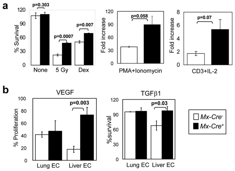
a, Increased proliferation and defective induction of apoptosis in Mx-Cre+ thymocytes. Thymocytes from 8-week old mice (n=3 per genotype) were plated and the indicated mitogens or apoptotic stimuli were applied. Experiment was performed twice with similar results; data from a representative experiment is shown. Survival % was calculated after 18hrs of treatment (5 Gy: γ-irradiation; Dex: 1 μM Dexamethasone). b, Analysis of viability of endothelial cells. MTT assays were performed on Mx-Cre− and Mx-Cre+ lung (n=2) and liver (n=3) ECs. Y axis, normalized response (%) per OD595. Results represent two independent experiments. For a and b, data are mean ± SEM.