Abstract
The initiation of sporulation in Bacillus species is controlled by the phosphorelay signal transduction system. Multiple regulatory elements act on the phosphorelay to modulate the level of protein phosphorylation in response to cellular, environmental, and metabolic signals. In Bacillus anthracis nine possible histidine sensor kinases can positively activate the system, while two response regulator aspartyl phosphate phosphatases of the Rap family negatively impact the pathway by dephosphorylating the Spo0F intermediate response regulator. In this study, we have characterized the B. anthracis members of the Spo0E family of phosphatases that specifically dephosphorylate the Spo0A response regulator of the phosphorelay and master regulator of sporulation. The products of four genes were able to promote the dephosphorylation of Spo0A∼P in vitro. The overexpression of two of these B. anthracis Spo0E-like proteins from a multicopy vector consistently resulted in a sporulation-deficient phenotype. A third gene was found to be not transcribed in vivo. A fourth gene encoded a prematurely truncated protein due to a base pair deletion that nevertheless was subject to translational frameshift repair in an Escherichia coli protein expression system. A fifth Spo0E-like protein has been structurally and functionally characterized as a phosphatase of Spo0A∼P by R. N. Grenha et al. (J. Biol. Chem. 281:37993-38003, 2006). We propose that these proteins may contribute to maintain B. anthracis in the transition phase of growth during an active infection and therefore contribute to the virulence of this organism.
The Bacillus anthracis exotoxins play a central role in the pathogenesis of this organism. The protective antigen, in association with either lethal factor or edema factor, can cause lethality and edema, respectively, to infected hosts (3, 5, 25, 26, 45). Lately it has become evident that the production of these toxins is under control of the pathway regulating sporulation initiation (39). In fact, AtxA, an essential activator of toxin gene transcription, is negatively regulated by the transition phase regulator AbrB, whose transcription is, in turn, regulated by the Spo0A response regulator. Spo0A is the master regulator of sporulation initiation and the final acceptor of activating phosphoryl groups in the phosphorelay signal transduction pathway that regulates the initiation of the developmental process, as it has been well characterized in Bacillus subtilis (13, 37). In this organism, five histidine sensor kinases may provide the phosphoryl group inputs into the system by autophosphorylating on a histidine residue in response to activating signals; this event is followed by transfer of the phosphoryl group to the intermediate response regulator Spo0F (17, 18, 20). From Spo0F the phosphoryl group is transferred to Spo0A through the Spo0B phosphotransferase (7). Once activated by phosphorylation, Spo0A promotes directly or indirectly the transcription of a myriad of genes required for sporulation while it represses genes whose products are not necessary during development (3, 4, 27, 44). Negative inputs into the phosphorelay are provided by aspartyl phosphate phosphatases that specifically dephosphorylate the Spo0F response regulator (Rap proteins) or the Spo0A transcription factor (Spo0E proteins) (31, 32). The Spo0F, Spo0B, and Spo0A proteins of B. subtilis are essentially identical to their counterparts in B. anthracis, indicating that the phosphorelay pathway is functionally identical in controlling sporulation initiation in the two organisms (42).
Recently, we have identified nine genes in the B. anthracis genome whose products may function as sporulation histidine sensor kinases (6). Furthermore, we have characterized two of the six Rap proteins of B. anthracis as aspartyl phosphate phosphatases of Spo0F with a possible role in the pathogenesis of this organism (5). Here we report the characterization of the Spo0E family of proteins and their role in negatively regulating the sporulation pathway of B. anthracis.
MATERIALS AND METHODS
Bacterial strains and growth conditions.
The B. anthracis Sterne strain 34F2 (pXO1+ pXO2−) was used in this study, and transformation was carried out by the electroporation method described by Koehler et al. (19). The B. subtilis JH642 (trpC2 phe-1) strain used in this study was transformed by the method of Anagnostopoulos and Spizizen (1).
Strains were grown in Schaeffer's sporulation medium (SM) (40) with antibiotics at the following concentrations: erythromycin, 5 μg/ml, and lincomycin, 25 μg/ml (MLS resistance); kanamycin, 7.5 μg/ml for B. anthracis and 2 μg/ml for B. subtilis.
Escherichia coli DH5α was used for plasmid construction and propagation. Cells were grown in LB medium supplemented with ampicillin (100 μg/ml) or kanamycin (30 μg/ml). E. coli SCS110 (Stratagene) was used as a source of unmethylated plasmid DNA.
Plasmid construction.
The vectors used in this study were as follows: pHT315 (2), a multicopy shuttle vector replicating in E. coli, B. subtilis, and B. anthracis (copy number in gram positives, ∼15) carrying ampicillin and MLS resistance markers; pJM115 (30), a transcriptional lacZ fusion vector conferring kanamycin resistance and integrating by double crossover at the amyE locus of the B. subtilis chromosome; pTCVlac (38), a transcriptional lacZ fusion vector that replicates in E. coli and B. anthracis and confers resistance to kanamycin.
The BA1877, BA0046, BA2416, and BAYisI coding sequences and their promoter regions (approximately 200 to 300 bp upstream the start site of translation) were amplified by PCR from chromosomal DNA of strain 34F2 using the oligonucleotide pairs shown in Table S1 of the supplemental material. The 5′ oligonucleotide introduced either an EcoRI site or a KpnI site, while the 3′ primer introduced a BamHI site. The fragments obtained were cloned in pHT315 digested with KpnI and BamHI or EcoRI and BamHI.
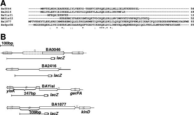
The lacZ transcriptional fusion constructs were generated in pJM115 and pTCVlac as follows. The region upstream of BA0046 (extending 268 bp upstream from the starting codon) was amplified with oligonucleotide primers BA00465′Kpn and BA00463′Bam2, and the fragment was digested with BamHI and cloned in the vectors digested with SmaI and BamHI. The BA1877 promoter (extending 328 bp upstream of the starting codon) was amplified with oligonucleotide primers BA18775′EcoRI and BA18773′Bam2 and cloned in EcoRI-BamHI-digested vectors. The BAYisI fragment (extending 203 bp upstream from the starting codon) obtained with oligonucleotide primers BAYisI5′Eco and BAYisI3′Bam was digested with EcoRI and XmnI (this restriction site naturally occurs within the gene), and the fragment was cloned in the vectors digested with EcoRI and SmaI. The BA2416 promoter region (extending 186 bp upstream of the starting codon) was generated by EcoRI digestion of plasmid pHT315-2416 (the EcoRI site at the 5′ end originated from the vector multiple cloning site, while the EcoRI at the 3′ end was naturally occurring within the BA2416 coding sequence), and it was cloned in the EcoRI-digested vectors. These fragments are schematically represented in Fig. 1B.
FIG. 1.
The B. anthracis Spo0E-like family. A. Amino acid sequence alignment of the B. anthracis Spo0E-like proteins. Proteins were aligned with the ClustalW program. Asterisks indicate identical residues in all five sequences; colons denote conserved substitutions. The four B. anthracis (Ba) proteins analyzed in this study are aligned against the B. subtilis Spo0E protein (35). The proteins resulting from the frameshift mutation in the BAYisI gene are indicated with the numbers 1 and 2. B. Schematic representation of the chromosomal regions containing the B. anthracis spo0E-like genes. Open reading frames and their orientations are shown by arrows. The regions forming stem-loop structures are indicated by the Ω-like symbol. The lines represent the fragments cloned in the pJM115 and pTCVlac vectors generating lacZ transcriptional fusion plasmids. The dotted bar represents the 51 codons possibly extending the BA0046 gene at the 5′ end.
To generate the BA0046-lacZ fusion plasmid for isotopic integration, a KpnI-NsiI fragment of approximately 4 kb containing the BA0046 putative promoter region and the entire lacZ gene was recovered from plasmid pJM115-0046 and cloned in pORI-Cm (6) digested with KpnI and PstI. The resulting plasmid was introduced into strain 34F2 by electroporation. The plasmid was forced to integrate into the chromosome at the BA0046 locus by growing the cells at nonpermissive temperature (37°C). The correct integration by single crossover was checked by diagnostic PCR.
The BAYisI translational fusions to the lacZ gene of E. coli were first constructed in plasmid pJM115 by cloning the EcoRI-SalI-digested fragments generated by PCR amplification using oligonucleotide primers BAYisI5′Eco and BaYisI3′Sal1 or BAYisI3′Sal2 in the EcoRI-SalI-digested vector. Because of the presence of two SalI sites in pTCVlac, the fragments were then recovered as EcoRI-SacI (the latter naturally occurring in the lacZ gene) and recloned in EcoRI-SacI-digested pTCVlac.
A markerless deletion of BA1877 was obtained by the method of Janes and Stibits (15) using the temperature-sensitive plasmid pORI-Cm (6) modified to contain the I-SceI homing endonuclease recognition site. Two regions flanking the BA1877 gene were amplified by PCR using oligonucleotides BA18775′SacII-BA18773′SalI and BA18775′SalI-BA18773′Xho and sequentially cloned in the pORI-Cm-SceI vector. The plasmid was introduced by electroporation in strain 34F2, and the protocol of Janes and Stibits (15) was followed to generate the markerless gene deletion. Diagnostic PCR was carried out to ensure that the entire coding sequence had been correctly deleted. Attempts to generate a similar markerless or a marker replacement construct for the deletion of the BA2416 gene consistently resulted in the loss of antibiotic resistance expression in B. anthracis, thus hampering our efforts to obtain a deletion mutant. Fidelity of all PCR amplifications and fragment orientation were checked by DNA sequence analysis, and this confirmed the presence of the frameshift mutation in the BAYisI gene of strain 34F2.
Protein expression and purification.
The coding sequence of each B. anthracis Spo0E-like protein was amplified by PCR using oligonucleotide primers that introduced an NdeI or NheI site at the 5′ end and a BamHI site at the 3′ end (see Table S1 in the supplemental material). After digestion with NdeI and BamHI or NheI and BamHI, the fragments were cloned in a similarly digested pET28 vector (Novagen), generating a fusion to six-histidine codons at the 5′ end of the genes. The plasmids obtained were transformed in E. coli BL21(DE3), and protein expression was obtained by growing the cells in LB broth in the presence of kanamycin. Expression was induced with isopropyl-β-d-thiogalactopyranoside at 1 mM and an optical density at 600 nm of 0.7, and growth was continued for approximately 3 h. Due to solubility problems, the proteins were purified under denaturing conditions using a buffer containing 50 mM Tris-HCl pH 8.0, 100 mM KCl, 1 mM β-mercaptoethanol, 5 mM imidazole, and 7 M urea. After binding to a Ni-nitrilotriacetic acid-agarose column (QIAGEN), proteins were eluted using the same buffer with 50 mM imidazole. The eluate was dialyzed in buffer containing a sequentially lower molarity of urea (6, 5, 4, 3, and 1.5 M); the proteins were then concentrated and stored at −20°C in a buffer containing 50 mM Tris-HCl pH 8.0, 100 mM KCl, 1 mM dithiothreitol, 1.5 M urea, and 20% glycerol. The presence of urea was necessary to maintain the proteins in solution.
In vitro assays.
Phosphorylation and dephosphorylation reactions were carried out essentially as previously described (5, 33). The reactions were initiated by the addition of [γ-32P]ATP (specific activity, 6,000 Ci/mmol), and aliquots were taken at the time points indicated in the figures.
Sporulation assays.
Strains were grown in SM liquid medium at 37°C for the times indicated in the tables in the presence of erythromycin and lincomycin. Serial dilutions were plated in duplicate before and after treatment with chloroform (10% [vol/vol]). The percentage of sporulation was calculated as the ratio between viable counts (before CHCl3 treatment) and spore counts (after CHCl3 treatment).
β-Galactosidase assay.
Cultures for transcriptional and translational assays were grown in SM liquid medium. Kanamycin was added for the B. anthracis cultures carrying pTCVlac derivatives. Duplicate samples were collected at hourly intervals. β-Galactosidase activity determinations in B. subtilis samples were carried out essentially as described elsewhere (10, 24). The B. anthracis samples were treated with lysozyme for 1 h at 37°C, and Triton was used at a 5% final concentration. The data shown are representative of at least two independent experiments.
RESULTS
Identification of B. anthracis Spo0E-like proteins.
The Spo0E protein of B. subtilis (35) was used as query in a BLAST search (tblastn) against the first available B. anthracis genome (A2012). Four genes were identified as encoding putative Spo0E-like proteins based on two main criteria: (i) protein size ranging between 40 and 90 amino acids (aa), and (ii) the presence of the Spo0E signature sequence, SQELD (31). Two genes were annotated as Bant0046 and Bant1877. A third gene was not identified in strain A2012 but was annotated as BA2416 when the genome of the Ames strain became available. The fourth gene is still not annotated in any of the available B. anthracis genomes because of a base pair deletion that results in a frameshift of the corresponding open reading frame. If the frame were to be corrected, the gene product would be most similar to B. subtilis YisI (33% identity); we called this gene and its product BAYisI. A summary of the gene names used in this study and in the most commonly used B. anthracis strains is reported in Table 1, and their amino acid alignment is shown in Fig. 1A.
TABLE 1.
Nomenclature of the Spo0E family of proteins in B. anthracis strains
| Protein name in:
| |||
|---|---|---|---|
| This worka | Strain A2012b | Strain Sterneb | Strain Amesb |
| NZ_AAAC02000001 | NC_005945 | NC_003997 | |
| BA0046 | Bant0047 | BAS4809 | BA5174 |
| BA2416 | NAc | NA | BA2416 |
| BA1877 | Bant1968 | BAS1251 | NA |
| BAYisI | NAd | NAd | NAd |
| 1617906-1617731 | 1121865-1121690 | 1121973-1121798 | |
As of 05/02/2003, when the first BLAST search was carried out.
As of 11/15/06.
NA, not annotated.
The frameshifted open reading frame is not annotated, but it is located within the nucleotides indicated.
The BA1877 and BAYisI genes appear to be monocistronic because they are preceded by a gene divergently transcribed and they are followed by a strong stem-loop structure potentially acting as a transcriptional terminator. The BA2416 gene is followed by a strong potential terminator, but its initial codon is located 157 bp downstream from the stop codon of a hypothetical protein with no similarity with proteins of known function. The BA1877, BAYisI, and BA2416 genes are preceded by plausible ribosome binding sites. The BA0046 locus is less defined, as it could have a 51-codon extension at the amino-terminal end. Neither start codon seems to be preceded by a plausible ribosome binding site, raising the possibility that, if transcribed, this gene may not be translated. Between the two possible starting codons, there is a sequence that can form a strong stem-loop structure with an energy of −16 Kcal/mol; this structure may act as a terminator of transcription and prevent readthrough from an upstream promoter. No possible stem-loop structure was detected at the 3′ end of the gene, which is 55 bp upstream from the start site of the downstream gene. A schematic representation of the chromosomal regions carrying the Spo0E-encoding genes is given in Fig. 1B.
A fifth gene annotated as BA1655 in the genome of the Ames strain (NC_003997) encoding a Spo0E-like protein with a divergent signature motif (SRDLD) has been functionally and structurally characterized by Grenha et al. (11).
In vivo analysis of B. anthracis Spo0E-like proteins.
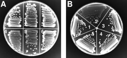
In order to determine whether the products of the Spo0E-like protein-encoding genes identified in B. anthracis could affect the pathway of sporulation initiation, the four spo0E-like loci were PCR amplified from B. anthracis Sterne strain 34F2 using the oligonucleotide primers listed in Table S1 (see the supplemental material) and cloned in the multicopy shuttle vector pHT315. Overexpression of a negative regulator of sporulation from a multicopy vector generally results in inhibition of sporulation, as previously shown by studies on Rap and Spo0E phosphatases of B. subtilis (16, 31). The resulting plasmids were transformed into B. subtilis wild-type strain JH642 and, after passage through the dam E. coli strain SCS110, electroporated into B. anthracis 34F2. The phenotype of the transformants obtained was analyzed on Schaeffer's sporulation agar medium and compared to the parental strain carrying the vector alone. As shown in Fig. 2A, expression of BA1877 strongly inhibited sporulation of B. anthracis and some inhibition was also observed with the expression of BA2416, while no effect was seen in the strains expressing BA0046 and BAYisI. When the same plasmids were transformed in B. subtilis wild-type strain JH642, sporulation inhibition was observed only by the overexpression of BA1877 protein (Fig. 2B). A quantitative analysis of the sporulation efficiency of the strains constructed was also carried out in Schaeffer's liquid medium (Table 2). The results indicated that BA1877 expression is the most drastic in inhibiting sporulation, even in liquid cultures in both organisms, while none of the other Spo0E-like proteins significantly affected sporulation efficiency in liquid. The inhibition of sporulation by overexpression of BA1877 was comparable to that observed with the YisI protein of B. subtilis (31). BA2416, which visibly affected sporulation under solid growth conditions, did not significantly affect the efficiency of spore formation in liquid culture, suggesting that perhaps different transcriptional regulatory mechanisms exist between the two modes of cell growth.
FIG. 2.
Sporulation phenotypes of B. anthracis (A) and B. subtilis (B) strains expressing the Spo0E-like proteins from the multicopy plasmid pHT315. Streaks in panel A are strains carrying the following plasmids: sector 1, pHT315; sector 2, pHT315-YisI; sector 3, pHT315-0046; sector 4, pHT315-1877; sector 5, pHT315-2416; sector 6, pHT315. Strains in panel B carry the following plasmids: sector 1, pHT315; sector 2, pHT315-1877; sector 3, pHT315-2416; sector 4, pHT315-YisI; sector 5, pHT315-0046. Sporulation on agar plates results in a level of opacity of the streak that is associated with the efficiency of spore formation. The more opaque the streak is, the whiter it appears in a black-and-white picture. Sporulation-deficient, and thus transparent, streaks, as in sector 5 in panel A and sector 2 in panel B, have a gray appearance. Strains were streaked on SM plates and incubated at 37°C for 48 h.
TABLE 2.
Sporulation efficiency of B. subtilis JH642 and B. anthracis 34F2 strains carrying spo0E-like genes on the multicopy plasmid pHT315
| Species and plasmid | Viable count | Spore count | % of sporesa |
|---|---|---|---|
| B. subtilis | |||
| pHT315 | 3.4 × 10⁸ | 1.3 × 10⁸ | 38.2 (±2.0) |
| pHT315-1877 | 2.4 × 10⁸ | 1.7 × 10³ | 0.0007 (±0.001) |
| pHT315-0046 | 4.3 × 10⁸ | 1.4 × 10⁸ | 32.5 (±2.8) |
| pHT315-2416 | 3.3 × 10⁸ | 1.3 × 10⁸ | 39.4 (±0.9) |
| pHT315-YisI | 3.7 × 10⁸ | 1.4 × 10⁸ | 37.8 (±5.8) |
| B. anthracis | |||
| pHT315 | 1.8 × 10⁸ | 5.0 × 10⁷ | 27.7 (±4.6) |
| pHT315-1877 | 8.5 × 10⁷ | 3.6 × 10⁴ | 0.04 (±1.5) |
| pHT315-0046 | 2.5 × 10⁸ | 7.2 × 10⁷ | 28.8 (±2.1) |
| pHT315-2416 | 2.9 × 10⁸ | 6.8 × 10⁷ | 23.4 (±3.6) |
| pHT315-YisI | 2.3 × 10⁸ | 6.6 × 10⁷ | 28.6 (±6.2) |
Strains were grown for 48 h at 37°C in Schaeffer's sporulation medium. The results shown are the averages of two independent experiments. Deviations from the mean are shown in parentheses.
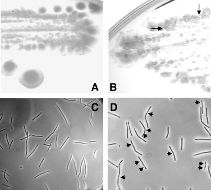
Deletion of genes encoding negative regulators of sporulation and Spo0E-like proteins, in particular, generally results in subtle phenotypes that are not necessarily quantifiable (31). Nevertheless, we generated a markerless deletion of the BA1877 gene in strain 34F2 and analyzed the phenotype of the resultant mutant strain on sporulation agar plates and liquid medium. On a solid surface, the B. anthracis 34F2ΔBA1877 strain did not differ from the parental 34F2 strain after 24 h of growth at 37°C. However, after 48 h, structures resembling papillae were detectable in the BA1877 deletion strain but not in the parental strain (Fig. 3A and B). The formation of papillae containing a sporulation-deficient revertant of hypersporulating B. subtilis strains was previously described as the phenotype of strains containing a deletion of the aspartyl phosphate phosphatase genes rapA and spo0E or expressing Spo0A hyperactive mutants (sof) (34, 36, 41). Microscopically, we could detect phase-bright spores in liquid cultures of the deletion strain 34F2ΔBA1877 after 9 h of growth, while 11 h were necessary for the spores to become apparent in the parental culture (Fig. 3C and D). Due to the persistence of long chains of cells even after spore formation at early growth times, an accurate quantitation of the early sporulation phenomenon could not be obtained. By the time the chains of cells broke into single cells, the efficiency of sporulation of the parental and mutant strains was essentially undistinguishable.
FIG. 3.
Phenotype of the B. anthracis BA1877 deletion strain. The parental strain 34F2 (A and C) and the 34F2ΔBA1877 deletion strain (B and D) were grown on SM agar plates for 48 h (A and B) or in SM liquid medium for 9 h (C and D) at 37°C. (A and B) Plates were photographed with an AlphaInnotech imaging system. Two representative papillae are indicated by the arrows. (C and D) Samples were analyzed with a phase-contrast microscope. The images shown are representative of multiple images taken for both strains. Phase-bright spores (some indicated by the arrows) were found at a higher frequency (at least 20-fold) in fields of 34F2ΔBA1877 cells than in fields of 34F2 cells.
These observations in solid and liquid media suggested that the deletion of the BA1877 gene causes the sporulation process to initiate earlier than in the parental strain. This is consistent with the BA1877 protein being a negative regulator of sporulation.
In vitro analysis of B. anthracis Spo0E-like proteins.
In order to determine whether the products of the four genes were all capable of dephosphorylating the Spo0A response regulator, in vitro assays were carried out using purified components. The four coding sequences of the B. anthracis Spo0E-like genes were PCR amplified from strain 34F2 and cloned in the E. coli expression vector pET28 (Novagen) in order to create a fusion to six-histidine codons at the N-terminal end. All four proteins formed inclusion bodies and therefore were purified using nickel-nitrilotriacetic acid-agarose columns (QIAGEN) under denaturing conditions. After being renatured, the proteins were assayed in a phosphorelay dephosphorylation reaction using the KinA, Spo0F, Spo0B, and Spo0A proteins of B. subtilis. Note that, despite the presence of a frameshift mutation, the E. coli strain carrying the pET28-BaYisI plasmid expressed a protein whose molecular mass was approximately 9,200 Da as determined by denaturing gel analysis and matrix-assisted laser desorption ionization-time of flight mass spectrometry (see Fig. S1 in the supplemental material), in good agreement with the theoretical molecular mass deduced from the amino acid sequence if the frame were corrected for the base pair deletion. This indicated that the frameshift mutation had been translationally repaired in E. coli and a full-length protein of the expected size was produced (45), despite the persistence of the mutation in the nucleotide sequence of the clone.
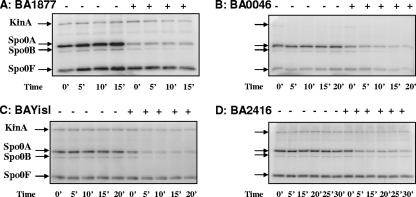
The results of the in vitro assay, shown in Fig. 4, indicated that upon addition of the Spo0E proteins, the phosphorylation level of each component of the pathway decreased, suggesting that all four proteins were active. Full dephosphorylation of the system is rarely achieved in in vitro assays, possibly because conformational differences in the purified proteins may result in forms more resistant to dephosphorylation than others. Nevertheless, all four proteins were capable of dephosphorylating the phosphorelay, and the reaction rates observed were relatively fast as dephosphorylation occurred already within the time frame of the zero time value. Since in an assay mixture containing only KinA and Spo0F no dephosphorylation was observed upon addition of the Spo0E-like proteins (see Fig. S2 in the supplemental material), by extrapolation from the genetic and biochemical evidence so far accumulated on the Spo0E family of proteins, we concluded that the four B. anthracis members of the Spo0E family all acted by dephosphorylating the Spo0A protein. The activity of BA1877 was approximately two- to threefold stronger than the activity of the other three proteins (see Fig. S3 in the supplemental material), but because of the purification of the proteins under denaturing conditions we did not further analyze the kinetics of the reactions. Nevertheless, this result is in agreement with the in vivo data, which indicated that BA1877 was more active than BA0046, BA2416, and BAYisI.
FIG. 4.
Phosphorelay dephosphorylation assay. Purified Spo0E-like proteins of B. anthracis were tested in vitro for their ability to dephosphorylate the B. subtilis phosphorelay. KinA (0.2 μM), Spo0F (2.5 μM), Spo0B (0.2 μM), and Spo0A (2.5 μM) were incubated in the presence of [γ-32P]ATP for 30 min. The reaction mixture was then divided in two aliquots: one was incubated with the corresponding Spo0E protein buffer (−), and the other was incubated with buffer plus the Spo0E protein (+). Aliquots were withdrawn at the indicated times. BA1877 (A), BA0046 (B), BAYisI (C), and BA2416 (D) proteins purified from E. coli were used at a 5 μM final concentration. The position of the phosphorylated proteins is indicated by the arrows.
Transcriptional and translational analyses.
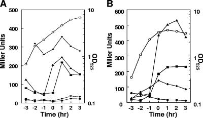
In order to determine whether the different effects, or lack thereof, observed in vivo were due to differences in the transcriptional level of each gene, a fragment presumably containing the promoter regions of the four B. anthracis spo0E-like genes was cloned in front of the promoterless E. coli lacZ gene in the transcriptional fusion vectors pJM115 and pTCVlac (Fig. 1B). The pJM115 derivatives were transformed in Bacillus subtilis JH642, selecting for kanamycin resistance, and integrated by double crossover at the amyE region. The pTCV lac derivatives were introduced by electroporation into strain 34F2, selecting for kanamycin resistance. Transcriptional analysis was carried out by means of β-galactosidase assays in Schaeffer's sporulation medium. The results shown in Fig. 5A indicated that in B. anthracis the BA2416 gene was transcribed at the highest levels, compared to the other genes, and throughout vegetative growth and sporulation, while BA1877 and BAYisI were induced 1 hour before or around the transition from vegetative growth to stationary phase (T0), respectively.
FIG. 5.
Transcriptional analysis of B. anthracis spo0E-like genes in B. anthracis (A) and B. subtilis (B). Cultures were grown in SM liquid medium. Symbols: •, BA0046; ▪, BAYisI; ▴, BA1877; ⧫, BA2416; ▾, pTCVlac vector. The growth curve of strains carrying the BA0046 construct is shown as representative of all growth curves (○).
The promoter of BA0046 was inactive, at least under the growth conditions tested, as it never exceeded the level of activity detected in the control strain carrying a promoterless vector, pTCVlac. In order to test the possibility that BA0046 could be part of an operon and therefore its transcription could depend on a promoter located upstream the region cloned in the pTCVlac vector, we subcloned the fragment and the lacZ gene from the transcriptional fusion vector in the temperature-sensitive replicative vector pORI-Cm (6). The resulting plasmid was electroporated into B. anthracis strain 34F2 and integrated isotopically via a single crossover event by growing the cells at a nonpermissive temperature (37°C) (see Materials and Methods). Analysis of β-galactosidase production did not detect any transcriptional activity reading through from upstream sequences, confirming that, at least under the assay conditions used in this study, the BA0046 gene was not transcribed (data not shown).
In B. subtilis (Fig. 5B) the BA1877 promoter was the most active and, as in B. anthracis, the transcription was strongly induced approximately 1 hour before the transition phase. The promoter of BAYisI was also induced at approximately the same time but at a lower level, while the BA2416 promoter was expressed throughout growth, with the highest level reached at T0. BA0046 was transcribed at a low level in vegetative phase but by transition phase its transcription was essentially at the background level, suggesting that a vegetative promoter active only in B. subtilis was present in the fragment used for this analysis. The results helped to explain why, despite the fact that all four Spo0E-like proteins were active in vitro, only BA1877 suppressed sporulation in vivo in liquid cultures. BA0046 did not appear to be transcribed, at least under the assay conditions used in this study, consistent with a lack of phenotype; BA2416 was weakly active in vitro, and it may also be weakly active in vivo since its level of expression was higher than BA1877 in B. anthracis but its inhibition of sporulation in vivo was less dramatic. In B. subtilis, BA2416 did not cause a sporulation-deficient phenotype, consistent with a lower level of expression compared to the BA1877 gene.
BAYisI was transcriptionally regulated in a manner similar to BA1877; however, it was inactive in vivo, presumably because of a frameshift mutation in the sequence that results in a truncated protein. Since significant translational frameshift repair was found to occur when BAYisI was expressed in E. coli, allowing us to purify the full-length protein, we analyzed the possibility of a similar repair occurring in B. anthracis by means of a translational lacZ fusion constructed as described in Materials and Methods. The results shown in Fig. S4 of the supplemental material suggested that some translational frameshift repair occurred even in B. anthracis but it did not result in a sufficient level of protein to generate a visible sporulation phenotype. Accordingly, we were not able to detect the full-length BAYisI protein when overexpressed from the pHT315 vector with a His tag at the carboxy-terminal end that provided the epitope for Western blot analysis (data not shown).
DISCUSSION
In this study we have characterized the Spo0E family of phosphatases in the anthrax-causing organism B. anthracis. In in vivo studies we showed that of the four genes encoding Spo0E-like proteins, two, BA1877 and BA2416, were expressed to produce proteins active in inhibiting the sporulation process; BA0046 was not transcribed under the assay conditions used in this study, but it could potentially produce a Spo0A-like targeting protein. A fourth gene, BAYisI, was found to contain a frameshift mutation and therefore did not produce an active protein. Nevertheless, in vitro studies showed that all four proteins were able to dephosphorylate the Spo0A response regulator of the sporulation phosphorelay. Extensive translational frameshift repair was observed in E. coli for the BAYisI gene, which allowed us to purify and test a full-length protein in vitro. Although the differential level of activity observed among the four proteins could be a result of the purification procedures, we cannot exclude that in vivo the proteins may actually have different affinities for Spo0A∼P.
The B. anthracis members of the Spo0E family of phosphatases were identified based on the sizes of the proteins and the presence of the Spo0E signature motif, SQELD, which is generally preceded by a hydrophobic residue 2 amino acids upstream of the serine residue and followed by two hydrophobic residues located 1 amino acid downstream of the aspartate residue (31).
A fifth gene (BA1655) encoding a Spo0E-like protein active in dephosphorylating Spo0A∼P in vitro and preventing sporulation when overexpressed from the pHT315 multicopy vector in vivo is also present on the chromosome of B. anthracis, and its characterization has been reported by Grenha et al. (11). BA1655 contains a slightly divergent signature motif (SRDLD), indicating that variations may exist within the Spo0E family of proteins without affecting the specificity for target recognition.
Pair score analysis indicated that the B. anthracis Spo0E proteins characterized in this study share between 21% (BA1877) and 32% (BA0046) identical residues with the B. subtilis Spo0E protein. Among each other, the B. anthracis proteins share between 25 and 51% identical residues, with the highest score being between BA0046 and BA2416, clearly indicative of a relatively recent gene duplication event. Noticeable in the alignment in Fig. 1A is the length of BA1877 (93 aa), which resembles the length of the B. subtilis Spo0E (85 aa) whose carboxy-terminal 25 amino acids are known to be inhibitory to its phosphatase activity (29). Whether a similar C-terminal inhibitory effect exists on BA1877 remains to be determined. Interestingly, the gene immediately downstream of the BA1877 gene is annotated as a homologue of the B. subtilis kinD sporulation histidine kinase (BA1878 in strain A2012 and BAS1351 in strain Sterne). This is reminiscent of the chromosomal localization of the B. subtilis spo0E gene immediately upstream of kinD. BAS1351 was indeed inferred to have a role in B. anthracis sporulation for its ability to prevent sporulation in wild-type B. subtilis when expressed from a multicopy plasmid, a phenotype due to the ability of the kinase to act as a phosphatase when overproduced in the absence of activating signals (6). The other three proteins (BA0046 [56 aa], BA2416 [49 aa], and BAYisI [potentially 59 aa]) resemble in size the YisI and YnzD proteins of B. subtilis (56 and 57 aa, respectively), with identities ranging between 28 and 37%. The contention that the BAYisI gene is the homologue of B. subtilis yisI is supported by the observation that the two genes are located in the same region of the chromosome in both organisms and they are both followed by the gerP germination operon, while the unknown yisK gene is divergently transcribed upstream of them (Fig. 1B). The chromosomal location of BA1655, BA0046, and BA2416 is not conserved with the ynzD gene of B. subtilis, indicating that they are unique to the B. anthracis species.
When expressed from a multicopy vector, the most active protein at dephosphorylation in vitro, BA1877, was also the most active in vivo in inhibiting sporulation in both B. anthracis and B. subtilis. Although expressed at the highest level in B. anthracis, BA2416 was only moderately active in inhibiting sporulation in B. anthracis and inactive in B. subtilis. The results are consistent with a weak activity in vitro of the purified protein. BA0046 and BAYisI did not affect sporulation in vivo, consistent with the absence of gene transcription and the frameshifted open reading frame, respectively. Consistent with the inhibitory effect observed upon overexpression of BA1877, the chromosomal deletion of its encoding gene resulted in an early sporulation phenotype that readily segregated sporulation-deficient revertants visible in the form of papillae when cells were grown in solid medium. The formation of Spo−-containing papillae was previously described as a phenotype of deletions in the rapA and spo0E genes of B. subtilis which, like BA1877, encode negative regulators of sporulation, and in sof mutants expressing hyperactive Spo0A mutant proteins (34, 36, 41). Thus, in B. anthracis as in B. subtilis, the timing of sporulation initiation must be precisely regulated, and any mutation that may affect it, such as the deletion of the Spo0E-like-encoding gene BA1877, results in a deleterious effect that the cell tries to overcome by reverting into vegetative growth.
The initiation of sporulation is a critical phase in the life cycle of sporulating bacteria. In bacilli, a complex signal transduction pathway, called phosphorelay, works to integrate a myriad of signals that the cell receives from the environment as well as its own metabolism to ensure that the decision to sporulate or to remain in vegetative growth is appropriate for survival of the bacterium. Aspartyl phosphate phosphatases of the Rap or Spo0E families modulate the phosphorylation level of the phosphorelay to ensure that initiation of sporulation does not occur at inappropriate times. Each member of the Rap and Spo0E family of phosphatases of B. subtilis has been found to be transcriptionally regulated by physiological conditions antagonistic to sporulation, such as growth or competence to DNA transformation (28, 31, 34). Typical among the members of the Rap and Spo0E families of proteins are a variety of transcription patterns indicative of differential regulatory controls. This seemed to be the case for the Spo0E proteins of B. anthracis, although one gene (BA0046) was not transcribed, at least under the assay conditions used in this study. A certain correlation was observed in transcriptional patterns between B. anthracis and B. subtilis, although the lower level of BA2416 observed in B. subtilis compared to B. anthracis and with respect to the transcription level of BA1877 suggests that different transcriptional controls exist in these two organisms. Further studies will be necessary in order to unravel these transcriptional control systems and establish whether they have any relevance to the pathogenesis of B. anthracis.
The transition phase, defined as the growth phase between vegetative growth and stationary phase, is a critical time in a bacterium life cycle. For a number of spore-forming bacteria, the transition phase is characterized by the production of secondary metabolites, virulence factors, and toxins. The key regulator of this phase is the Spo0A response regulator. One of the genes repressed by Spo0A at the beginning of the transition phase is abrB, whose gene product generally acts as a preventer of stationary-phase gene product expression (43, 44). Repression of abrB by Spo0A∼P is required for production of peptide antibiotics in B. subtilis and Bacillus brevis and proteases in B. subtilis (9, 22, 23). Directly or indirectly, Spo0A is required for crystal toxin production in Bacillus thuringiensis and Bacillus sphaericus, the enterotoxin of Clostridium perfringens, or the production of solvent in Clostridium acetobutylicum (8, 12, 14, 21). Recently, the role of Spo0A in repressing abrB transcription was found to be a requirement for full expression of toxin genes in B. anthracis (39). Thus, it has become even more clear how important it is for cell survival and pathogenicity to precisely regulate the level of Spo0A∼P so that secondary metabolites and toxins are produced only at the appropriate time. In B. anthracis, as in B. subtilis, the Spo0E-like proteins are probably the last checkpoint on the phosphorelay to ensure the appropriate timing of toxin production or sporulation. Furthermore, during an infection, the Spo0E-like proteins may contribute to maintain the level of Spo0A∼P such that abrB is repressed, and thus toxins are produced, but sporulation does not progress further. We have recently proposed that such a scenario may occur in B. anthracis cells within an infected body by hypothesizing a mechanism for pathogenesis-induced prevention of sporulation (6). Formation of a spore is a dead-end pathway that does not contribute to infection, and macrophages have the ability to phagocytose and destroy germinating spores while toxin-producing encapsulated bacteria escape this defense mechanism (26). Weak or inhibited sporulation histidine sensor kinases together with a pXO1 plasmid-encoded Rap phosphatase have been shown to prevent spore formation and affect the level of AbrB in the cell (5, 6). The Spo0E-like proteins identified in this study may provide an additional level of control to ensure that spores are not formed while an infection is in full development. The differential levels of expression observed in our transcriptional studies in batch cultures are suggestive of differential regulatory mechanisms that can modulate the timing and level of gene expression under different growth conditions and perhaps during an active infection.
Supplementary Material
Acknowledgments
This work was supported in part by grants GM055594 from the National Institute of General Medical Sciences and AI055860 from the National Institute of Allergy and Infectious Diseases, NIH. The Stein Beneficial Trust supported in part the oligonucleotide synthesis and DNA sequencing.
This is manuscript number 18152-MEM from The Scripps Research Institute.
Footnotes
Published ahead of print on 26 January 2007.
Supplemental material for this article may be found at http://jb.asm.org/.
REFERENCES
- 1.Anagnostopoulos, C., and J. Spizizen. 1961. Requirements for transformation in Bacillus subtilis. J. Bacteriol. 81:741-746. [DOI] [PMC free article] [PubMed] [Google Scholar]
- 2.Arantes, O., and D. Lereclus. 1991. Construction of cloning vectors for Bacillus thuringiensis. Gene 108:115-119. [DOI] [PubMed] [Google Scholar]
- 3.Baldus, J. M., C. M. Buckner, and C. P. Moran, Jr. 1995. Evidence that the transcriptional activator Spo0A interacts with two sigma factors in Bacillus subtilis. Mol. Microbiol. 17:281-290. [DOI] [PubMed] [Google Scholar]
- 4.Bird, T. H., J. K. Grimsley, J. A. Hoch, and G. B. Spiegelman. 1993. Phosphorylation of Spo0A activates its stimulation of in vitro transcription from the Bacillus subtilis spoIIG operon. Mol. Microbiol. 9:741-749. [DOI] [PubMed] [Google Scholar]
- 5.Bongiorni, C., R. Stoessel, D. Shoemaker, and M. Perego. 2006. Rap phosphatase of virulence plasmid pXO1 inhibits Bacillus anthracis sporulation. J. Bacteriol. 188:487-498. [DOI] [PMC free article] [PubMed] [Google Scholar]
- 6.Brunsing, R. L., C. La Clair, S. Tang, C. Chiang, L. E. Hancock, M. Perego, and J. A. Hoch. 2005. Characterization of sporulation histidine kinases of Bacillus anthracis. J. Bacteriol. 187:6972-6981. [DOI] [PMC free article] [PubMed] [Google Scholar]
- 7.Burbulys, D., K. A. Trach, and J. A. Hoch. 1991. The initiation of sporulation in Bacillus subtilis is controlled by a multicomponent phosphorelay. Cell 64:545-552. [DOI] [PubMed] [Google Scholar]
- 8.El-Bendary, M., F. G. Priest, J. F. Charles, and W. J. Mitchell. 2005. Crystal protein synthesis is dependent on early sporulation gene expression in Bacillus sphaericus. FEMS Microbiol. Lett. 252:51-56. [DOI] [PubMed] [Google Scholar]
- 9.Ferrari, E., D. J. Henner, M. Perego, and J. A. Hoch. 1988. Transcription of Bacillus subtilis subtilisin and expression of subtilisin in sporulation mutants. J. Bacteriol. 170:289-295. [DOI] [PMC free article] [PubMed] [Google Scholar]
- 10.Ferrari, E., S. M. H. Howard, and J. A. Hoch. 1985. Effect of sporulation mutations on subtilisin expression, assayed using a subtilisin-β-galactosidase gene fusion, p. 180-184. In J. A. Hoch and P. Setlow (ed.), Molecular biology of microbial differentiation. American Society for Microbiology, Washington, D.C.
- 11.Grenha, R., N. Rzechorzek, J. A. Brannigan, R. N. de Jong, E. Ab, T. Diercks, A. Bouvin, V. Truffault, J. C. Ladds, M. J. Fogg, M. Perego, R. Kaptein, K. S. Wilson, G. E. Folkers, and A. J. Wilkinson. 2006. Structural characterization of Spo0E-like protein aspartic acid phosphatases that regulate sporulation in bacilli. J. Biol. Chem. 281:37993-38003. [DOI] [PubMed] [Google Scholar]
- 12.Harris, L. M., N. E. Welker, and E. T. Papoutsakis. 2002. Northern, morphological, and fermentation analysis of spo0A inactivation and overexpression in Clostridium acetobutylicum ATCC 824. J. Bacteriol. 184:3586-3597. [DOI] [PMC free article] [PubMed] [Google Scholar]
- 13.Hoch, J. A. 2000. Two-component and phosphorelay signal transduction. Curr. Opin. Microbiol. 3:165-170. [DOI] [PubMed] [Google Scholar]
- 14.Huang, I. H., M. Waters, R. R. Grau, and M. R. Sarker. 2004. Disruption of the gene (spo0A) encoding sporulation transcription factor blocks endospore formation and enterotoxin production in enterotoxigenic Clostridium perfringens type A. FEMS Microbiol. Lett. 233:233-240. [DOI] [PubMed] [Google Scholar]
- 15.Janes, B. K., and S. Stibitz. 2006. Routine markerless gene replacement in Bacillus anthracis. Infect. Immun. 74:1949-1953. [DOI] [PMC free article] [PubMed] [Google Scholar]
- 16.Jiang, M., R. Grau, and M. Perego. 2000. Differential processing of propeptide inhibitors of Rap phosphatases in Bacillus subtilis. J. Bacteriol. 182:303-310. [DOI] [PMC free article] [PubMed] [Google Scholar]
- 17.Jiang, M., W. Shao, M. Perego, and J. A. Hoch. 2000. Multiple histidine kinases regulate entry into stationary phase and sporulation in Bacillus subtilis. Mol. Microbiol. 38:535-542. [DOI] [PubMed] [Google Scholar]
- 18.Kobayashi, K., K. Shoji, T. Shimizu, K. Nakano, T. Sato, and Y. Kobayaski. 1995. Analysis of a suppressor mutation ssb (kinC) of sur0B20 (spo0A) mutation in Bacillus subtilis reveals that kinC encodes a histidine protein kinase. J. Bacteriol. 177:176-182. [DOI] [PMC free article] [PubMed] [Google Scholar]
- 19.Koehler, T. M., Z. Dai, and M. Kaufman-Yarbray. 1994. Regulation of the Bacillus anthracis protective antigen gene: CO2 and a trans-acting element activate transcription from one of two promoters. J. Bacteriol. 176:586-595. [DOI] [PMC free article] [PubMed] [Google Scholar]
- 20.LeDeaux, J. R., and A. D. Grossman. 1995. Isolation and characterization of kinC, a gene that encodes a sensor kinase homologous to the sporulation sensor kinases KinA and KinB in Bacillus subtilis. J. Bacteriol. 177:166-175. [DOI] [PMC free article] [PubMed] [Google Scholar]
- 21.Lereclus, D., H. Agaisse, C. Grandvalet, S. Salamitou, and M. Gominet. 2000. Regulation of toxin and virulence gene transcription in Bacillus thuringiensis. Int. J. Med. Microbiol. 290:54. [DOI] [PubMed] [Google Scholar]
- 22.Marahiel, M. A., M. M. Nakano, and P. Zuber. 1993. Regulation of peptide antibiotic production in Bacillus. Mol. Microbiol. 7:631-636. [DOI] [PubMed] [Google Scholar]
- 23.Marahiel, M. A., P. Zuber, G. Czekay, and R. Losick. 1987. Identification of the promoter for a peptide antibiotic biosynthesis gene from Bacillus brevis and its regulation in Bacillus subtilis. J. Bacteriol. 169:2215-2222. [DOI] [PMC free article] [PubMed] [Google Scholar]
- 24.Miller, J. H. 1972. Experiments in molecular genetics, p. 352-355. Cold Spring Harbor Laboratory, Cold Spring Harbor, N.Y.
- 25.Moayeri, M., and S. H. Leppla. 2004. The roles of anthrax toxin in pathogenesis. Curr. Opin. Microbiol. 7:19-24. [DOI] [PubMed] [Google Scholar]
- 26.Mock, M., and A. Fouet. 2001. Anthrax. Annu. Rev. Microbiol. 55:647-671. [DOI] [PubMed] [Google Scholar]
- 27.Molle, V., M. Fujita, S. T. Jensen, P. Eichenberger, J. E. Gonzalez-Pastor, J. S. Liu, and R. Losick. 2003. The Spo0A regulon of Bacillus subtilis. Mol. Microbiol. 50:1683-1701. [DOI] [PubMed] [Google Scholar]
- 28.Mueller, J. P., G. Bukusoglu, and A. L. Sonenshein. 1992. Transcriptional regulation of Bacillus subtilis glucose starvation-inducible genes: control of gsiA by the ComP-ComA signal transduction system. J. Bacteriol. 174:4361-4373. [DOI] [PMC free article] [PubMed] [Google Scholar]
- 29.Ohlsen, K. L., J. K. Grimsley, and J. A. Hoch. 1994. Deactivation of the sporulation transcription factor Spo0A by the Spo0E protein phosphatase. Proc. Natl. Acad. Sci. USA 91:1756-1760. [DOI] [PMC free article] [PubMed] [Google Scholar]
- 30.Perego, M. 1993. Integrational vectors for genetic manipulation in Bacillus subtilis, p. 615-624. In A. L. Sonenshein, J. A. Hoch, and R. Losick (ed.), Bacillus subtilis and other gram-positive bacteria: biochemistry, physiology, and molecular genetics. American Society for Microbiology, Washington, D.C.
- 31.Perego, M. 2001. A new family of aspartyl-phosphate phosphatases targeting the sporulation transcription factor Spo0A of Bacillus subtilis. Mol. Microbiol. 42:133-144. [DOI] [PubMed] [Google Scholar]
- 32.Perego, M., and J. A. Brannigan. 2001. Pentapeptide regulation of aspartyl-phosphate phosphatases. Peptides 22:1541-1547. [DOI] [PubMed] [Google Scholar]
- 33.Perego, M., S. P. Cole, D. Burbulys, K. A. Trach, and J. A. Hoch. 1989. Characterization of the gene for a protein kinase which phosphorylates the sporulation-regulatory proteins Spo0A and Spo0F of Bacillus subtilis. J. Bacteriol. 171:6187-6196. [DOI] [PMC free article] [PubMed] [Google Scholar]
- 34.Perego, M., C. G. Hanstein, K. M. Welsh, T. Djavakhishvili, P. Glaser, and J. A. Hoch. 1994. Multiple protein aspartate phosphatases provide a mechanism for the integration of diverse signals in the control of development in Bacillus subtilis. Cell 79:1047-1055. [DOI] [PubMed] [Google Scholar]
- 35.Perego, M., and J. A. Hoch. 1987. Isolation and sequence of the spo0E gene: its role in initiation of sporulation in Bacillus subtilis. Mol. Microbiol. 1:125-132. [DOI] [PubMed] [Google Scholar]
- 36.Perego, M., and J. A. Hoch. 1991. Negative regulation of Bacillus subtilis sporulation by the spo0E gene product. J. Bacteriol. 173:2514-2520. [DOI] [PMC free article] [PubMed] [Google Scholar]
- 37.Perego, M., and J. A. Hoch. 2002. Two-component systems, phosphorelays and regulation of their activities by phosphatases, p. 473-481. In A. L. Sonenshein, J. A. Hoch, and R. Losick (ed.), Bacillus subtilis and its closest relatives: from genes to cells. ASM Press, Washington, D.C.
- 38.Poyart, C., and P. Trieu-Cuot. 1997. A broad-host-range mobilizable shuttle vector for the construction of transcriptional fusions to β-galactosidase in gram-positive bacteria. FEMS Microbiol. Lett. 156:193-198. [DOI] [PubMed] [Google Scholar]
- 39.Saile, E., and T. M. Koehler. 2002. Control of anthrax toxin gene expression by the transition state regulator abrB. J. Bacteriol. 184:370-380. [DOI] [PMC free article] [PubMed] [Google Scholar]
- 40.Schaeffer, P., J. Millet, and J. P. Aubert. 1965. Catabolic repression of bacterial sporulation. Proc. Natl. Acad. Sci. USA 54:704-711. [DOI] [PMC free article] [PubMed] [Google Scholar]
- 41.Spiegelman, G. B., E. van Hoy, M. Perego, J. Day, K. A. Trach, and J. A. Hoch. 1990. Structural alterations in the Bacillus subtilis Spo0A regulatory protein which suppress mutations at several spo0 loci. J. Bacteriol. 172:5011-5019. [DOI] [PMC free article] [PubMed] [Google Scholar]
- 42.Stephenson, K., and J. A. Hoch. 2002. Evolution of signaling in the sporulation phosphorelay. Mol. Microbiol. 46:297-304. [DOI] [PubMed] [Google Scholar]
- 43.Strauch, M. A., and J. A. Hoch. 1993. Transition state regulators: sentinels of Bacillus subtilis post-exponential gene expression. Mol. Microbiol. 7:337-342. [DOI] [PubMed] [Google Scholar]
- 44.Strauch, M. A., V. Webb, G. Spiegelman, and J. A. Hoch. 1990. The Spo0A protein of Bacillus subtilis is a repressor of the abrB gene. Proc. Natl. Acad. Sci. USA 87:1801-1805. [DOI] [PMC free article] [PubMed] [Google Scholar]
- 45.Weiss, R. B., D. M. Dunn, J. F. Atkins, and R. F. Gesteland. 1990. Ribosomal frameshifting from −2 to +50 nucleotides. Prog. Nucleic Acid Res. Mol. Biol. 39:159-183. [DOI] [PubMed] [Google Scholar]
Associated Data
This section collects any data citations, data availability statements, or supplementary materials included in this article.


