



Abstract
Aim
To investigate the safety and efficacy of β ray brachytherapy in treatment of vasoproliferative tumours of the retina (VTR).
Methods
35 consecutive patients with symptomatic VTR were treated with a ruthenium‐106 (106Ru) plaque. Three tumours had been treated previously (two with cryotherapy; one with transpupillary thermotherapy). 32 VTR (91.4%) were located in the lower half of the retina and all of them were found between the mid‐periphery and the ora serrata. The mean tumour thickness was 2.8 mm. An exudative retinal detachment was present in 25 eyes (71.4%) and in 15 cases (42.9%) hard exudates were found in the macula. The major symptom was loss of vision (77.1%).
Results
Brachytherapy was well tolerated by every patient. The mean applied dose was 416 Gy at the sclera and 108 Gy at the tumour apex. In all but four eyes (88.6%), it was possible to control the VTR activity. The median follow up time was 24 months. Three of the above mentioned four eyes with treatment failure had had secondary glaucoma before therapy. There was no case of radiation induced neuropathy or retinopathy. Cataract surgery was necessary for five patients. The development of epiretinal gliosis was the most common event during follow up (n = 10, 28.6%). The mean visual acuity decreased slightly (0.33 before and 0.29 after brachytherapy). Multivariate analysis showed that the presence of macular pathology before treatment was associated with a 6.1‐fold risk of vision of 0.25 or better (p = 0.03).
Conclusions
β ray brachytherapy with 1106Ru plaques was able to control the activity of VTR and retain vision. Cases with secondary glaucoma before treatment had a very poor prognosis.
Keywords: vasoproliferative tumour, retina, brachytherapy, ocular tumour, radiation
Vasoproliferative tumours of the retina (VTR) are very rare intraocular lesions. These tumours consist of glial and vascular proliferation to a varied extent.1 Owing to the variable mixture of glial and vascular components, these tumours have a heterogeneous clinical appearance; which is the reason why they have been given several different names—for example, angioma‐like mass,2 presumed acquired retinal haemangioma,3 massive retinal gliosis4 and others, probably describing the same tumour entity. Though no exclusive characteristics of this tumour exist, most of the VTR are located in the lower periphery of the retina with a sort of pink or yellow colour.5 VTR are benign lesions but they may cause severe damage to the eye ranging from visual loss caused by exudates in the macula to painful secondary glaucoma and loss of the eye.6
For years, several treatments including laser photocoagulation, cryotherapy, radiotherapy, photodynamic therapy (PDT), or even surgical removal, have been reported.5,6 The aim of such treatment is to stop leakage from tumour vessels. Brachytherapy has also been reported as a potentially effective method of treating these tumours.1 The rationale for this approach goes back to the early days of brachytherapy with ruthenium‐106 (106Ru), when the pioneers discovered the possibility of inducing a chorioretinal scar and stopping blood supply to the tumour. In this study we report our experience with 106Ru brachytherapy in 35 patients with symptomatic VTR.
Patients and methods
Patient and tumour characteristics
Between June 1997 and September 2003, 35 consecutive patients with VTR were treated with brachytherapy at the University Hospital Essen. The group consisted of 11 male and 24 female patients. The mean age at diagnosis was 53.5 years. All patients were referred to us with various diagnoses—tumour of unknown origin (n = 11), choroidal melanoma (n = 7), Coats' disease (n = 6), angioma (n = 3), choroidal metastases (n = 2), and others. Diagnosis was made by conventional eye examination based on the typical characteristics of VTR. Sonography and, whenever possible, angiography were performed in order to document tumour size and tumour related retinal findings. One patient presented with dense vitreous haemorrhage. In this case vitrectomy and transretinal biopsy were performed. If the biopsy did not provide any information, the clinical features were decisive for VTR. Uveitis and retinitis pigmentosa had been present in three patients before diagnosis of VTR. Four patients had had cataract surgery and one patient retinal surgery for rhegmatogenous retinal detachment before initial presentation at our hospital. At the first visit the patients reported on the following major symptoms: visual loss (n = 27), visual impairment combined with metamorphopsia (n = 3), metamorphopsia alone (n = 3), and no symptoms (n = 5).
All but three tumours (91.4%) were located in the lower half of the retina and mainly in the inferior temporal quadrant (fig 1). All VTR were located between the mid‐periphery and ora serrata (fig 1). The baseline characteristics are summarised in table 1. Three patients presented with secondary glaucoma which had already been treated with cyclophotocoagulation and/or iridectomy.

Figure 1 Distribution of the 35 vasoproliferative tumours of the retina in ocular fundus. Each star marks the centre of the lesion.
Table 1 Baseline characteristics of the 35 VTR.
| Mean tumour thickness (range) | 2.8 mm (1.4–4.8) | |
| Mean visual acuity | 0.33 (SD 0.34) | |
| Exudative retinal detachment | 25/35 (71.4%) | |
| surrounding the VTR* | 15 | |
| less than half of the retina | 8 | |
| more than half of the retina | 2 | |
| Hard exudates surrounding the VTR | 30/35 (85.7%) | |
| Reactive hypertrophy of the RPE† | 11/35 (31.4%) | |
| Intraretinal haemorrhage in the VTR area | 14/35 (40%) | |
| Vitreous haemorrhage | 7/35 (20%) | |
| Anterior chamber flare | 5/35 (14.3%) | |
| Macular pathology | 28/35 (80%) | |
| hard exudates | 15 | |
| cystoid macula oedema | 11 | |
| epiretinal membranes | 2 | |
| Secondary glaucoma | 3/35 (8.6%) |
*VTR, vasoproliferative tumour of the retina; †RPE, retinal pigment epithelium.
Brachytherapy
All patients treated with brachytherapy were symptomatic because of VTR. Only three VTR had previously been treated: two with cryotherapy and one with transpupillary thermotherapy (TTT). Brachytherapy was combined with vitrectomy in three cases; in two cases because of vitreous haemorrhage and in one case because of a posterior epiretinal membrane which was removed. In all patients a 106Ru plaque (Bebig Company, Berlin, Germany, www.bebig.com) was used. Written informed consent had been obtained from all patients before treatment. Institutional ethics committee approval was not required. During surgery, the tumour margins were visualised using scleral indentation, in a first step. After that, a plaque completely covering the lesion was chosen and sutured over the tumour. The plaque overlapped the tumour with a safety margin of at least 1 mm. The plaque position was checked by indentation and transillumination. Whenever necessary, the muscles were temporarily detached. The dose applied to the sclera varied according to tumour thickness. Prescribing the dose was guided by following rules: (i) the minimal scleral contact dose should be 200 Gy and (ii) the apex dose should be ideally between 80–100 Gy. If the scleral contact dose exceeded 400 Gy, an apex dose lower than 80 Gy would be accepted; there was no minimal acceptable apex dose prescription.
Statistical evaluation
We processed patient and tumour data into a databank using the File Maker Pro 7.0 software package. The following baseline parameters were assessed: sex, age (at the time of diagnosis), location of the lesion, tumour thickness, presence and extent of tumour associated exudative retinal detachment, presence or absence of hard exudates, retinal haemorrhage, alterations of retinal pigment epithelium around the tumour, vitreous haemorrhage, hard exudates in the macula, other macular pathology (oedema, gliosis), visual acuity, and ocular history. The visual acuity was specified in decimal form. When no letters could be distinguished, we considered hand movements as 0.01 and just light perception as 0. The mean values were calculated accordingly. A decrease of at least 0.2 was defined as a significant loss. With regard to brachytherapy, the radiation dose applied at the sclera and at the tumour apex was assessed. As for the follow up variables, we included tumour thickness assessed by a‐sonography and b‐sonography and visual acuity at each visit, the development of side effects, and additional surgery.
Comparing the associations between the various parameters, we used both, Pearson's χ2 and Spearman's rho test for nominal and categorical values, respectively. Cox regression analysis was performed to evaluate the influence of various parameters on satisfactory visual outcome: visual acuity better than 0.25 (50/200) determines the borderline for reading ability. We calculated hazard ratios (RR) with 95% confidence intervals (95% CI). All p values were presented two sided without adjustment for multiple testing. The statistical analysis was performed with the SPSS 11.0 software package.
Results
The median follow up time was 24 months (range 6–69 months). The mean applied dose at the sclera was 416 Gy (range 189–770 Gy) and 108 Gy (range 50–229 Gy) at the tumour apex. Brachytherapy was well tolerated in all cases. The findings are summarised in table 2. The tumours shrank over time in all patients and so the mean tumour thickness at the last visit was 1.5 mm compared to 2.8 mm before treatment (fig 2). Treatment failure occurred in four cases (11.4%) and so far two of these four eyes have been enucleated. The first patient presented with a 4.5 mm thick VTR, painful secondary glaucoma with iris neovascularisation, band keratopathy, and a residual visual acuity of hand movements. Cyclocryotherapy had been performed before. After brachytherapy there was no relief of the symptoms and the patient agreed to enucleation of the eye. The second patient presented with a 4.0 mm thick VTR, secondary glaucoma with iris neovascularisation, subtotal retinal detachment, and a visual acuity of hand movements. After brachytherapy the tumour regressed completely but there occurred a rhegmatogenous retinal detachment. After vitreoretinal surgery with silicon fill and lens removal the eye pressure decompensated and a painful glaucoma developed. The patient agreed to the enucleation although the visual acuity had improved to 0.05. The third eye had a 3.0 mm VTR, a complete retinal detachment, and a visual acuity of hand movements. After brachytherapy the tumour regressed (2.5 mm) but the retinal detachment still existed and vision was lost completely. The last case of treatment failure presented with secondary glaucoma and a residual vision of only light perception. An iridectomy had been performed before. After brachytherapy the tumour flattened completely but secondary glaucoma progressed with irregular pain attacks. Bivariate statistical analysis revealed that treatment failure was associated with increasing tumour thickness (p = 0.02), tumour location closer to ora serrata (p = 0.03), and presence of secondary glaucoma before brachytherapy (p = 0.001).
Table 2 Findings after 106Ru brachytherapy.
| Median follow up time (range) | 24 months (6–69) | |
| Mean tumour thickness (range) | 1.5 mm (0–2.7) | |
| Mean visual acuity | 0.29 (SD 0.29) | |
| Tumour shrinkage (sonographically) | 35/35 (100%) | |
| Enucleation | 2/35 (5.7%) | |
| Vitreous haemorrhage | 4/35 (11.4%) | |
| Rhegmatogenous retinal detachment | 1/35 (2.9%) | |
| Macular gliosis | 10/35 (28.6%) | |
| Vitreoretinal surgery performed | 7/35 (20%) | |
| Cataract surgery performed | 5/35 (14.3%) |
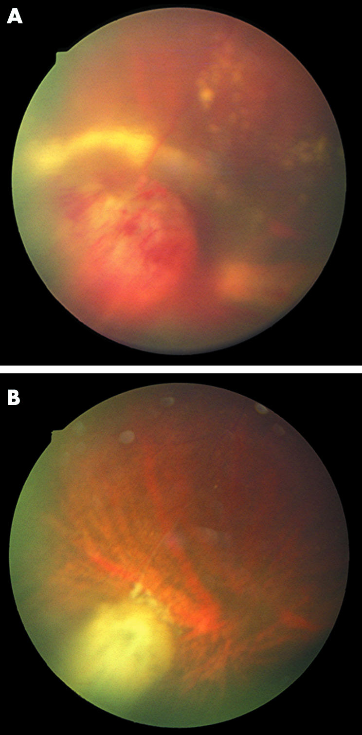
Figure 2 A case of a symptomatic vasoproliferative tumours of the rtiuce (VTR) (A) before treatment and (B) 6 months after brachytherapy with a 106Ru plaque. Note the visible chorioretinal scar around the VTR, the fibrotic regression of the lesion, and the resolved exudates.
The most common finding following brachytherapy was the development of epiretinal gliosis in the macular region in 10 eyes (28.6%). There was no correlation between the development of epiretinal gliosis and any of the tumour features examined. None of the patients developed secondary glaucoma after brachytherapy.
The mean visual acuity was 0.33 (SD 0.34) before treatment and 0.29 (SD 0.29) at the last visit. Twenty patients (57.1%) retained their visual acuity and eight of them experienced an improvement during follow up. Of the remaining 15 patients, only five (14.3%) had a loss of more than 0.2, whereas 10 patients showed only a slight decrease in their visual acuity. The significant loss of visual acuity—that is, more than 0.2, was associated with the development of epiretinal gliosis (p = 0.005).
The visual outcome was better than 0.25 in 17 patients (48.6%). This was associated inversely with the tumour thickness (p = 0.008), the presence of exudative retinal detachment before treatment (p = 0.02), the presence of macular pathology before therapy (p = 0.02)—that is, oedema, hard exudates, gliosis—and the performance of retinal surgery during follow up (p = 0.04). Univariate Cox regression analysis showed that both the presence of exudative retinal detachment (RR: 2.8, 95% CI: 1.2 to 6.2; p = 0.01) and the presence of macular pathology (RR: 7, 95% CI: 1.8 to 28.8; p = 0.004) correlated with a loss of vision of 0.25 or better. Multivariate analysis with both parameters showed that only the presence of macular pathology was predictive of a visual outcome worse than 0.25 (RR: 6.1, 95% CI: 1.2 to 31.3; p = 0.03; model fitting: p = 0.005).
Discussion
The treatment of VTR is still a challenge and so far only reports on small series with different treatment approaches have been published. The present work is the first with a single treatment modality applied to a relatively large series of symptomatic VTR. The authors have already had some experience with 106Ru brachytherapy for vascular tumours of the ocular fundus.1,7 The main advantage of β ray brachytherapy using 106Ru is the limited range of the electrons used to deliver the dose to a very restricted volume. It allows a high dose in contact with the plaque and a very steep dose gradient at 5 mm depth. The resulting dose distribution has the following clinical gains: (i) the ability to treat the entire lesion even in bigger tumours not eligible for laser or cryotherapy, (ii) the ability to induce a scar gently and prevent side effects like bleeding and massive exudation, and (iii) to protect the healthy structures of the eye. On the other hand, brachytherapy is a much more invasive procedure than laser or cryotherapy and has many side effects with potentially negative impact on visual outcome. The major problems of brachytherapy affecting vision are neuropathy of the optic nerve, maculopathy, retinopathy, cataract, and iris neovascularisation with subsequent secondary glaucoma.8 The follow up in the present study has been long enough to evaluate late toxicity after irradiation. There has not been a single case of radiation induced neuropathy, maculopathy, retinopathy, or secondary glaucoma. A possible explanation may be the fact that the majority of the tumours were located in the retinal periphery and the tumour base was small compared to melanomas of the fundus periphery. Thus, only a relatively small area of the retina was affected and this was in most cases far off the optic nerve or the macular region. Owing to the proximity to the lens, however, an increased cataract risk was expected and, so far, five eyes have undergone cataract surgery.
The applied dose was responsible for side effects as well as for the success of the treatment. It was not possible to determine the optimal dose of radiation by means of a phase I trial because of the rarity of this condition. The techniques have evolved during the long course of the study. The patients in the present series, however, have been treated quite homogeneously; nevertheless, the results obtained did not allow optimisation of the dose by establishing a dose‐effect relation.
The mean visual acuity decreased slightly after brachytherapy. Almost half of the patients (17/35) retained a final visual acuity of 0.25 or better. The presence of pathological features in the macular region before therapy was associated with a 6.1‐fold risk of loss of such good vision. Since macular pathology reflects grade and time of tumour activity, our observation indirectly supports the idea to treat VTR at an early stage, as already proposed by others.9 The main reason for a significant loss of vision after brachytherapy was the development of epiretinal gliosis. The presence of epiretinal gliosis in association with VTR has already been described.10 The incidence of epiretinal gliosis to this extent (cumulative risk: 19.5% at 2 years) after brachytherapy has not been reported so far. However, it can only be speculated whether this is a side effect of brachytherapy or just a secondary effect of VTR, or a combination of both.
In conclusion, brachytherapy with 106Ru plaques led to regression of VTR. Eyes harbouring VTR, which had already developed secondary glaucoma could not be treated effectively with brachytherapy. The presence of macular pathology before treatment was a predictive factor for poor visual prognosis and radiation induced cataract was the only clearly related treatment complication within the follow up time of the present series.
Acknowledgements
This work was supported by the Deutsche Forschungsgemeinschaft (KFO‐109) and the Kulturstiftung Essen. We thank Mr Lehnart for his excellent assistance with the data bank upgrade. The authors of this work have no competing interests.
Abbreviations
PDT - photodynamic therapy
RPE - retinal pigment epithelium
TTT - transpupillary thermotherapy
VTR - vasoproliferative tumours of the retina
References
- 1.Heimann H, Bornfeld N, Vij O.et al Vasoproliferative tumours of the retina. Br J Ophthalmol 2000841162–1169. [DOI] [PMC free article] [PubMed] [Google Scholar]
- 2.Barr C C, Rice T A, Michels R G. Angioma‐like mass in a patient with retrolental fibroplasia. Am J Ophthalmol 198089647–650. [DOI] [PubMed] [Google Scholar]
- 3.Shields J A, Decker W L, Sanborn G E.et al Presumed acquired retinal hemangiomas. Ophthalmology 1983901292–1300. [DOI] [PubMed] [Google Scholar]
- 4.Sahel J A, Frederick A R, Jr, Pesavento R.et al Idiopathic retinal gliosis mimicking a choroidal melanoma. Retina 19888282–287. [DOI] [PubMed] [Google Scholar]
- 5.Shields C L, Shields J A, Barrett J.et al Vasoproliferative tumors of the ocular fundus. Classification and clinical manifestations in 103 patients. Arch Ophthalmol 1995113615–623. [DOI] [PubMed] [Google Scholar]
- 6.Jain K, Berger A R, Yucil Y H.et al Vasoproliferative tumours of the retina. Eye 200317364–368. [DOI] [PubMed] [Google Scholar]
- 7.Kreusel K M, Bornfeld N, Lommatzsch A.et al Ruthenium‐106 brachytherapy for peripheral retinal capillary hemangioma. Ophthalmology 19981051386–1392. [DOI] [PubMed] [Google Scholar]
- 8.Foerster M H, Bornfeld N, Schulz U.et al Complications of local beta radiation of uveal melanomas. Graefes Arch Clin Exp Ophthalmol 1986224336–340. [DOI] [PubMed] [Google Scholar]
- 9.Laqua H, Wessing A. Peripheral retinal telangiectasis in adults simulating a vascular tumor or melanoma. Ophthalmology 1983901284–1291. [DOI] [PubMed] [Google Scholar]
- 10.McDonald H R, Schatz H, Johnson R N.et al Vitrectomy in eyes with peripheral retinal angioma associated with traction macular detachment. Ophthalmology 1996103329–335. [DOI] [PubMed] [Google Scholar]