

1. Introduction
A promising new system for 3D dosimetry combines a radiochromic plastic dosimeter, PRESAGE [1–3], with an optical-computed-tomography system (optical-CT) capable of reading the dose recorded in the dosimeter [4,5]. Optical-CT is also the method of choice for reading out the dose recorded in the more established polymer gel dosimetry system, for many applications [6–9]. Achieving accurate dosimetry depends critically on the performance characteristics of the optical-CT imaging system. Several systems have been developed [6,10–12], and two are commercially available at the present time (MGS Research Inc, and Modus Medical Devices Inc). Common issues of quality assurance (QA) become significant for all these systems to ensure correct initial commissioning of optical-CT 3D dosimetry and correct continued functioning. Here we present a QA phantom and procedure designed for efficient evaluation of the basic imaging performance of any optical-CT scanning system. Example results are presented from two optical-CT systems, an in-house CCD based system and the OCTOPUS™ system from MGS Research.
2. Methods
The optical-CT QA phantom consists of a ‘layer cake’ model (figure 1) where each layer contains special features designed to test different aspects of the imaging system. The phantom is constructed out of polyurethane and contains a range of inserts. It is also constructed to be optically stable in time, so that repetitious scanning yields information on the temporal stability of the scanning system.
Figure 1.
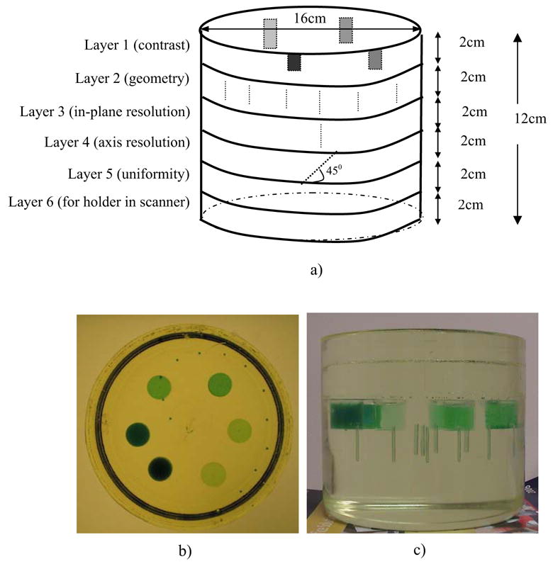
a) Schematic of the optical-CT QA phantom, and photographs of the optical-CT QA phantom b) top view and c) side view
3. Results
Sample images of select layers of the optical-CT QA phantom and derived imaging performance obtained with the OCTOPUS™ scanner are shown in figure 2 to 4. Good geometrical accuracy (within 0.5 mm) and linearity of reconstructed attenuation coefficients (r2 = 0.9917 in figure 4a) were observed.
Figure 2.
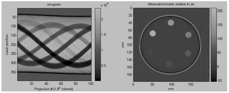
Contrast-Detail layer
Figure 4.
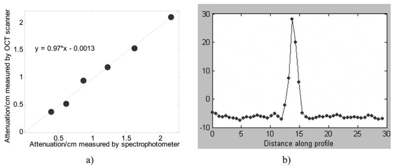
a) linearity of attenuation coefficients, b) point-spread-function (uncorrected for wire diameter).
4. Conclusions
The optical-CT QA phantom presented here represents an important development for ensuring correct implementation and ongoing performance of an optical-CT scanning system. Our initial results with the OCTOPUS scanning system from MGS Inc are encouraging. Repeat scanning has revealed excellent consistency of performance over many months.
Figure 3.
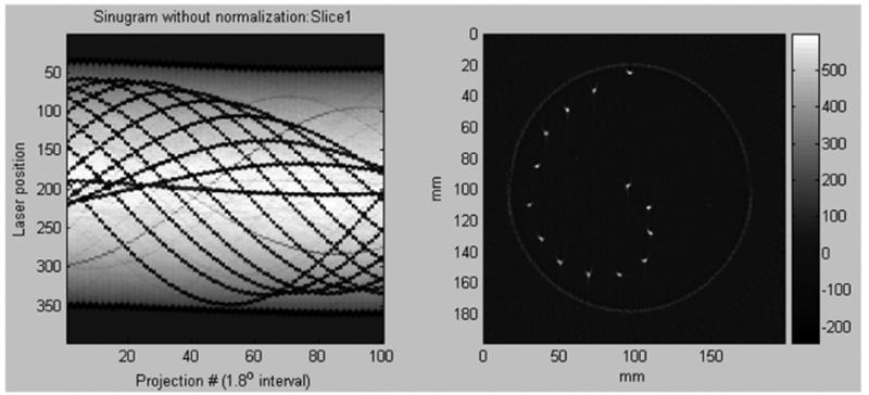
Geometrical accuracy layer
Contributor Information
Pengyi Guo, Dept of Radiation Oncology, Duke University Medical Center, Durham, NC, USA.
John Adamovics, Dept of Chemistry and Biology, Rider University, Lawrenceville, NJ, USA.
Mark Oldham, Dept of Radiation Oncology, Duke University Medical Center, Durham, NC, USA.
References
- 1.Adamovics J, Maryanski MJ. OCT scanning properties of PRESAGE - A 3D radiochromic solid polymer dosimeter. Med Phys. 2004;31:1906. [Google Scholar]
- 2.Guo P, Adamovics J, Oldham M. PRESAGE - A promising new material for 3D dosimetry. Int J Radiat Oncol Biol Phys. 2005;63:S206. [Google Scholar]
- 3.Guo P, Adamovics J, Oldham M. Characterization of a new radiochromic three-dimensional dosimeter. Med Phys. 2006 May;33(5):1338–45. doi: 10.1118/1.2192888. [DOI] [PMC free article] [PubMed] [Google Scholar]
- 4.Guo P, Adamovics J, Oldham M. A practical three-dimensional dosimetry system for radiation therapy. Med Phys. 2006 Oct;33(10):3962–72. doi: 10.1118/1.2349686. [DOI] [PMC free article] [PubMed] [Google Scholar]
- 5.Heard M, De La Mora A, Adamovics J, Ibbott G. Evaluation of a new 3D polyurethane dosimeter for IMRT verification. Med Phys. 2005;32:2167. [Google Scholar]
- 6.Oldham M, Siewerdsen JH, Shetty A, Jaffray DA. High resolution gel-dosimetry by optical-CT and MR scanning. Med Phys. 2001;28:1436–1445. doi: 10.1118/1.1380430. [DOI] [PubMed] [Google Scholar]
- 7.Oldham M. Optical-CT scanning of polymer gels. J Phys: Conf Ser. 2004;3:122–135. doi: 10.1088/1742-6596/3/1/011. [DOI] [PMC free article] [PubMed] [Google Scholar]
- 8.Islam KT, Dempsey JF, Ranade MK, Maryanski MJ, Low DA. Initial evaluation of commercial optical CT-based 3D gel dosimeter. Med Phys. 2003;30:2159–2168. doi: 10.1118/1.1593636. [DOI] [PubMed] [Google Scholar]
- 9.Xu Y, Wuu CS, Maryanski MJ. Performance of a commercial optical CT scanner and polymer gel dosimeters for 3-D dose verification. Med Phys. 2004;31:3024–3033. doi: 10.1118/1.1803674. [DOI] [PubMed] [Google Scholar]
- 10.Doran SJ, Koerkamp KK, Bero MA, Jenneson P, Morton EJ, Gilboy WB. A CCD-based optical CT scanner for high-resolution 3D imaging of radiation dose distributions: equipment specifications, optical simulations and preliminary results. Phys Med Biol. 2001;46:3191–3213. doi: 10.1088/0031-9155/46/12/309. [DOI] [PubMed] [Google Scholar]
- 11.Gore JC, Ranade M, Maryanski MJ, Schulz RJ. Radiation dose distributions in three dimensions from tomographic optical density scanning of polymer gels: I. Development of an optical scanner. Phys Med Biol. 1996;41:2695–2704. doi: 10.1088/0031-9155/41/12/009. [DOI] [PubMed] [Google Scholar]
- 12.Kelly RG, Jordan KJ, Battista JJ. Optical CT reconstruction of 3D dose distributions using the ferrous-benzoic-xylenol (FBX) gel dosimeter. Med Phys. 1998;25:1741–1750. doi: 10.1118/1.598356. [DOI] [PubMed] [Google Scholar]