A simple model for teaching indirect ophthalmoscopy
Indirect ophthalmoscopy is a useful technique to allow a wide angle view of the fundus, to screen for retinal disease, and to examine the peripheral retina. It provides a better view of the fundus in patients with lens opacities than is allowed by direct ophthalmoscopy. As the threshold at which cataracts are operated in developing countries is changing we have found it important that junior doctors and other eye health workers who select cataract patients for operation can use the instrument.
We use a simple model to teach the technique of indirect ophthalmoscopy. An hour or two with this model gets the beginner “over the hump” of learning how to position and move the light and lens. When the first real patient is examined, the learner can immediately concentrate on the fundus, minimising frustration and time spent shining an annoying light into a patient's eye.
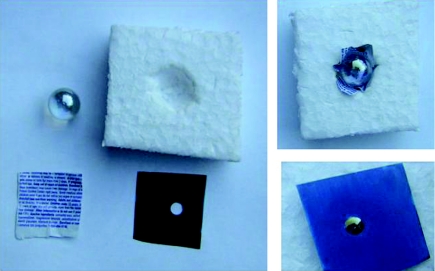
The model is shown in the figure 1. The “eyeball” is a clear glass marble, set into anything that will keep it from rolling, such as a small bottle cap or a hole made in a piece of Styrofoam. Behind the marble, moulded around it, a piece of paper is placed with the smallest print available—the package inserts from prescription medicines work well. Finally, a hole is punched in a scrap of paper to use as a pupil. The optics of the system are excellent for practising indirect ophthalmoscopy and the student can be tested by how many words, letters, or lines he can see compared to the instructor.
Figure 1 Model for practising indirect ophthalmoscopy.
Other models have been described before, which are more sophisticated or allow for scleral indentation but the materials may be hard to obtain.1,2,3 A model equally simple to make as that described here has been proposed4; it does not require a glass marble and is good for helping the beginner get oriented, but the marble model provides more of a challenge at learning to view the retina outside the posterior pole.
Footnotes
Thanks to Dr Paul Meyer, Addenbrookes Hospital, Cambridge, UK, for this idea.
References
- 1.Dodaro N R, Maxwell D P. An eye for an eye. A simplified model for teaching. Arch Ophthalmol 1995113824–826. [DOI] [PubMed] [Google Scholar]
- 2.Chew D, Gray R H. A model eye to practice indentation during indirect ophthalmoscopy. Eye 19937599–600. [DOI] [PubMed] [Google Scholar]
- 3.Bartner H, Paton D. An improved model for instruction in binocular indirect ophthalmoscopy. Arch Ophthalmol 197185530–533. [DOI] [PubMed] [Google Scholar]
- 4.Ing E B, Ing T G E. A method of teaching indirect ophthalmoscopy to beginning residents. Can J Ophthlamol 199227166–167. [PubMed] [Google Scholar]


