Abstract
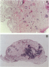
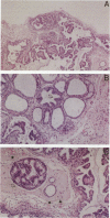
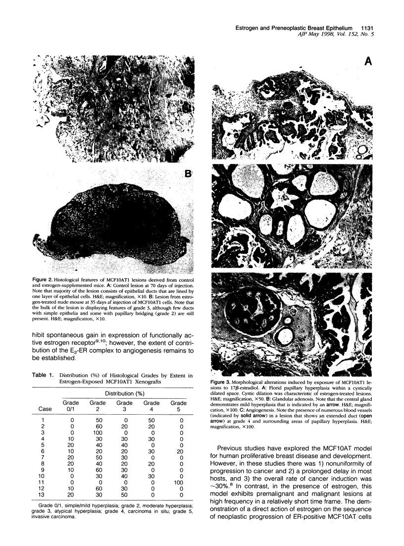
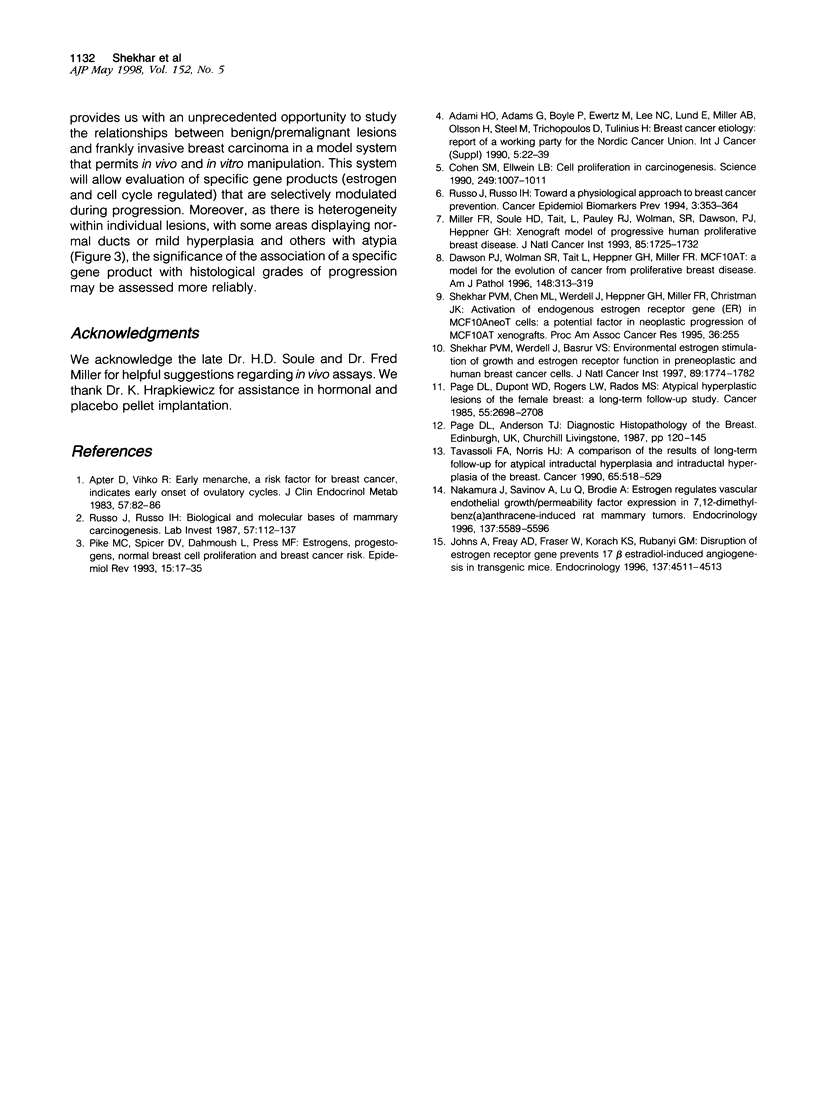
We have used the MCF10AT xenograft model of human proliferative breast disease to examine the early effects of estradiol exposure on morphological progression of preneoplastic lesions and to define the step(s) in the morphological sequence at which estrogen may act. The effects of estradiol on neoplastic progression of estrogen-receptor-positive MCF10AT cells in the orthotopic site were examined in ovariectomized female nude mice that received subcutaneous administration of implants of 17beta-estradiol or placebo pellets. At 10 weeks, histological analysis of the lesions derived from the estrogen-supplemented group revealed that 92% of lesions displayed histological features of atypical hyperplasia, carcinoma in situ, or invasive carcinoma, and the remaining 8% exhibited histological features of moderate hyperplasia. These highly proliferative lesions are in marked contrast to the control group in which 60% of samples displayed no evidence of hyperplasia. In contrast with control xenografts, estrogen-exposed xenografts demonstrated extensive areas of papillary growth, adenosis-like areas, prominent host inflammatory infiltration, and angiogenesis. Our results suggest that estrogen exerts a growth-promoting effect on benign or premalignant ductal epithelium by enhancing 1) the frequency of lesion formation, 2) the size of lesions, 3) the speed of transformation from normal/mild hyperplasia to those with atypia, 4) the degree of dysplasia, and 5) angiogenesis.
Full text
PDF
Images in this article
Selected References
These references are in PubMed. This may not be the complete list of references from this article.
- Adami H. O., Adams G., Boyle P., Ewertz M., Lee N. C., Lund E., Miller A. B., Olsson H., Steel M., Trichopoulos D. Breast-cancer etiology. Report of a working party for the Nordic Cancer Union. Int J Cancer Suppl. 1990;5:22–39. [PubMed] [Google Scholar]
- Apter D., Vihko R. Early menarche, a risk factor for breast cancer, indicates early onset of ovulatory cycles. J Clin Endocrinol Metab. 1983 Jul;57(1):82–86. doi: 10.1210/jcem-57-1-82. [DOI] [PubMed] [Google Scholar]
- Cohen S. M., Ellwein L. B. Cell proliferation in carcinogenesis. Science. 1990 Aug 31;249(4972):1007–1011. doi: 10.1126/science.2204108. [DOI] [PubMed] [Google Scholar]
- Dawson P. J., Wolman S. R., Tait L., Heppner G. H., Miller F. R. MCF10AT: a model for the evolution of cancer from proliferative breast disease. Am J Pathol. 1996 Jan;148(1):313–319. [PMC free article] [PubMed] [Google Scholar]
- Johns A., Freay A. D., Fraser W., Korach K. S., Rubanyi G. M. Disruption of estrogen receptor gene prevents 17 beta estradiol-induced angiogenesis in transgenic mice. Endocrinology. 1996 Oct;137(10):4511–4513. doi: 10.1210/endo.137.10.8828515. [DOI] [PubMed] [Google Scholar]
- Miller F. R., Soule H. D., Tait L., Pauley R. J., Wolman S. R., Dawson P. J., Heppner G. H. Xenograft model of progressive human proliferative breast disease. J Natl Cancer Inst. 1993 Nov 3;85(21):1725–1732. doi: 10.1093/jnci/85.21.1725. [DOI] [PubMed] [Google Scholar]
- Nakamura J., Savinov A., Lu Q., Brodie A. Estrogen regulates vascular endothelial growth/permeability factor expression in 7,12-dimethylbenz(a)anthracene-induced rat mammary tumors. Endocrinology. 1996 Dec;137(12):5589–5596. doi: 10.1210/endo.137.12.8940388. [DOI] [PubMed] [Google Scholar]
- Page D. L., Dupont W. D., Rogers L. W., Rados M. S. Atypical hyperplastic lesions of the female breast. A long-term follow-up study. Cancer. 1985 Jun 1;55(11):2698–2708. doi: 10.1002/1097-0142(19850601)55:11<2698::aid-cncr2820551127>3.0.co;2-a. [DOI] [PubMed] [Google Scholar]
- Pike M. C., Spicer D. V., Dahmoush L., Press M. F. Estrogens, progestogens, normal breast cell proliferation, and breast cancer risk. Epidemiol Rev. 1993;15(1):17–35. doi: 10.1093/oxfordjournals.epirev.a036102. [DOI] [PubMed] [Google Scholar]
- Russo J., Russo I. H. Biological and molecular bases of mammary carcinogenesis. Lab Invest. 1987 Aug;57(2):112–137. [PubMed] [Google Scholar]
- Russo J., Russo I. H. Toward a physiological approach to breast cancer prevention. Cancer Epidemiol Biomarkers Prev. 1994 Jun;3(4):353–364. [PubMed] [Google Scholar]
- Shekhar P. V., Werdell J., Basrur V. S. Environmental estrogen stimulation of growth and estrogen receptor function in preneoplastic and cancerous human breast cell lines. J Natl Cancer Inst. 1997 Dec 3;89(23):1774–1782. doi: 10.1093/jnci/89.23.1774. [DOI] [PubMed] [Google Scholar]
- Tavassoli F. A., Norris H. J. A comparison of the results of long-term follow-up for atypical intraductal hyperplasia and intraductal hyperplasia of the breast. Cancer. 1990 Feb 1;65(3):518–529. doi: 10.1002/1097-0142(19900201)65:3<518::aid-cncr2820650324>3.0.co;2-o. [DOI] [PubMed] [Google Scholar]


