Eosinophilic gastroenteritis is a heterogeneous and uncommon disorder characterised by eosinophilic inflammation of the gastrointestinal tissues. The location and depth of infiltration determine its various manifestations and later serve as the basis for its classification as mucosal, muscular, and serosal forms of eosinophilic gastroenteritis. The gold standard for diagnosis is an endoscopic biopsy showing prominent tissue eosinophilia. Gastrointestinal mucosal involvement causes malabsorption, protein losing enteropathy, and diarrhoea. Infiltration of the muscular layer of the bowel wall may cause gastric outlet or small bowel obstruction. Serosal involvement causes exudative ascites rich in eosinophils; this is the least common form and is usually diagnosed by laparoscopic examination and biopsy of the whole intestinal wall.1,2
We present the case of an 18 year old male student with a history of allergic rhinitis presenting with abdominal pain, nausea, and low grade fever, which started a few weeks before his admission. Medical history was unremarkable for any other serious disease. Laboratory findings showed leucocytosis with a high percentage of eosinophils (81%); skin prick testing showed sensitivity to Dermatophagoides pteronyssinus and ragweed pollen; total immunoglobulin E in serum was three times the normal level (311 kI/l); and a bone marrow aspiration specimen showed increased numbers of mostly mature eosinophils (62%) in the differential cell count. These findings suggested an atopic constitution.
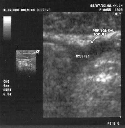
Endoscopic examination of the whole gastrointestinal tract, histological analysis, and abdominal computed tomography showed no pathological features. Sonographic abnormalities were discovered mostly in the right lower quadrant of the abdomen where a small amount of ascites was found together with nodular deposits on the parietal peritoneum in the same region, indicating peritoneal inflammation (fig 1). Cytological examination of ascites revealed numerous eosinophils (differential count 91% eosinophils) and a few mesothelial cells in the highly cellular cytospin preparation. Sonographic visualisation of nodular peritoneal deposits associated with eosinophilic ascites, peripheral blood eosinophilia, and atopic constitution permitted a diagnosis of the serosal form of eosinophilic gastroenteritis.
Figure 1 Initial sonographic examination. A peritoneal nodule (4 mm, arrow) in the region of the terminal ileum and ascites are typical of the serosal form of eosinophilic gastroenteritis.
Two weeks of oral prednisolone 20 mg/day brought relief to the patient's symptoms, with normalisation of laboratory parameters and sonographic findings. Two months later the patient suffered a relapse with the same abdominal symptoms. Again, the same diagnostic procedure was done, and therapy with oral prednisolone was effective. During the next relapse of the serosal form of eosinophilic enteritis, which was accompanied by a relapse of rhinitic symptoms, we introduced leukotriene receptor antagonists (montelukast 10 mg/day) with the aim of reducing corticosteroid therapy. After four weeks of this therapy the patient achieved complete clinical and laboratory remission, which continued for the next six months of leukotriene inhibitor therapy at the same dose.
We conclude that leukotriene receptor antagonists may be useful as steroid sparing agents in patients with the serosal form of eosinophilic gastroenteritis. Eosinophils are potent sources of leukotrienes, including leukotriene C4 and leukotriene D4, molecules known to induce eosinophil chemotaxis, smooth muscle contraction, mucous secretion, and mucosal oedema. Many factors may play a role in directing eosinophils to a site of inflammation. These data should challenge our thinking about the treatment of eosinophilic gastrointestinal diseases with new antileukotriene agents, such as leukotriene inhibitors, that provide a more targeted approach to eosinophil derived products.3
Footnotes
Conflict of interest: None declared.
References
- 1.Lee C, Changchien C, Chen P.et al Eosinophilic gastroenteritis. 10 years experience. Am J Gastroenterol 19938870. [PubMed] [Google Scholar]
- 2.Rothenberg M E. Eosinophilic gastrointestinal disorders (EGID). J Allergy Clin Immunol 200411311–28. [DOI] [PubMed] [Google Scholar]
- 3.Schmid‐Grendelmeier P, Altznauer F, Fischer B. Eosinophils express functional IL‐13 in eosinophilic inflammatory diseases. J Immunol 20021691021–1027. [DOI] [PubMed] [Google Scholar]


