Answer
From question on page 1560
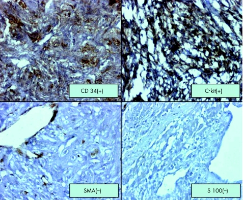
Multidetector computed tomography (MDCT) showed a mass at the jejunum with high density in the early arterial phase (fig 1). Angiography showed a hypervascular mass that was supplied by jejunal branches (fig 2). The patient underwent jejunal segmental resection. Histopathological assessment revealed a gastrointestinal stromal tumour (GIST) expressing C‐kit and CD 34 (fig 3).
Figure 3 Immunohistochemical staining revealed a gastrointestinal stromal tumour expressing C‐kit and CD 34.
GISTs, frequently manifesting as gastrointestinal bleeding, are defined as mesenchymal neoplasms originating from the interstitial cells of Cajal that express the c‐kit proto‐oncogene protein (CD117) which is a cell membrane receptor with tyrosine kinase activity. The disease occurs mainly in the stomach and small intestine. MDCT angiography is useful for detecting the causes of obscure gastrointestinal bleeding1,2 and the vascularity of GIST that is related to malignancy.3 To date, however, the sensitivity and specificity of MDCT for diagnosis of the causes of obscure gastrointestinal bleeding are not known, and further studies are needed.1 Approximately 20–30% of GIST show malignant behaviour. Tumour size of >5 cm and >5 mitotic activities in 50‐fold high power fields are useful predictors of malignancy. Surgery is the main modality of therapy and imatinib, a selective inhibitor of tyrosine kinases, can be used in metastatic or unresectable patients as neoadjuvant treatment.
References
- 1.Miller F H, Hwang C M. An initial experience: using helical CT imaging to detect obscure gastrointestinal bleeding. Clin Imaging 200428245–251. [DOI] [PubMed] [Google Scholar]
- 2.Nishida T, Kumano S, Sugiura T.et al Multidetector CT of high‐risk patients with occult gastrointestinal stromal tumors. AJR Am J Roentgenol 2003180185–189. [DOI] [PubMed] [Google Scholar]
- 3.Fukuta N, Kitano M, Maekawa K.et al Estimation of the malignant potential of gastrointestinal stromal tumors: the value of contrast‐enhanced coded phase‐inversion harmonics US. J Gastroenterol 200540247–255. [DOI] [PubMed] [Google Scholar]


