Abstract
Mesenchymal hamartoma of the liver (MHL) is an uncommon tumour composed of architecturally abnormal bile ducts in an uncommitted myxoid stroma. Most MHL are diagnosed in childhood and few cases have been reported in adults. This report describes a case of a well defined solid mass in the right lobe of the liver in a 51 year old man. Preoperative radiological examination revealed a large completely solid mass. Biopsy showed a dense fibrous stroma with hyalinisation and some bile ducts. A provisional diagnosis of MHL was made. Surgical excision was impossible and liver transplantation was undertaken. Definitive pathology confirmed the diagnosis. Review of published reports shows this to be the fourth case of MHL treated by liver transplantation.
Keywords: mesenchymal hamartoma, liver, transplantation
Mesenchymal hamartoma of the liver (MHL) is an uncommon benign tumour that mostly appears in childhood. It has often been confused with other benign lesions such as vascular hamartomas. It makes up about 3–8% of all primary liver tumours in children.1,2,3,4,5 It is well recognised that in most cases the diagnosis is made during the first two years of life, with a clinical presentation dominated by absence of symptoms, a remarkable abdominal mass or progressive enlargement of the abdomen, and on rare occasions there may be symptoms linked to compression of other organs. In some isolated cases these lesions have been reported in adults. Histologically, there is a mixture of mesodermal and endodermal structures in loose connective tissue stroma, almost always with fluid filled spaces lacking an endothelial lining. Bile ducts, liver cells, and angiomatous elements are present with the appearance of well differentiated ductal structures surrounded by loose fibroblasts containing mesenchymal tissue.6
The pathophysiology remains unknown. For years, it has been accepted that this disease is the result of a failure of the ductal plate to develop normally in utero, a reaction to biliary obstruction, or even the result of a regional ischaemic process.7 More recently, some immunohistochemical and flow cytometric studies have suggested that the MHL is a true neoplasm rather than a developmental anomaly, and it may be the initial step or a stage in the development of an embryonal sarcoma of the liver.8,9,10,11
Fewer than 5% of cases of MHL occur in patients older than five years, and the condition is extraordinarily rare in adults (older than 18 years). Under 20 adult cases have been reported.12,13,14,15,16,17,18,19,20,21,22,23,24 In all of these, there have been clear differences compared with the manifestations of the disease in childhood.
Case report
The patient was a 51 year old man with a previous medical history of renal colic and the expulsion of stones on several occasions. He recently suffered from the new symptom of intense right upper quadrant abdominal pain, which was initially interpreted as a new episode of renal colic. Abdominal ultrasonography was undertaken, and a large, solid, rounded tumour mass in the right lobe of the liver was found for the first time. The patient was referred and admitted to our institution. The only positive finding on physical examination was the palpable tumour mass, of hard consistency and slightly painful. The edge was approximately 4 cm below the right costal margin in the mid‐clavicular line. There was no vascular bruit over the liver and no free abdominal fluid.
Initial laboratory test results were unremarkable (table 1). The α‐fetoprotein level was in the normal range and serology for HIV, hepatitis C virus, and hepatitis B virus was negative.
Table 1 Laboratory results for the case patient.
| Study | Result | Normal range |
|---|---|---|
| Full blood count | ||
| Haemoglobin | 12.8 g/dl | 12 to 16 |
| Packed cell volume | 40% | 40 to 52 |
| Leucocyte count | 4.6×10⁹/l | 4.3 to 10.8 |
| Neutrophils | 45.7% | 45 to 74 |
| Lymphocytes | 44.4% | 16 to 45 |
| Basophils | 1% | 0 to 7 |
| Eosinophils | 4.2% | 0 to 7 |
| Platelet count | 125×109/l | 150 to 350 |
| ESR | 3 mm/h | 0 to 20 |
| Blood chemistry | ||
| Aspartate aminotransferase | 14 u/l | <40 |
| Alanine aminotransferase | 10 u/l | <50 |
| γ‐Glutamyl transferase | 26 u/l | <50 |
| Alkaline phosphatase | 97 u/l | <20 |
| Total bilirubin | 14 μmol/l | <17 |
| Total proteins | 56 g/l | 45 to 80 |
| Albumin | 38 g/l | 35 to 60 |
| Glucose | 5.18 mmol/l | 3.3 to 6.0 |
| Uric acid | 114 μmol/l | <400 |
| Creatinine | 83 μmol/l | <120 |
ESR, erythrocyte sedimentation rate.
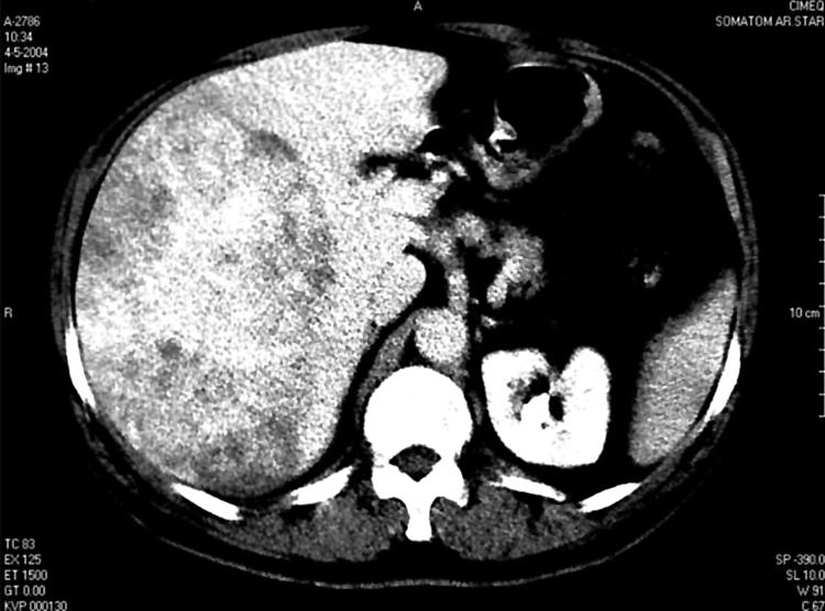
Repeat abdominal ultrasonography confirmed the presence of the solid tumour in the same location. Intravenous contrast enhanced computed tomography (CT) scan supported the ultrasound findings. A well defined complex solid mass of 19×13 cm of diameter was demonstrated, which occupied almost the whole of the right lobe of the liver and showed heterogeneous enhancement. The right kidney was displaced downwards because of the tumour (fig 1).

Figure 1 Computed tomographic scan showing a complex solid tumour, which filled the right lobe of the liver and showed heterogeneous enhancement.
Laparoscopy was undertaken and showed enlargement of the right lobe of the liver towards the right flank; the surface had a pale red hue and both lobes were smooth. A prominent plate‐like area tinted rose to orange arose from the right anterior and superior segments of the right lobe. Liver biopsy showed a mesenchymal tumour of benign appearance composed of collagenous fibrous tissue containing isolated bile ducts.
The possibility to complete resection of the tumour by right hemihepatectomy was anticipated, and so hepatic angiography was undertaken to complete the study. This showed that the tumour was hypervascularised, with anomalous arterial vessels inside the lesion originated from the origin of the right hepatic artery. It was shown that owing to the size of the tumour, the middle and right hepatic vein and the right branch of the portal vein were markedly constricted and distorted. It was concluded that surgery was impossible because of the technical difficulties which would impose a serious risk for the patient during the operation and the postoperative period. Because of this the patient was placed on the waiting list for liver transplantation.
One month later, the orthotopic liver transplantation was undertaken without complications. Thirteen days after surgery the patient was discharged from hospital. One year after surgery, he was well.
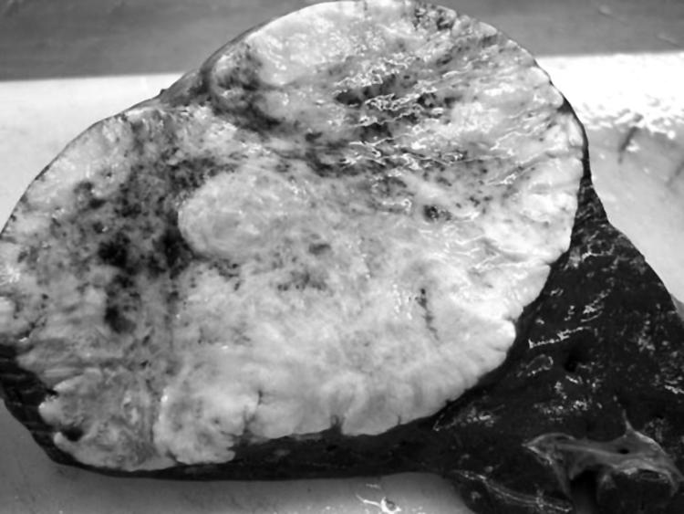
The excised liver contained a large solid rounded tumour. It was well demarcated and there was no capsule. It occupied the entire right lobe. The cut surface showed solid grey shining tissue with a slightly nodular appearance and isolated small haemorrhage areas (fig 2).

Figure 2 Cut surface of the tumour.
Microscopy showed disrupted architecture, no fibrous capsule, and heavily hyalinised stroma containing fibroblasts and occasional irregular bile ducts (fig 3). In some areas, there was a paucicellular appearance. There was no evidence of malignancy, hepatic parenchyma within the stromal areas, lymphocytic or plasma cell inflammatory infiltrate, or extramedullary haematopoiesis. As there was no cystic component to the tumour, a diagnosis of MHL without cystic component was made.

Figure 3 Microscopy of the tumour showing a heavily hyalinised stroma with fibroblasts and irregular bile ducts.
Discussion
The first reported case of MHL was by Maresch in 1903. Until few years ago, this disease was known by different names such a cavernous lymphadenomatoid tumour, bile cell fibroadenoma, and benign mesenchymoma. Edmundson coined the term mesenchymal hamartoma in 1956 for the first time. He used the term “hamartoma”, proposed by Alberh, to explain that the lesion was not really a malignant tumour.25 Later, the clinicopathological features of this lesion in children were well documented.26
There are few published reports of this disease in adults, in whom the characteristics of the tumour differ those found in childhood. First, abdominal pain is the most important symptom in adults, while in children the occurrence of pain is very rare. Second, in adults the disease is more common in females, while in children the sex distribution is equal. Features in common for both adults and children include a predilection to involve the right lobe,27 massive growth of the tumour, and a cystic predominance (the tumours are cystic in most childhood cases and in approximately 63% of adult cases). Fewer than half the adult cases are predominantly solid. Features of the current cases and previously reported cases of MHL in adults are summarised in table 2.
Table 2 Reported cases of MHL in adults.
| Case | Reference | Age (y) | Sex | Location | Size | Gross appearance | Pain at presentation |
|---|---|---|---|---|---|---|---|
| 1 | 12 | 21 | F | Right lobe | 17×10 cm | Cystic | Yes |
| 2 | 13 | 69 | F | Left lobe | 29×20×11.5 cm | Cystic | No |
| 3 | 14 | 30 | F | Right lobe | 18 cm | Cystic | No |
| 4 | 15 | 19 | F | Left lobe | 24×19×8 cm | Cystic | Yes |
| 5 | 16 | 32 | F | Left lobe | 14×11 cm | Cystic | No |
| 6 | 17 | 53 | M | Right lobe | 20×14×10 cm | Cystic | Yes |
| 7 | 18 | 20 | F | Left lobe | 6×8 cm | Cystic | No |
| 8 | 19 | 28 | F | Right lobe | 30×20×14 | Solid | No |
| 9 | 20 | 57 | F | Left lobe | 6×4×3.5 cm | Solid | Yes |
| 10 | 21 | 56 | F | Both lobes | 7.5 cm | Cystic | Yes |
| 11 | 22 | 62 | M | Left lobe | 8 cm | Solid | No |
| 12 | 23 | 46 | F | Right lobe | 6×4×5 cm | Cystic | Yes |
| 13 | 23 | 63 | F | Left lobe | 11×16×24 cm | Solid | Yes |
| 14 | 23 | 66 | F | Right lobe | 5×4×2 cm | Cystic | No |
| 15 | 24 | 38 | M | Right lobe | 8×5 cm | Solid | No |
| 16 | This report | 51 | M | Right lobe | 19×13 cm | Solid | Yes |
F, female; M, male; y, years.
The tumour mass may blend with the adjacent hepatic tissue or can be encapsulated by a fibrous wall.28 The tumour is usually large, and the cut surface shows the presence of cysts or, less often, is completely solid. Adjacent hepatic tissue is usually compressed, fibrosed, or atrophied.
Imaging methods will usually establish the diagnosis, ultrasound being the most informative examination and the one to use as the initial test in any patient with abdominal enlargement or a palpable mass. CT supports the ultrasound findings by showing the size of the hepatic mass and its relation to the adjacent structures.
The aetiology of this disease has been much discussed. Traditionally, it was considered to be an anomaly of biliary development.29 Another hypothesis proposed that it developed in ischaemic zones.30 Recently there has been increasing interest in the possibility that this tumour is a form on undifferentiated (embryonal) sarcoma.9,11 This is based on published reports of malignant transformation of MHL and descriptions of specific translocations in chromosome 19, along with immunohistochemical and flow cytometric alterations.31,32,33
Histological studies show a variable mixtures of tissues from which the liver is normally formed. The predominant component is mesenchymal and consists of loose, oedematous connective tissue with cyst‐like collections of fluid, dilated lymphatics and blood vessels, and multiple, branched and tortuous bile ducts. The haematopoietic element is considered to be a component of fetal hepatic haematopoiesis. The cystic predominance of MHL may mimic a lymphangioma, both macroscopically and microscopically. In adults the stromal component is variable and is similar to the stroma of MHL occurring in children. In most areas, however, the stroma had a densely hyalinised and paucicellular appearance. Also, in adults small calibre vessels are present within the stromal areas, and haemosiderin laden macrophages are present focally within areas of granulation tissue associated with intralesional haemorrhage. Along most of the interface between tumour tissue and normal hepatic parenchyma, there is a proliferation of small bile ducts and thin walled vessels. Satellite nodules may be observed in some cases, consisting of dilated and irregularly branching bile ducts in fibrous stroma
Once the diagnosis has been established, the chosen treatment depends on the state of the patient, the size of the tumour, and its relation to surrounding structures. Sometimes the tumour can be enucleated with a minimal loss of normal hepatic tissue. The best surgical option has been the excision with resection of the normal hepatic tissue edge that surround the tumour.34,35 If the tumour is too large, with associated complications such as pulmonary hypertension and kidney damage, endovascular embolisation or ligature of the hepatic artery should be considered, especially in very sick patients, before definitive resection.36 When excision is impossible because of the size of the tumour or because it compromises vital structures, liver transplantation should be considered. In these cases life expectancy is good in the long term.37 This option may be hard for the patient to accept. MHL is certainly a very rare indication for liver transplant, with only three cases having reported so far.37,38
Conclusions
MHL is primarily a benign disease almost exclusively seen in children and very infrequently in adults. The case described here is one of only 15 adult cases so far reported. This tumour was also unusual in being solid, as the majority of these tumours are cystic. MHL is often misdiagnosed in adults because it clinically resembles a malignant tumour on account of its growth behaviour. The diagnosis is usually made by abdominal ultrasound and CT, and confirmed by histological analysis. Recommended treatment is surgical, aimed at total tumour excision, but that is not technically possible orthotopic liver transplantation can be considered, as in this case.
Abbreviations
MHL - mesenchymal hamartoma of the liver
References
- 1.DeMaioribus C A, Lally K P, Sim K.et al Mesenchymal hamartoma of the liver. A 35‐year review. Arch Surg 1990125598–600. [DOI] [PubMed] [Google Scholar]
- 2.Srouji M N, Chatten J, Schulman W M.et al Mesenchymal hamartoma of the liver in infants. Cancer 1978422484–2489. [DOI] [PubMed] [Google Scholar]
- 3.Lai F M, Jayakumar C R, Saw L.et al Hepatic mesenchymal hamartoma: a case report and radiological findings. Singapore Med J 199637226–228. [PubMed] [Google Scholar]
- 4.Barnhart D C, Hirschl R B, Garver K A.et al Conservative management of mesenchymal hamartoma of the liver. J Pediatr Surg 1997321495–1498. [DOI] [PubMed] [Google Scholar]
- 5.Stocker J T. Hepatic tumor in children. Clin Liver Dis 20015259–281. [DOI] [PubMed] [Google Scholar]
- 6.Stocker J T, Ishak K G. Mesenchymal hamartoma of the liver: report of 30 cases and review of the literature. Pediatr Pathol 19831245–267. [DOI] [PubMed] [Google Scholar]
- 7.Desmet V J. Ludwig symposium on biliary disorders – part I. Pathogenesis of ductal plate abnormalities. Mayo Clin Proc 19987380–89. [DOI] [PubMed] [Google Scholar]
- 8.Bove K E, Blough R I, Soukup S. Third report of t (19q) (13.4) in mesenchymal hamartoma of liver with comments on link to embryonal sarcoma. Pediatr Dev Pathol 19981438–442. [DOI] [PubMed] [Google Scholar]
- 9.Mascarello J T, Krous H F. Second report of a translocation involving 19q13.4 in a mesenchymal hamartoma of the liver. Cancer Genet Cytogenet 199258141–142. [DOI] [PubMed] [Google Scholar]
- 10.O'Sullivan M J, Swanson P E, Knoll J.et al Undifferentiated embryonal sarcoma with unusual features arising within mesenchymal hamartoma of the liver: report of a case and review of the literature. Pediatr Dev Pathol 20014482–489. [DOI] [PubMed] [Google Scholar]
- 11.Speleman F, De Telder V, De Potter K R.et al Cytogenetic analysis of a mesenchymal hamartoma of the liver. Cancer Genet Cytogenet 19894029–32. [DOI] [PubMed] [Google Scholar]
- 12.Papastratis G, Margaris H, Zografos G N.et al Mesenchymal hamartoma of the liver in an adult: a review of the literature. Int J Clin Pract 200054552–554. [PubMed] [Google Scholar]
- 13.Drachenberg C B, Papadimitriou J C, Rivero M A.et al Distinctive case – adult mesenchymal hamartoma of the liver: report of a case with light microscopic, FNA cytology, immunohistochemistry, and ultrastructural studies and review of the literature. Mod Pathol 19914392–395. [PubMed] [Google Scholar]
- 14.Gutierrez O H, Burgener F A. Mesenchymal hamartoma of the liver in an adult: radiologic diagnosis. Gastrointest Radiol 198813341–344. [DOI] [PubMed] [Google Scholar]
- 15.Grases P J, Matos‐Villalobos M, Arcia‐Romero F.et al Mesenchymal hamartoma of the liver. Gastroenterology 1979761466–1469. [PubMed] [Google Scholar]
- 16.Jennings C M, Merrill C R, Slater D N. The computed tomographic appearances of benign hepatic hamartoma. Clin Radiol 198738103–104. [DOI] [PubMed] [Google Scholar]
- 17.Chau K Y, Ho J W, Wu P C.et al Mesenchymal hamartoma of liver in a man: Comparison with cases in infants. J Clin Pathol 199447864–866. [DOI] [PMC free article] [PubMed] [Google Scholar]
- 18.Alanen A, Katevuo K, Toikkanen S. A non‐cystic mesenchymal hamartoma of the liver – an unusual case of an unusual entity: case report and review of the literature. Bildgebung 198756181–184. [PubMed] [Google Scholar]
- 19.Gramlich T L, Killough B W, Garvin A J. Mesenchymal hamartoma of the liver: report of a case in a 28‐year‐old. Hum Pathol 198819991–992. [DOI] [PubMed] [Google Scholar]
- 20.Chung J H, Cho K J, Choi D W.et al Solid mesenchymal hamartoma of the liver in adult. J Korean Med Sci 199914335–337. [DOI] [PMC free article] [PubMed] [Google Scholar]
- 21.Megremis S, Sfakianaki E, Voludaki A.et al The ultrasonographic appearance of a cystic mesenchymal hamartoma of the liver observed in a middle‐aged woman. J Clin Ultrasound 199422338–341. [DOI] [PubMed] [Google Scholar]
- 22.Wada M, Ohashi E, Jin H.et al Mesenchymal hamartoma of the liver: report of an adult case and review of the literature. Intern Med 1992311370–1375. [DOI] [PubMed] [Google Scholar]
- 23.Cook J R, Pfeifer J D, Dehner L P. Mesenchymal hamartoma of the liver in the adult: Association with distinct clinical features and histological changes. Hum Pathol 200233893–898. [DOI] [PubMed] [Google Scholar]
- 24.Brkic T, Hrstic I, Vucelic B.et al Benign mesenchymal liver hamartoma in an adult male: a case report and review of the literature. Acta Med Austriaca 200330134–137. [PubMed] [Google Scholar]
- 25.Edmundson H. Differential diagnosis of tumors and tumorlike lesions of liver in infancy and childhood. Am J Dis Child 195691168–186. [DOI] [PubMed] [Google Scholar]
- 26.Dehner L P, Ewing S L, Sumner H W. Infantile mesenchymal hamartoma of the liver: histologic and ultrastructural observations. Arch Pathol 197599379–382. [PubMed] [Google Scholar]
- 27.Vandendriessche L, Bonhomme A, Breysem L.et al Mesenchymal hamartoma: radiological differentiation from other possible liver tumors in childhood. J Belg Radiol 19967974–75. [PubMed] [Google Scholar]
- 28.Kishikwa T, Toda T, Arian M.et al Mesenchymal hamartoma of liver of an infant. J Pediatric Surg 198419315–317. [DOI] [PubMed] [Google Scholar]
- 29.Desmet V J. Ludwig symposium on biliary disorders. Part I. Pathogenesis of ductal plate abnormalities. Mayo Clin Proc 19987380–89. [DOI] [PubMed] [Google Scholar]
- 30.Lennington W J, Gray G F, Page D L. Mesenchymal hamartoma of liver: a regional ischemic lesion of a sequestered lobe. Am J Dis Child 1993147193–196. [DOI] [PubMed] [Google Scholar]
- 31.Justrabo E, Martin L, Yaziji N.et al Hepatic mesenchymal hamartoma in children. Immunohistochemical, ultrastructural and flow cytometric case study. Gastroentol Clin Biol 199822964–968. [PubMed] [Google Scholar]
- 32.Otal T M, Hendricks J B, Pharis B.et al Mesenchymal hamartoma of the liver. Cancer 1994741237–1242. [DOI] [PubMed] [Google Scholar]
- 33.Abdulkader I, Fraga M, Perez‐Becerra E.et al Mesenchymal hamartoma of the liver; Clinicopathological, immunohistochemical and flow cytometric study of two cases. Hepatol Res 200428216–219. [DOI] [PubMed] [Google Scholar]
- 34.Ehren H, Mahour G H, Isaacs H. Benign liver tumors in infancy and childhood. Report of 48 cases. Am J Surg 1983145325–329. [DOI] [PubMed] [Google Scholar]
- 35.Yen J B, Kong M S, Lin J N. Hepatic mesenchymal hamartoma. J Paediatr Child Health 200339632–634. [DOI] [PubMed] [Google Scholar]
- 36.Mulrooney D A, Carpenter B, Georgieff M.et al Hepatic mesenchymal hamartoma in neonate: A case report and review of the literature. J Pediatr Hematol Oncol 200123316–317. [DOI] [PubMed] [Google Scholar]
- 37.Tepetes K, Selby R, Webb M.et al Orthotopic liver transplantation for benign hepatic neoplasms. Arch Surg 1995130153–156. [DOI] [PubMed] [Google Scholar]
- 38.Bejarano P A, Serrano M F, Casillas J.et al Concurrent infantile hemangioendothelioma and mesenchymal hamartoma in a developmentally arrested liver of an infant requiring hepatic transplantation. Pediatr Dev Pathol 20036552–557. [DOI] [PubMed] [Google Scholar]


