Abstract
An embryonal rhabdomyosarcoma (sarcoma botryoides) of the cervix occurring in a 30‐year‐old woman is described. In addition to typical areas of the embryonal rhabdomyosarcoma, including cartilaginous elements, the neoplasm was characterised by the presence of foci composed of highly pleomorphic cells. The significance of this finding is uncertain. These foci may represent areas of dedifferentiation in an embryonal rhabdomyosarcoma.
Embryonal rhabdomyosarcoma of the cervix, in contrast with the corresponding tumour in the vagina, usually occurs in women in their late teens and early 20s.1,2,3,4,5,6 We report a case of cervical embryonal rhabdomyosarcoma in a 30‐year‐old woman. An unusual feature, and the prime reason for reporting this case, was the focal presence of highly pleomorphic cells, which to our knowledge has been described previously only in a single case of cervical embryonal rhabdomyosarcoma.7 We discuss the differential diagnosis and the possible importance of the foci of pleomorphic cells.
Case report
A 30‐year‐old nulliparous woman was referred to colposcopy because of a 4 cm diameter cervical polyp that was seen at a cervical smear. This was avulsed. The clinical impression was that of a benign endocervical polyp. After removal of the polyp, clinical examination and a magnetic resonance imaging scan showed no residual tumour in the cervix or uterus. The case is recent, with no significant follow‐up. Chemotherapy is currently being considered.
Pathological findings
The surgical specimen comprised a 4 cm soft, friable, focally myxoid polyp.
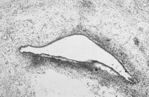
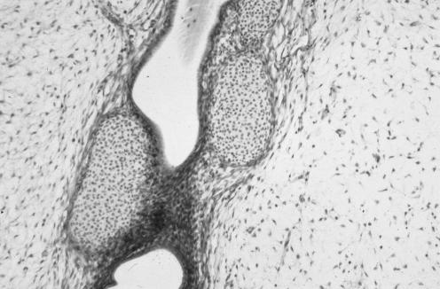
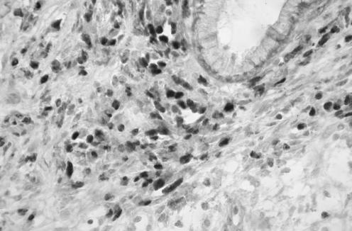
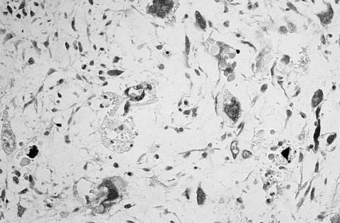
Histological examination of multiple sections showed a polypoid lesion covered by both mucinous glandular and squamous epithelium that was also present in the core of the polyp. No cervical intraepithelial neoplasia or cervical glandular intraepithelial neoplasia was observed. The stroma was oedematous and myxoid, and was largely composed of mildly atypical ovoid to spindle‐shaped cells with hyperchromatic nuclei that condensed subepithelially, creating a cambium layer (fig 1). Many mitotic figures (2–3 in a single high‐power field) were present in the cellular areas around the epithelium. Occasional cells with abundant eosinophilic cytoplasm and cross striations were present, although these were sparse. There were occasional foci of cellular cartilage (fig 2). Focal surface ulceration with inflammatory slough was observed. Immunohistochemically, the stromal cells were positive for desmin and showed nuclear positivity with the skeletal muscle marker myogenin (fig 3). Embryonal rhabdomyosarcoma was diagnosed. An additional unusual feature was the presence of a few small microscopic foci where the tumour cells were extremely large and pleomorphic with multilobated giant nuclei, associated with atypical mitotic figures (fig 4). In these areas, hyaline globules were present. These large tumour cells were positive for desmin.
Figure 1 Epithelial elements embedded in myxoid stroma. The stroma has an increased cellularity around the epithelial elements, forming a cambium layer.
Figure 2 Foci of cellular cartilage are present within the polyp.
Figure 3 Positive immunohistochemical staining with the skeletal muscle marker myogenin.
Figure 4 Foci where tumour cells contain multilobated nuclei with marked atypia and atypical mitotic figures.
Discussion
Embryonal rhabdomyosarcoma of the cervix is rare, most commonly occurring in the late teens and early 20s, and usually presenting as a cervical polyp.1,2,3,4,5,6 Sometimes there are multiple polyps. The neoplasm we describe is in keeping with a sarcoma botryoides, a variant of embryonal rhabdomyosarcoma that has a polypoid appearance and originates below a mucous membrane‐covered surface, forming a cambium layer. The patient in this case was older than most of those previously reported with cervical embryonal rhabdomyosarcoma, although examples have been reported in a 75‐year‐old patient and in three patients in their 40s.2,3,4,5 The corresponding tumour in the vagina usually occurs in infants and young children and, although establishing the diagnosis may be difficult, the pathologist generally keeps this possibility in mind. In an older age group without clinical suspicion of malignancy and in whom cervical polyps are common, a diagnosis of embryonal rhabdomyosarcoma does not immediately come to mind and the combination of epithelial elements, squamous and glandular, in an oedematous stroma may result in misdiagnosis as a benign polyp. The initial clue to the diagnosis is often the gross appearance, in that the polyp may be unusually large and myxoid or there may be multiple polyps. Other clues to the diagnosis include the characteristic cambium layer, with increased cellularity around the epithelial elements, and the associated nuclear atypia and mitotic activity in these areas. Rhabdomyoblasts with cross striations confirm the diagnosis, although they may be sparse and in some cases are not identified. Cartilaginous elements are present in a high percentage of cases and seem much more common in cervical embryonal rhabdomyosarcoma than in this neoplasm in other organs. Positive staining with skeletal muscle antibodies helps in confirming the diagnosis, although some cases are negative and embryonal rhabdomyosarcoma can be diagnosed in the absence of staining with skeletal muscle markers.
As well as a usual endocervical polyp, other diagnoses that may be considered include a prolapsed endometrial polyp, fibroepithelial polyp, endometriosis, leiomyoma, endometrial stromal neoplasm, adenofibroma and adenosarcoma. Adenosarcoma is particularly worthy of mention, as the presence of a polypoid lesion with a cambium layer and associated nuclear atypia and mitotic activity may strongly suggest this diagnosis. Adenosarcoma may contain heterologous elements in the form of rhabdomyoblasts or cartilage.8 Of value in the distinction between the two neoplasms is that adenosarcoma is rare in the age group in which cervical embryonal rhabdomyosarcoma characteristically occurs. It has been suggested that cervical embryonal rhabdomyosarcoma may represent an adenosarcoma with a rhabdomyosarcomatous stroma.1 However, most regard the epithelial elements as being “entrapped”, a viewpoint with which we agree.
The prime reason for reporting this case is the presence of small foci containing highly pleomorphic multilobated tumour nuclei. This is very unusual in embryonal rhabdomyosarcoma in general9 and, to our knowledge, has been described only in a single case in the cervix.7 The cells resemble those found in pleomorphic rhabdomyosarcoma.10 Embryonal rhabdomyosarcoma of the cervix usually has a good prognosis, with an aggressive behaviour in a minority of cases.1 The only known adverse prognostic indicator is deep cervical invasion. The case we describe is recent, and we have no information regarding follow‐up. The importance of the small foci of pleomorphic cells is not clear, and it is not known whether this imparts a more unfavourable prognosis, although in a previous publication from the Intergroup Rhabdomyosarcoma Study,9 the presence of aggregates of such cells was associated with a less favourable outcome. In the previously reported case, it was suggested that the neoplasm should be termed an anaplastic (pleomorphic) subtype of embryonal rhabdomyosarcoma.7 The pleomorphic cells may represent areas of dedifferentiation in an embryonal rhabdomyosarcoma, similar to dedifferentiation in other soft‐tissue sarcomas.
Footnotes
Competing interests: None declared.
References
- 1.Daya D A, Scully R E. Sarcoma botryoides of the uterine cervix in young women: a clinicopathological study of 13 cases. Gynecol Oncol 198829290–304. [DOI] [PubMed] [Google Scholar]
- 2.Miyamoto T, Shiozawa T, Nakamura T.et al Sarcoma botryoides of the uterine cervix in a 46‐year‐old woman: case report and literature review. Int J Gynecol Pathol 20042378–82. [DOI] [PubMed] [Google Scholar]
- 3.Ober W B. Sarcoma botryoides of the cervix uteri: a case report in a 75 year old woman. Mt Sinai J Med 197138363–374. [PubMed] [Google Scholar]
- 4.Brand E, Berek J S, Niebery R K. Rhabdomyosarcoma of the uterine cervix. Sarcoma botryoides. Cancer 1987601552–1560. [DOI] [PubMed] [Google Scholar]
- 5.Vlahos N P, Matthews R, Veridiano N P. Cervical sarcoma botryoides. A case report. J Reprod Med. 199944306–308. [PubMed]
- 6.Williamson H D, McIver F A. Sarcoma botryoides of the cervix. Treated by vaginal hysterectomy and subtotal vaginectomy. Obstet Gynecol 196831690–692. [DOI] [PubMed] [Google Scholar]
- 7.Caruso R A, Napoli P, Villari D.et al Anaplastic (pleomorphic) subtype embryonal rhabdomyosarcoma of the cervix. Arch Gynecol Obstet 2004270278–280. [DOI] [PubMed] [Google Scholar]
- 8.Clement P B, Scully R E. Mullerian adenosarcoma of the uterus: a clinicopathologic analysis of 100 cases with a review of the literature. Hum Pathol 199021363–381. [DOI] [PubMed] [Google Scholar]
- 9.Kodet R, Newton W A, Hamoudi A B.et al Childhood rhabdomyosarcoma with anaplastic (pleomorphic) features: a report of the Intergroup Rhabdomyosarcoma Study. Am J Surg Pathol 199317443–453. [DOI] [PubMed] [Google Scholar]
- 10.McCluggage W G, Lioe T F, McClelland H R.et al Rhabdomyosarcoma of the uterus: report of two cases, including one of the spindle cell variant. Int J Gynecol Cancer 200212128–132. [DOI] [PubMed] [Google Scholar]


