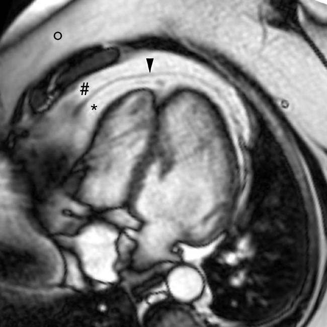
A 54‐year‐old woman with diabetes mellitus II and obesity (body mass index (BMI) 40 kg/m2) was referred to the cardiology outpatient clinic because of reduced exercise tolerance. Cardiac magnetic resonance imaging (MRI) (panel) was performed because the echocardiographic examination was insufficient. Although no structural abnormalities of the heart were found, an impressive amount of epicardial fat was seen.
Epicardial adipose tissue (EAT) is visceral fat deposited between the heart and the pericardium. EAT correlates well with abdominal visceral adipose tissue. Visceral obesity is closely linked to the metabolic syndrome, and appears to be a better predictor of cardiovascular risk than subcutaneous (peripheral) fat, BMI or waist circumference. Even in patients with a normal BMI visceral adipose tissue is a marker of cardiovascular events.
MRI is considered to be the gold standard for fat measurement. The distinction between epicardial and pericardial fat can easily be made on cardiac MRI. EAT can easily be assessed on routine cardiac MRI and may provide important additional information to the clinician.

Magnetic resonance single frame image from a breath‐hold gradient echo horizontal long axis image view of the heart revealing an impressive amount of epicardial fat (*). This is an important indicator of cardiovascular risk. O, subcutaneous fat; #, pericardial fat; ▾, pericardium.


