A 39 year old woman without history of prior cardiac disease presented with typical angina on exertion. Subsequent coronary angiography revealed high grade stenosis of the proximal right coronary artery (RCA). Within the scope of the first‐in‐human clinical trial, the patient was treated by implantation of a novel absorbable metal stent (Biotronik, Bülach, Switzerland). This novel stent consists of a magnesium‐based alloy which provides mechanical properties comparable to conventional stainless steel stents. At the same time, the magnesium alloy allows controlled complete absorption within approximately two months. Thereby, the stent provides temporary vessel scaffolding to prevent elastic recoil of the vessel wall in the first weeks after angioplasty, without remaining in the vessel life long. This allows adaptive vascular remodelling processes in the long term.
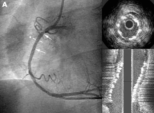
Panel A shows the satisfactory angiographic result after stent implantation without residual stenosis. Complete expansion of the stent is well visualised by intravascular ultrasound (IVUS) cross sectional images as well as in the longitudinal reconstruction.
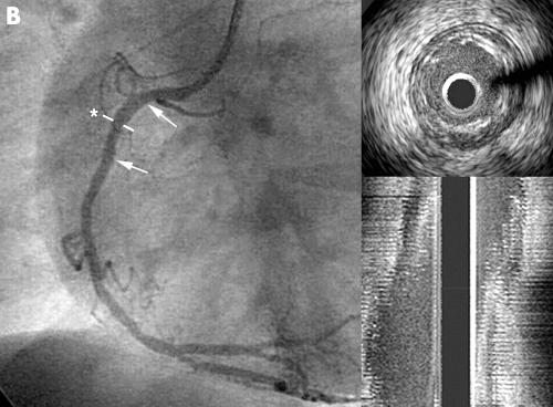
After 18 days the patient presented again with atypical, non‐exercise induced chest pain. The control angiography showed a good result without restenosis in the treated vessel segment (panel B). Interestingly, IVUS showed that the stent was already mostly absorbed in the first three weeks after implantation.
Left panel shows the angiographic result after stent implantation in the proximal part of the right coronary artery without residual stenosis (the position of the stent is indicated by the arrows). The intravascular ultrasound (IVUS) cross sectional image in the right upper panel shows a circular stent expansion with complete apposition of the stent struts to the vessel wall (asterisk indicates the site of the IVUS cross sectional image). The right lower panel shows a longitudinal reconstruction of the IVUS images in the stented segment with complete covering of the stenosis. The well apposed stent struts can be clearly identified in the longitudinal reconstruction.
Left panel shows the angiographic result three weeks after stent implantation without restenosis in the treated segment. The IVUS cross sectional image (upper right panel) and the longitudinal IVUS reconstruction (lower right panel) shows that the stent is mostly dissolved in the first three weeks without plaque progression.


