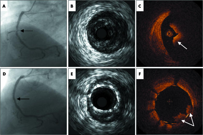
A 66 year old man presented with non‐ST elevation myocardial infarction and underwent early coronary angiography after medical stabilisation. This showed a non‐occlusive thrombus in the mid segment of the right coronary artery (panel A, indicated by broken black arrow). Intravascular ultrasound (IVUS) revealed the presence of a large thrombus at the site of the lesion (panel B) while optical coherence tomography (OCT) imaging (Lightlab Imaging, Inc) clearly showed the eccentric nature of the thrombus (panel C). The lesion was successfully stented with a paclitaxel eluting stent (Taxus), and then post‐dilated. The final result was angiographically excellent with post‐procedural TIMI 3 flow, and with no obvious in‐stent thrombus either on angiography (panel D) or on IVUS (panel E). However, OCT imaging showed that there was still remnant thrombus protruding out from between the stent struts at the site of the original lesion (panel F, double white arrows). OCT imaging utilises near‐infrared light waves to provide pictures of significantly superior resolution and clarity as compared to IVUS. Atheroembolism of thrombus is a major cause of slow or no reflow phenomenon after percutaneous coronary intervention, a complication that is associated with increased morbidity and mortality. These images remind us that thrombus often persists immediately after stenting, despite what may appear to be satisfactory angiographic appearances.
. 2006 Mar;92(3):409. doi: 10.1136/hrt.2005.069252
Optical coherence tomography imaging of thrombus protrusion through stent struts after stenting in acute coronary syndrome
1V Y Lim, L Buellesfeld, E Grube, vytlim@yahoo.com
Keywords: Images in cardiology
Copyright © 2006 BMJ Publishing Group and British Cardiovascular Society
PMCID: PMC1860835 PMID: 16501209


