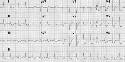
A 54 year old man was admitted to our hospital with recent onset anginal chest pain. His ECG at the time of admission (panel) showed pronounced diffuse ST segment depression in leads V2–V6, I, aVL, II and aVF. It also showed ST segment elevation measuring 2 mm in the lead aVR and measuring 1 mm in lead V1. His creatine phosphokinase MB concentration was 164 IU/dl. A bedside troponin T test was positive. He was regarded as being at high risk of acute coronary syndrome (ACS) and was treated with upstream eptifibatide infusion and an early invasive strategy. Coronary angiogram done subsequently showed 100% occlusion of the left main coronary artery. The patient was subjected to emergency coronary bypass surgery.
This ECG illustrates the features of left main coronary occlusion in the form of ST elevation in lead aVR (2 mm) > ST elevation in lead V1 (1 mm). Lead aVR ST segment elevation greater than the V1 ST segment elevation can predict left main stenosis in patients with ACS, and its early recognition can improve clinical outcomes in these patients. Another notable feature is the diffuse ST depression in nine of the 12 leads, suggesting a very severe circumferential ischaemia and a possible left main coronary occlusion. This ECG illustrates the utility of lead aVR in achieving a diagnosis and planning a treatment strategy for these patients.


