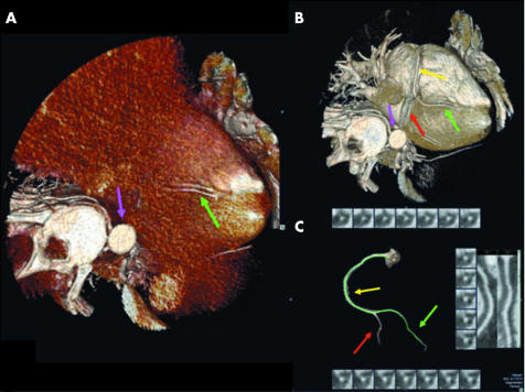
Figure 1 The performance of coronary half millimetre 32 detector row computed tomography angiography (32×0.5‐MDCTA) is shown in three dimensional cardiac volume renderings of the right coronary artery (RCA) (A) before (posterior descending artery, green arrow; descending aorta, purple arrow) and (B) after (RCA, yellow arrow; posterior descending artery, green arrow; posterolateral artery, red arrow; and descending aorta, purple arrow) the appropriate window level setting. (C) The isolated RCA is then traced by the vessel probe, with diagnosis based on visual assessment of true cross sectional and orthogonal curved multiplanar reformatted (CPR) images automatically provided, according to the protocol previously described.13

An official website of the United States government
Here's how you know
Official websites use .gov
A
.gov website belongs to an official
government organization in the United States.
Secure .gov websites use HTTPS
A lock (
) or https:// means you've safely
connected to the .gov website. Share sensitive
information only on official, secure websites.
